
Neuromuscular Fundamentals Anatomy and Physiology of Human Movement



- Slides: 68
Neuromuscular Fundamentals Anatomy and Physiology of Human Movement 420: 050 1
Outline l l l Introduction Structure and Function Muscle Actions Role of Muscles Neural Control 2
Introduction l l Responsible for movement of body and all of its joints Muscles also provide l l l Protection Posture and support Produce a major portion of total body heat Over 600 skeletal muscles comprise approximately 40 to 50% of body weight 215 pairs of skeletal muscles usually work in cooperation with each other to perform opposite actions at the joints which they cross Aggregate muscle action - muscles work in groups rather than independently to achieve a given joint motion 3
Muscle Tissue Properties l l Irritability or Excitability - property of muscle being sensitive or responsive to chemical, electrical, or mechanical stimuli Contractility - ability of muscle to contract & develop tension or internal force against resistance when stimulated Extensibility - ability of muscle to be passively stretched beyond it normal resting length Elasticity - ability of muscle to return to its original length following stretching 4
Outline l l l Introduction Structure and Function Muscle Actions Role of Muscles Neural Control 5
Structure and Function l l l Nervous system structure Muscular system structure Neuromuscular function 6
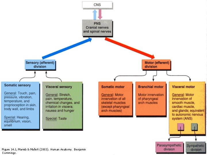
7 Figure 14. 1, Marieb & Mallett (2003). Human Anatomy. Benjamin Cummings.
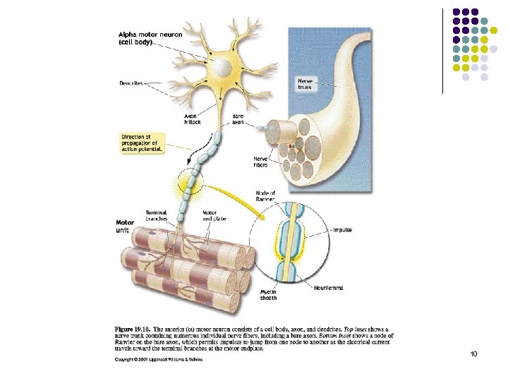
Nervous System Structure l Integration of information from millions of sensory neurons action via motor neurons Figure 12. 1, Marieb & Mallett (2003). Human Anatomy. Benjamin Cummings. 8
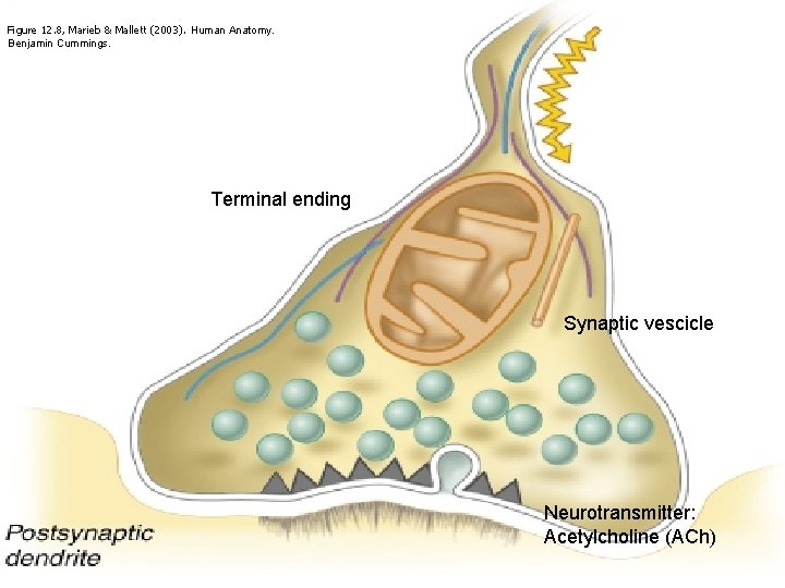
Figure 12. 8, Marieb & Mallett (2003). Human Anatomy. Benjamin Cummings. Terminal ending Synaptic vescicle Neurotransmitter: Acetylcholine (ACh) 9
10
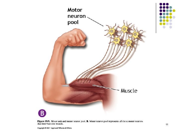
11
Structure and Function l l l Nervous system structure Muscular system structure Neuromuscular function 12
Classification of Muscle Tissue l Three types: 1. Smooth muscle 2. Cardiac muscle 3. Skeletal muscle 13
Skeletal Muscle: Properties l l Extensibility: The ability to lengthen Contractility: The ability to shorten Elasticity: The ability to return to original length Irritability: The ability to receive and respond to stimulus 14
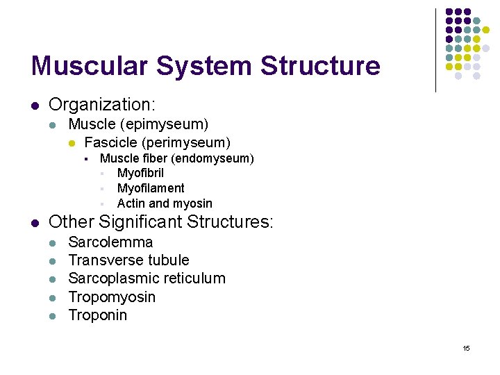
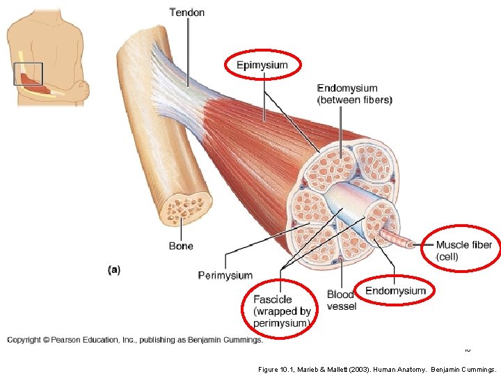
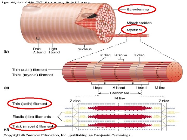
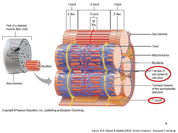
Muscular System Structure l Organization: l Muscle (epimyseum) l Fascicle (perimyseum) § l Muscle fiber (endomyseum) § Myofibril § Myofilament § Actin and myosin Other Significant Structures: l l l Sarcolemma Transverse tubule Sarcoplasmic reticulum Tropomyosin Troponin 15
16 Figure 10. 1, Marieb & Mallett (2003). Human Anatomy. Benjamin Cummings.
Figure 10. 4, Marieb & Mallett (2003). Human Anatomy. Benjamin Cummings. 17
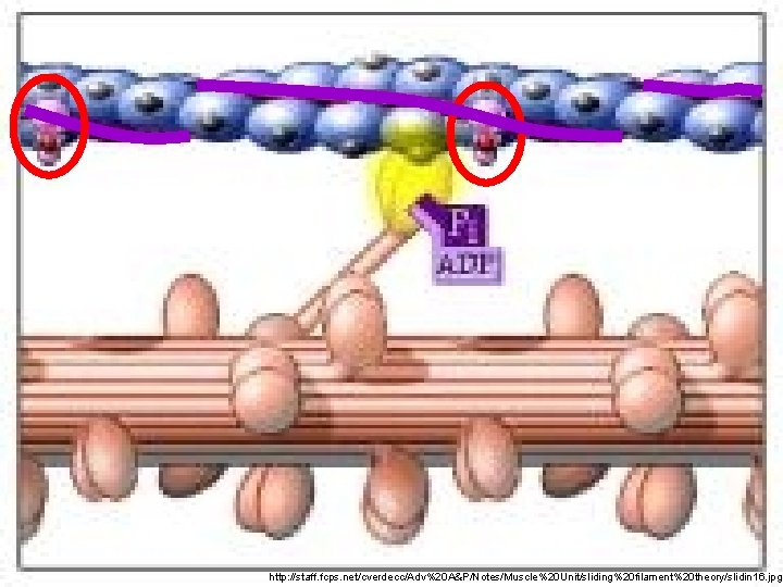
18 http: //staff. fcps. net/cverdecc/Adv%20 A&P/Notes/Muscle%20 Unit/sliding%20 filament%20 theory/slidin 16. jpg
19 Figure 10. 8, Marieb & Mallett (2003). Human Anatomy. Benjamin Cummings.
Structure and Function l l l Nervous system structure Muscular system structure Neuromuscular function 20
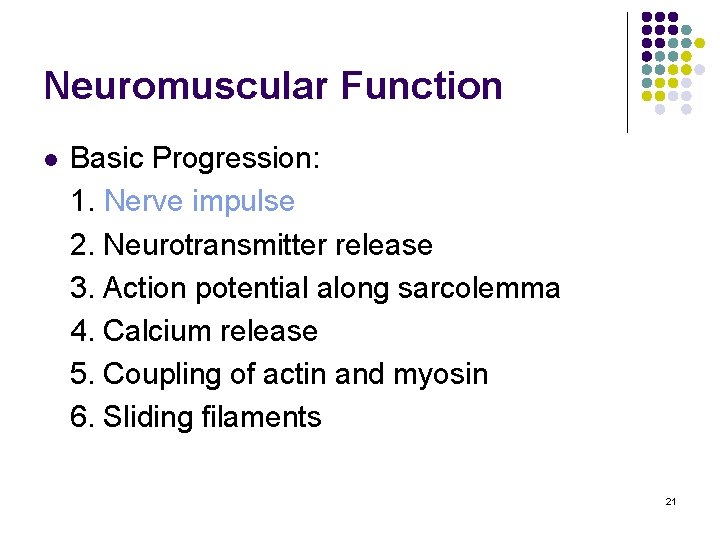
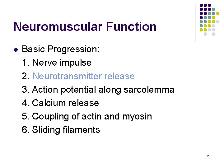
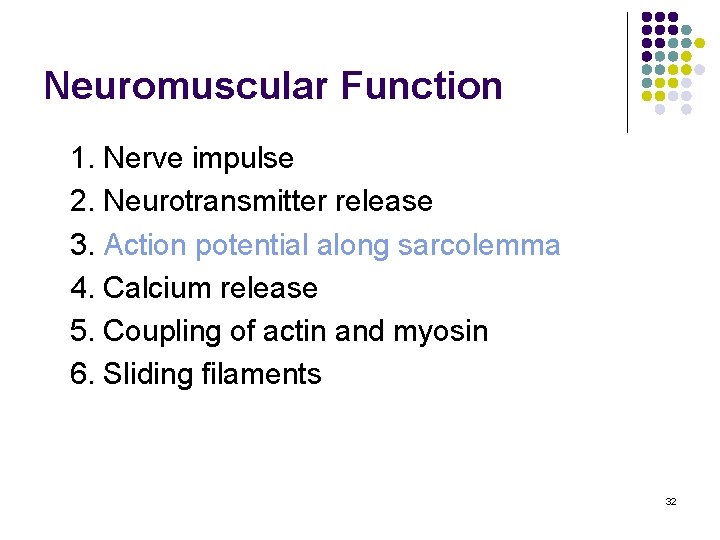
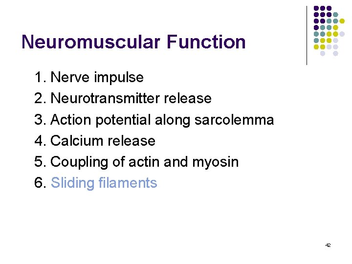
Neuromuscular Function l Basic Progression: 1. Nerve impulse 2. Neurotransmitter release 3. Action potential along sarcolemma 4. Calcium release 5. Coupling of actin and myosin 6. Sliding filaments 21
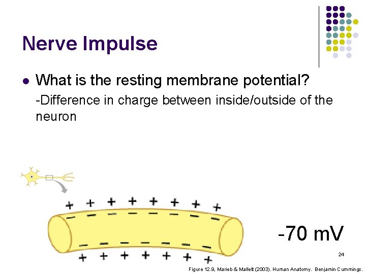
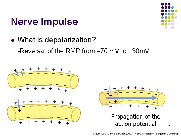
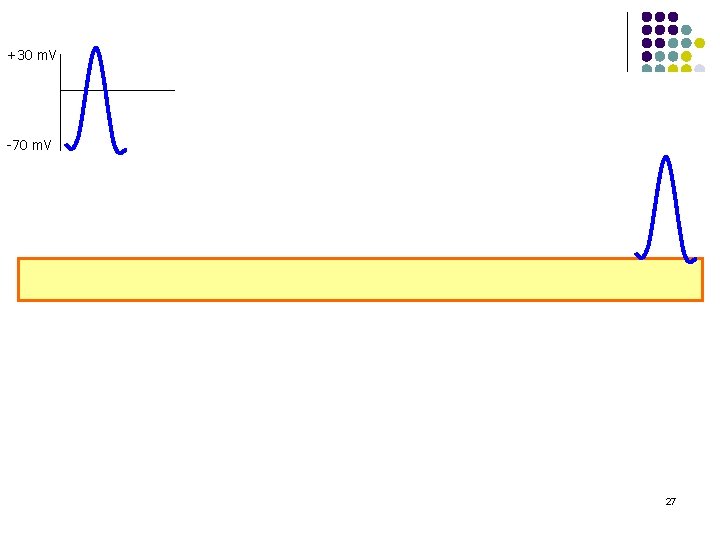
Nerve Impulse l What is a nerve impulse? -Transmitted electrical charge -Excites or inhibits an action -An impulse that travels along an axon is an ACTION POTENTIAL 22
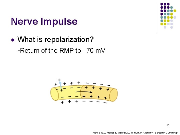
Nerve Impulse l How does a neuron send an impulse? -Adequate stimulus from dendrite -Depolarization of the resting membrane potential -Repolarization of the resting membrane potential -Propagation 23
Nerve Impulse l What is the resting membrane potential? -Difference in charge between inside/outside of the neuron -70 m. V 24 Figure 12. 9, Marieb & Mallett (2003). Human Anatomy. Benjamin Cummings.
Nerve Impulse l What is depolarization? -Reversal of the RMP from – 70 m. V to +30 m. V Propagation of the action potential 25 Figure 12. 9, Marieb & Mallett (2003). Human Anatomy. Benjamin Cummings.
Nerve Impulse l What is repolarization? -Return of the RMP to – 70 m. V 26 Figure 12. 9, Marieb & Mallett (2003). Human Anatomy. Benjamin Cummings.
+30 m. V -70 m. V 27
Neuromuscular Function l Basic Progression: 1. Nerve impulse 2. Neurotransmitter release 3. Action potential along sarcolemma 4. Calcium release 5. Coupling of actin and myosin 6. Sliding filaments 28
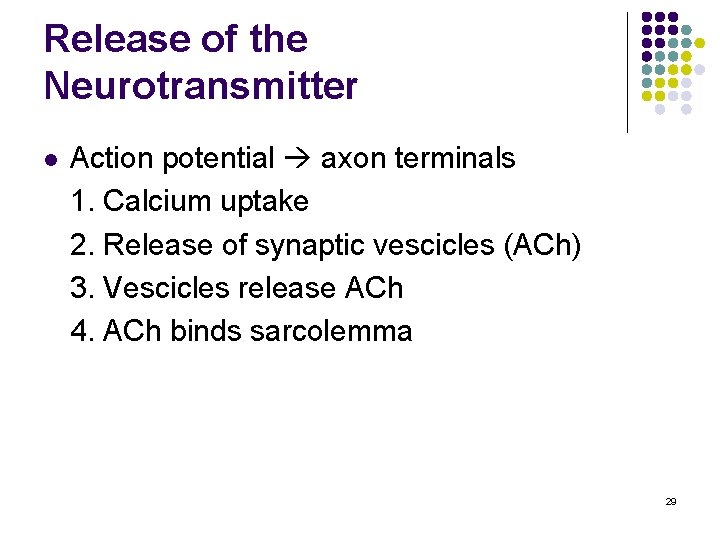
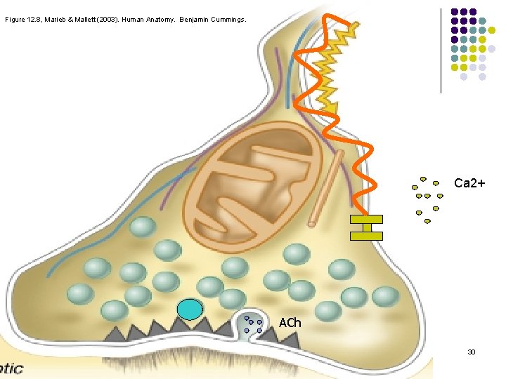
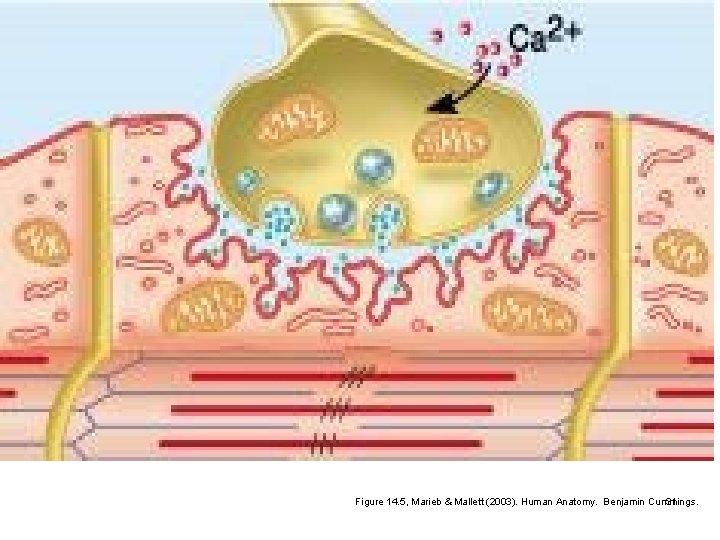
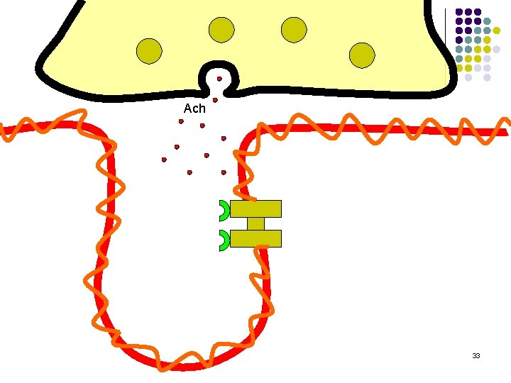
Release of the Neurotransmitter l Action potential axon terminals 1. Calcium uptake 2. Release of synaptic vescicles (ACh) 3. Vescicles release ACh 4. ACh binds sarcolemma 29
Figure 12. 8, Marieb & Mallett (2003). Human Anatomy. Benjamin Cummings. Ca 2+ ACh 30
Figure 14. 5, Marieb & Mallett (2003). Human Anatomy. Benjamin Cummings. 31
Neuromuscular Function 1. Nerve impulse 2. Neurotransmitter release 3. Action potential along sarcolemma 4. Calcium release 5. Coupling of actin and myosin 6. Sliding filaments 32
Ach 33
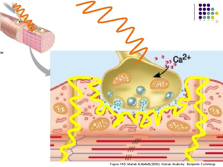
AP Along the Sarcolemma l Action potential Transverse tubules 1. T-tubules carry AP inside 2. AP activates sarcoplasmic reticulum 34
35 Figure 14. 5, Marieb & Mallett (2003). Human Anatomy. Benjamin Cummings.
Neuromuscular Function 1. Nerve impulse 2. Neurotransmitter release 3. Action potential along sarcolemma 4. Calcium release 5. Coupling of actin and myosin 6. Sliding Filaments 36
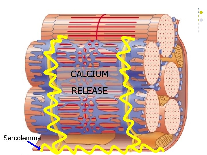
Calcium Release l AP T-tubules Sarcoplasmic reticulum 1. Activation of SR 2. Calcium released into sarcoplasm 37
CALCIUM RELEASE Sarcolemma 38
Neuromuscular Function 1. Nerve impulse 2. Neurotransmitter release 3. Action potential along sarcolemma 4. Calcium release 5. Coupling of actin and myosin 6. Sliding filaments 39
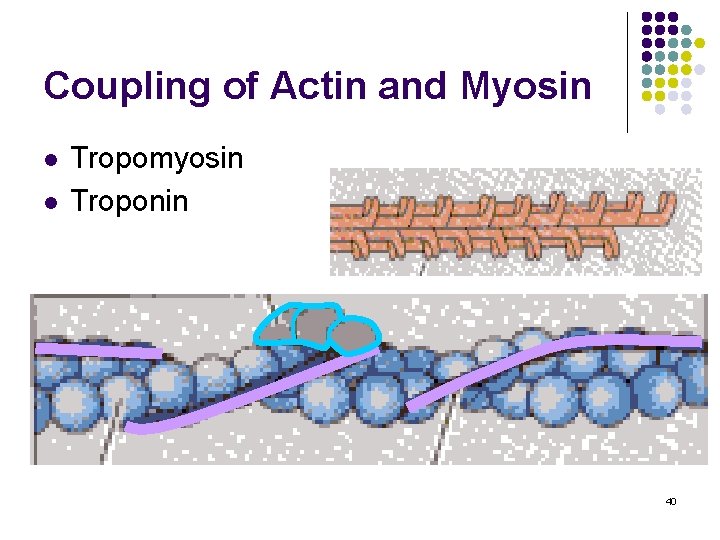
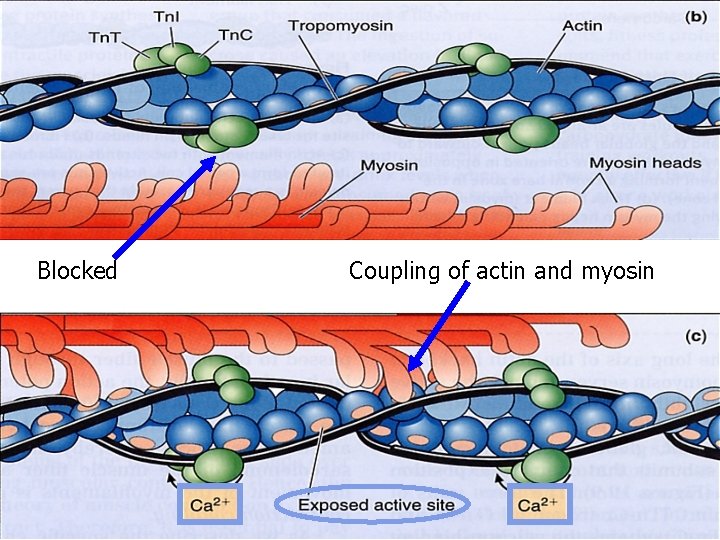
Coupling of Actin and Myosin l l Tropomyosin Troponin 40
Blocked Coupling of actin and myosin 41
Neuromuscular Function 1. Nerve impulse 2. Neurotransmitter release 3. Action potential along sarcolemma 4. Calcium release 5. Coupling of actin and myosin 6. Sliding filaments 42
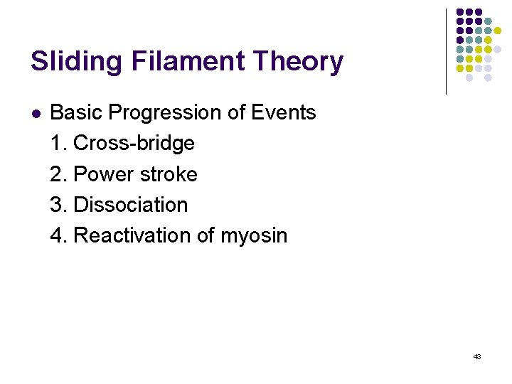
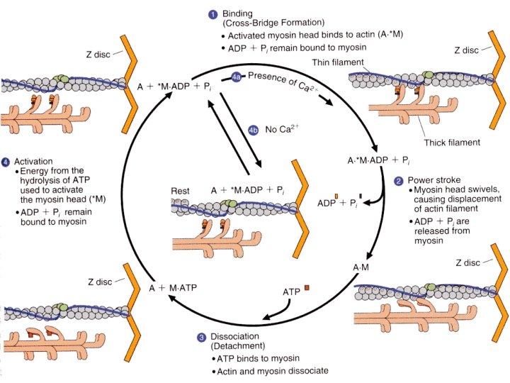
Sliding Filament Theory l Basic Progression of Events 1. Cross-bridge 2. Power stroke 3. Dissociation 4. Reactivation of myosin 43
Cross-Bridge l Activation of myosin via ATP -ATP ADP + Pi + Energy -Activation “cocked” position 44
Power Stroke l l l ADP + Pi are released Configurational change Actin and myosin slide 45
Dissociation l l New ATP binds to myosin Dissociation occurs 46
Reactivation of Myosin Head l ATP ADP + Pi + Energy l l l Reactivates the myosin head Process starts over Process continues until: -Nerve impulse stops -AP stops -Calcium pumped back into SR -Tropomyosin/troponin back to original position 47
48
Outline l l l Introduction Structure and Function Muscle Actions Role of Muscles Neural Control 49
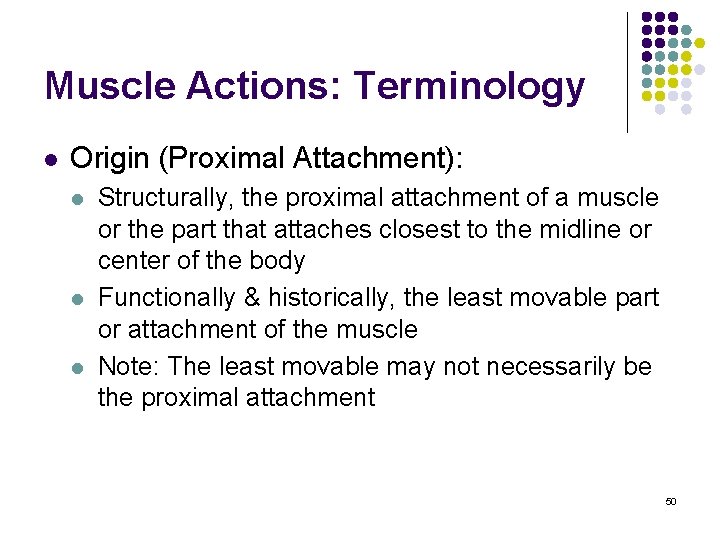
Muscle Actions: Terminology l Origin (Proximal Attachment): l l l Structurally, the proximal attachment of a muscle or the part that attaches closest to the midline or center of the body Functionally & historically, the least movable part or attachment of the muscle Note: The least movable may not necessarily be the proximal attachment 50
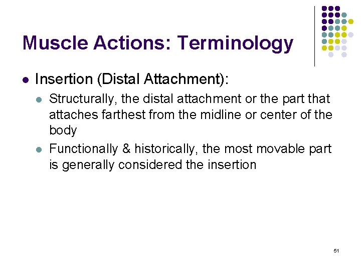
Muscle Actions: Terminology l Insertion (Distal Attachment): l l Structurally, the distal attachment or the part that attaches farthest from the midline or center of the body Functionally & historically, the most movable part is generally considered the insertion 51
Muscle Actions: Terminology l When a particular muscle is activated l l l It tends to pull both ends toward the center Actual movement is towards more stable attachment Examples: l l Bicep curl vs. chin-up Hip extension vs. RDL 52
Muscle Actions l l l Action - when tension is developed in a muscle as a result of a stimulus Muscle “contraction” term is exclusive in nature As a result, it has become increasingly common to refer to the various types of muscle contractions as muscle actions instead 53
Muscle Actions l Muscle actions can be used to cause, control, or prevent joint movement or l l l To initiate or accelerate movement of a body segment To slow down or decelerate movement of a body segment To prevent movement of a body segment by external forces 54
Types of Muscle Actions l Muscle action (under tension) l l Isometric Isotonic l l Concentric Eccentric 55
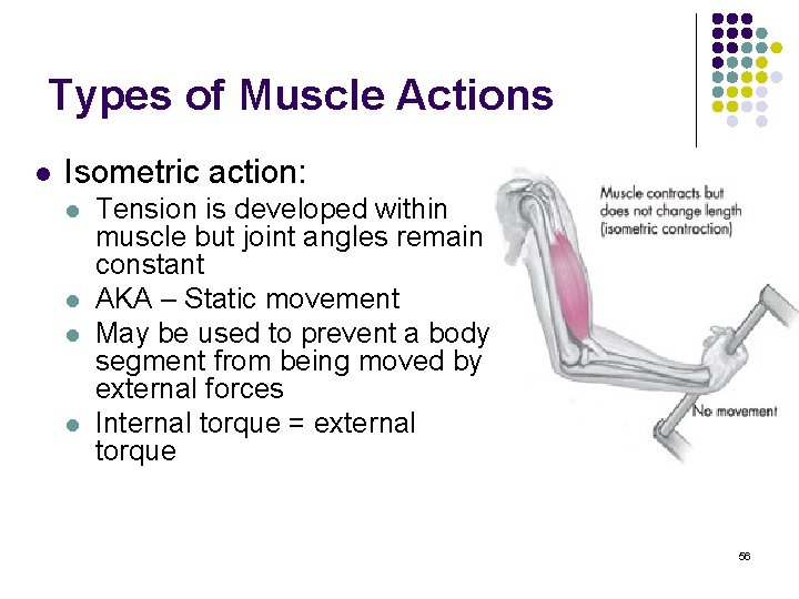
Types of Muscle Actions l Isometric action: l l Tension is developed within muscle but joint angles remain constant AKA – Static movement May be used to prevent a body segment from being moved by external forces Internal torque = external torque 56
Types of Muscle Actions l Isotonic (same tension) contractions involve muscle developing tension to either cause or control joint movement l l AKA – Dynamic movement Isotonic contractions are either concentric (shortening) or eccentric (lengthening) 57
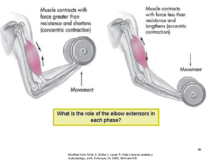
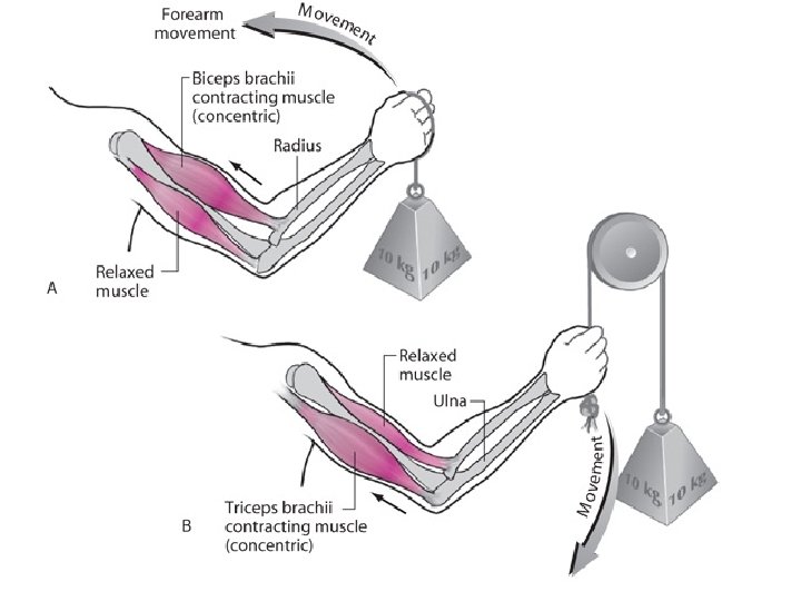
Types of Muscle Actions l Concentric contractions involve muscle developing tension as it shortens l l Internal torque > external torque Causes movement against gravity or other resistance Described as being a positive action Eccentric contractions involve the muscle lengthening under tension l l l External torque > internal torque Controls movement caused by gravity or other resistance Described as being a negative action 58
What is the role of the elbow extensors in each phase? 59 Modified from Shier D, Butler J, Lewis R: Hole’s human anatomy & physiology, ed 9, Dubuque, IA, 2002, Mc. Graw-Hill
Types of Muscle Actions l Movement may occur at any given joint without any muscle contraction whatsoever l l referred to as passive solely due to external forces such as those applied by another person, object, or resistance or the force of gravity in the presence of muscle relaxation 60
Outline l l l Introduction Structure and Function Muscle Actions Role of Muscles Neural Control 61
Role of Muscles l Agonist muscles l l The activated muscle group during concentric or eccentric phases of movement Known as primary or prime movers, or muscles most involved 62
Role of Muscles l Antagonist muscles l l Located on opposite side of joint from agonist Have the opposite concentric action Also known as contralateral muscles Work in cooperation with agonist muscles by relaxing & allowing movement l Reciprocal Inhibition 63
64
Role of Muscles l Stabilizers l l l Surround joint or body part Contract to fixate or stabilize the area to enable another limb or body segment to exert force & move Also known as fixators 65
Role of Muscles l Synergist l l Assist in action of agonists Not necessarily prime movers for the action Also known as guiding muscles Assist in refined movement & rule out undesired motions 66
Role of Muscles l Neutralizers l l Counteract or neutralize the action of another muscle to prevent undesirable movements such as inappropriate muscle substitutions Activation to resist specific actions of other muscles 67
Muscle Fiber Characteristics l Three basic types: 1. Type I: -Slow twitch, oxidative, red 2. Type IIb: -Fast twitch, glycolytic, white 3. Type IIa: -FOG 68