NEUROLOGICAL EXAMINATION OF SPINAL CORD INJURY Dr Osama


- Slides: 29
NEUROLOGICAL EXAMINATION OF SPINAL CORD INJURY Dr. Osama Neyaz Assistant Professor Department Of PMR
ANATOMY OF SPINE § 7 cervical vertebrae § 12 thoracic vertebrae § 5 lumbar vertebrae § 5 fused sacral vertebrae § 3 -4 small bones comprising the coccyx q Spinal cord ends as conus medullaris at level of first lumbar vertebra lumbar and sacral nerve roots exit below this and form the cauda equina
ANATOMY OF SPINE
ANATOMY OF SPINE
NEUROANATOMY 1&2 Posterior Columns: convey Ipsilateral information about two Point discrimination, proprioception And vibratory sense 5 Lateral Spinothalamic Tract: carries Pain and Temperature Information From contralateral extremity 4 Lateral Corticospinal Tract: Carries Motor Information from Contralateral Brain to Ipsilateral Extremity
MECHANISMS OF INJURY § Compression § Flexion Injury § Extension Injury § Rotation
COMPRESSION INJURY § Vertebral body fracture § Disc herniation § Epidural hematoma § Displacement of posterior wall of the vertebral body
JEFFERSON FRACTURE § A comminuted fracture of the ring of C 1. § Compression of base of skull against C 1 § Results in cracking the ring of C 1 § Best seen on open mouth x-ray
ATLANTOAXIAL AND DENS FRACTURES § The result of hyperflexion or hyperextension injuries 8% of Dens Fractures associated with C 1 fractures C 2 Fractures Dens Fracture : § Hyperflexion Injury Hangman Fracture : § Hyperextension Injury § Traumatic spondylolisthesis of the axis § Bilateral fractures through the pars interarticularis of the axis
FLEXION TEARDROP FRACTURE Hyperflexion of the subaxial cervical spine Retropulsion of the larger portion of a vertebral body into the spinal canal, detached from an anterior fragment (teardrop) Often associated with an anterior cord syndrome.
CLAY-SHOVELER’S FRACTURE § Avulsion fracture of the spinous process of C 6, C 7, or T 1. § It is not typically associated with neurologic injury.
THORACOLUMBAR TRAUMA q Mechanism of injury § Compression § Distraction § Rotation
CHANCE FRACTURE § Failure of all three columns due to flexion-distraction § Falls from a height § Strikes part of the torso on an immovable object § Injury pattern most likely to cause SCI
THE THREE-COLUMN CONCEPT OF SPINAL ANATOMY § The anterior column: ALL + anterior portion of the vertebral body + anterior portion of the disk. § The middle column: posterior portion of the vertebral body + the posterior portion of the disk + PLL § The posterior column: the pedicles facet joints + laminae + supraspinous ligament, interspinous ligament + facet joint capsule + ligamentum flavum.
STABLE Vs UNSTABLE FRACTURE § When the integrity of the middle and either the anterior or the posterior column is affected, the spine is likely to be unstable. q. The columns can be affected by: § Fracture § Ligamentous disruption § Gunshot wounds Because of the nature of the injury, can affect more than one column and the spine can still remain stable. SCI can occur without obvious radiographic findings.
CLINICAL SYNDROMES AFTER INCOMPLETE SPINAL CORD INJURY § Central Cord Syndrome § Brown-Sequard Syndrome § Anterior Cord Syndrome § Conus Medullaris Syndrome § Cauda Equina Syndrome
CENTRAL CORD SYNDROME § Motor>Sensory Loss § Upper>Lower Extremity Loss § Distal >Proximal Muscle Weakness § Classically occurs with hyperextension injuries of the cervical spine
BROWN-SEQUARD LESION q A burst fracture with posterior displacement of bone fragments compresses one side of the spinal cord. § Loss of Ipsilateral Proprioception, Light Touch and Motor Function § Loss of Contralateral Pain and Temperature Sensation § Due to hemisection of the cord due to penetrating injury § Incomplete lesions most common
ANTERIOR CORD SYNDROME q A large disk herniation compresses the anterior aspect of the spinal cord, leaving the dorsal columns intact. § Loss of Motor function, Pain and Temperature Sensation § Preservation of Light touch, Vibratory Sensation and Proprioception
CONUS MEDULLARIS SYNDROME A burst fracture of with posterior displacement of bone fragments compresses the conus medullaris. Injury to sacral cord, lumbar nerve roots causing § Areflexic bladder § Loss of control of bowels § Knee jerk relexes preserved, ankle jerk absent § Signs similar to cauda equina syndrome except more likely to be bilateral
CAUDA EQUINA SYNDROME q A central disk herniation at L 4 -L 5 level compresses the cauda equina. § Injury to nerve roots and not spinal cord itself § Muscle weakness and decreased sensation in affected dermatomes § Decreased bowel and bladder control
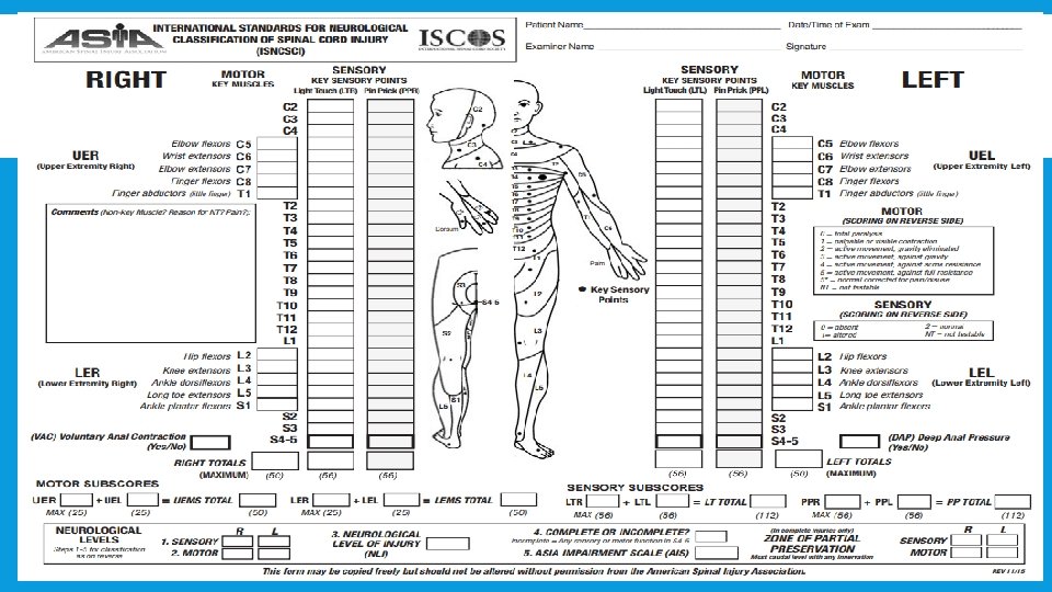
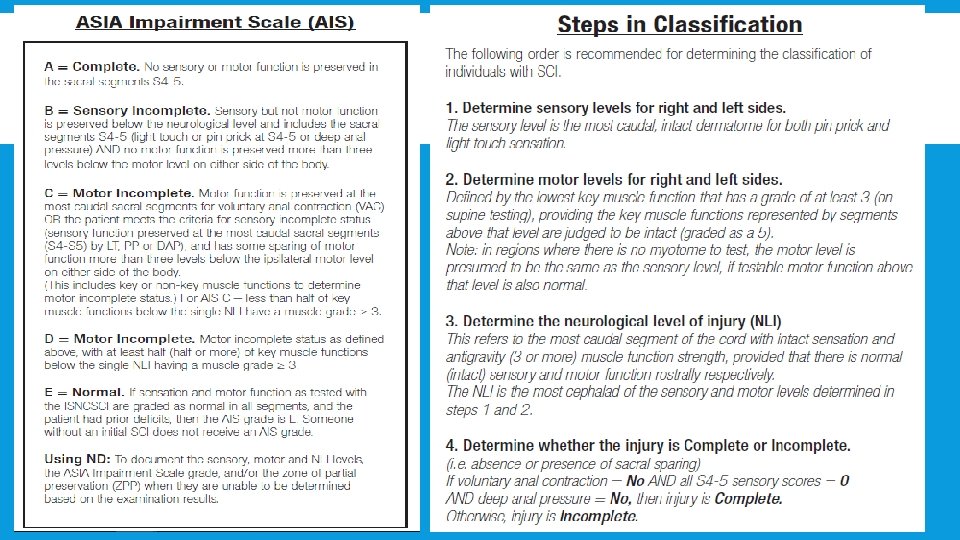
CLASSIFICATION OF SPINAL CORD INJURY Patients are classified according to the ASIA Impairment Scale (AIS) q Combined efforts from v. ASIA: American Spinal Injury Association v. ISCOS: International Spinal Cord Society
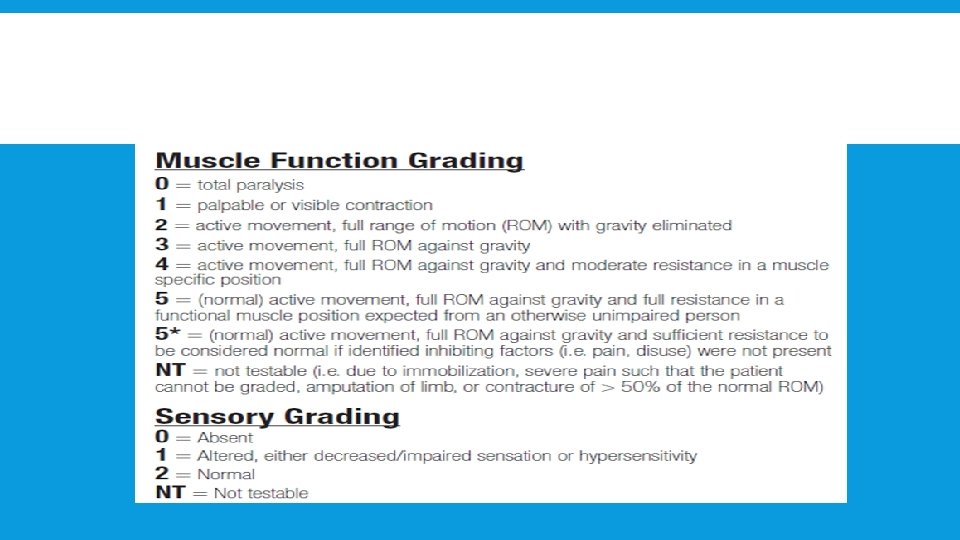
COMPONENTS OF THE TEST Three Main Parts to the Exam: § Strength Testing § Light Touch Sensation § Pinprick Sensation Lowest Level of motor control: § Voluntary Anal Contraction § Lowest Level of Sensation: § Deep Anal Pressure
NEUROLOGIC EXAM: DERMATOMES § C 5 - Deltoid § T 12 – Symphysis Pubis § C 6 – Thumb § L 4 – Medial aspect of leg § C 7 – Middle Finger § L 5 - Space between first and second toes § C 8 - Little Finger § T 4 – Nipple § T 8 – Xiphoid § T 10 - Umbilicus § S 1 – Lateral border of the foot § S 3 – Ischial Tuberosity § S 4 -5 – Perianal region
MYOTOMES § C 5 – Deltoid § L 2 - Hip flexion § C 6 – Wrist Extensors § L 3 - Knee Extension § C 7 – Elbow Extensor § L 4 - Ankle dorsiflexion § C 8 – Finger flexors § L 5 - Toe extension § T 1 – Little finger abduction § S 1 – Plantar flexion
Thank You