
NEUROLOGICAL EMERGENCIES Burcu Ugurel MD Phase III Department
NEUROLOGICAL EMERGENCIES Burcu Ugurel, MD Phase III Department of Neurology
NEUROLOGIC EMERGENCY OUTLINE • • • Change in Mental Status / Coma Stroke/TIA Syndromes Seizure & Status Epilepticus Head Trauma Infectious Vertigo/Headaches Peripheral Neuropathies Myastenia Acute myelopathies

THE NEUROLOGIC EXAM • Must do a complete thorough neuro exam to properly • identify and diagnose any neurologic abnormality. Exam should include 5 parts: – – – Mental status, level of alertness (GCS) Cranial nerve exam Motor / Sensory exam Reflexes Cerebellar Consider ; MMSE if Psych components
CHANGE IN MENTAL STATUS/COMA • Temperature – Hypothermia: causes coma when Temp<32. 0 C – Hyperthermia: causes coma when Temp>42. 0 C • Infection – Meningitis, Encephalitis, Sepsis • Endo/Exocrine, Electrolyte – – Hypo/Hyperglycemia Hypo/hyperthyroidism Hypo/hypernatremia Hepatic encephalopathy • Opiods/ OD / Alcohol – Heroin, Psych Meds (TCA’s, SSRI’s)

MENTAL STATUS/COMA PHYSICAL EXAM • Always attempt to get a complete history!! • LOOK at your patient! – Smell the breath (ketones, alcohol, fetid) – Observe respiratory rate & patterns (Cheyne-Stokes) – Look for abnormal posturing. • Decorticate (Flexion of UE with Extension of LE) • Decerebrate (Extension of all Ext. ) – Look for needle marks, cyanosis, signs of trauma • Obtain GCS Score! E 4 V 5 M 6 – If less than 8, IMMEDIATE airway stabilization FIRST priority!!

GLASGOW COMA SCALE • Scores range from 3 (Worst) – 15 (Best) • Important for classifying degree of alteration • (Head Trauma) • GCS < 8 = INTUBATE!!

GLASGOW COMA SCALE • Motor – – 6 = Obeys commands 5 = Localizes pain 4 = Withdraws to pain 3 = Decorticate (flexes to pain) – 2 = Decerebrate (extends to pain) – 1 = None • EYE Opening Response – – 4 = Spontaneous 3 = To Voice 2 = To Pain 1 = None • Verbal Response – – – 5 = Oriented and converses 4 = Confused but converses 3 = Inappropriate words 2 = Inappropriate sounds 1= None
ISCHEMIC STROKE SYNDROMES • Middle Cerebral Artery Occlusion (MCA) • Contralateral hemiplegia, hemianesthesia, and • • • homonymous hemianopsia Upper extremity deficit > Lower extremity Aphasia (if dominant hemisphere involved) Conjugate gaze impaired in the direction of the lesion
ISCHEMIC STROKE SYNDROMES • Anterior Cerebral Artery Occlusion (ACA) – – – Contralateral leg, arm, paralysis Lower Extremity deficit > Upper extremity Loss of frontal lobe control • Incontinence • Primitive grasp and suck reflexes enacted • Posterior Cerebral Artery Occlusion (PCA) – Ipsilateral CN III palsy, visual loss – Contralateral hemiparesis and hemisensory loss – Memory loss
ISCHEMIC STROKE SYNDROMES • Vertebrobasilar Artery Occlusion (VBA) • • Hallmark: Crossed Neurological Deficits Contralateral hemiplegia Ipsilateral CN palsy Cerebellar findings - Nausea/Vomiting - Vertigo, Nystagmus, - Ataxia, Dysarthia - Tinnitus, deafness CN and Cerebellar deficits that affect BOTH sides of the body
TIA (TRANSIENT ISCHEMIC ATTACKS) • A temporary loss of neurologic function, that resolves • • completely <24 h Main point: These patients at high risk for stroke if: – >50 – HT, DM, Smoker, Prior TIA in last month Treat as CVA : Head CT (CVA protocol) ASA 81 -325 mg po If cardiac arrythmia (atrial filbrillation) present, consider Heparin ONLY after Head CT and Neuro consultation
HEMORRHAGIC STROKE SYNDROMES • Hemorrhagic Strokes • Spontaneous rupture of berry aneurysm or AV malformation (Subarachnoid hemorrhage). secondary to: • Hypertension • Congenital abnormality • Blood dyscrasia / Anticoagulant usage • Infection • Neoplasm • Trauma (Epidural / Subdural Hematomas) • Hemorrhagic transformation of embolic stroke
HEMORRHAGIC STROKE SYNDROMES • Intracerebral – Hypertensive intracerebral hemorrhage MOST common cause – Traumatic, contusion, coup/contracoup – Rupture of small blood vessels with bleeding inside the brain parenchyma • Putamen • Cerebellar • Thalamus • Pontine ( 3 P’s – pinpoint pontine pupils)
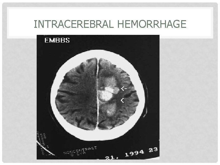
INTRACEREBRAL HEMORRHAGE
TREATMENT OF STROKE • AS ALWAYS – ABC’s FIRST • What’s the Serum Glucose? ? – Consider Thiamine 100 mg IV, D 50 bolus if hypoglycemic. – Treat Hyperglycemia if Serum Glucose > 300 mg/dl • Protect the “Penumbra” – Keep SBP >90 mm Hg – Goal keep CPP > 60 mm Hg (CPP=MAP-ICP) – Treat Fever ( Mild Hypothermia beneficial) • Acetaminophen 650 mg po or pr, cooling blanket – Oxygenate (Keep Sao 2 >95%) – Elevate head of bed 30 deg. (Clear c-spine) • Frequent repeat Neuro checks!! Reassess GCS!
THROMBOLYTIC THERAPY FOR ACUTE ISCHEMIC STROKE CHECKLIST • Answer to ALL must be YES: – Age 18 or older – Clinical diagnosis of Acute Ischemic Stroke causing a measurable NON improving neurologic deficit. – NO high clinical suspicion for SAH – Time of onset to treatment is <180 minutes.

THROMBOLYTIC THERAPY FOR ACUTE ISCHEMIC STROKE CHECKLIST • Answer to ALL MUST be NO: – Evidence of hemorrhage on CT – Active internal bleeding (GI/GU) within last 21 days. – Known bleeding diasthesis: • Platelets<100, 000 • Heparin within last 48 hours with elevated PTT • Warfarin use with PT > 15 seconds – Within 3 months of IC injury, prior surgery or prior ischemic stroke. – Within 14 days of serious trauma, major surgery – Recent AMI, arterial puncture/LP within 7 days – History of prior ICH, AVM, tumor, or aneurysm or seizure at stroke – Systolic BP >185 mm. Hg, or Diastolic BP >110 Hg
SEIZURES & STATUS EPILEPTICUS • Background: – 1 – 2% of the general population has seizures – Primary • Idiopathic epilepsy: onset ages 10 -20 – Secondary • Precipitated by one of the following: • Intracranial pathology – Trauma, Mass, Abscess, Infarct • Extracranial Pathology – Toxic, metabolic, hypertensive, eclampsia
APPROACH FOR 1 ST SEIZURE, NEW SEIZURE, OR SUBSTANCE/ TRAUMA INDUCED SEIZURE • • As always ABC’s First IV, O 2, Monitor – – – Send blood for CBC Anticonvulsant levels Prolactin levels / Lactate level • Head CT • Is patient actively seizing? Post ictal? Pseudoseizure? – Consider treatment options • Complete History and Physical Exam – Including detailed Neuro Exam – Repeat Neuro evaluations a must!
APPROACH TO BREAKTHROUGH SEIZURE • History, History!! • Main causes of Breakthrough Seizure: – Noncompliance with anticonvulsant regimen – Start of new medication (level alteration) • Antibiotics, OCP’s – Infection • Fever – Changes in body habitus, eating patterns – Supratherapeutic level
STATUS EPILEPTICUS • Seizure lasting greater than 5 minutes OR two • seizures between which there is incomplete recovery of consciousness Treatment algorhythm: – As before ABC’s – IV, O 2, Monitor – Consider ALL potential causes • INH (pyridoxime/B-6 deficiency) • Eclampsia • Alcoholic (thiamine/B-1 deficiency) • Other Tox ingestion (TCA’s, sulfonylurea OD) • Trauma
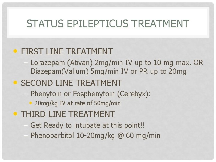
STATUS EPILEPTICUS TREATMENT • FIRST LINE TREATMENT – Lorazepam (Ativan) 2 mg/min IV up to 10 mg max. OR Diazepam(Valium) 5 mg/min IV or PR up to 20 mg • SECOND LINE TREATMENT – Phenytoin or Fosphenytoin (Cerebyx): • 20 mg/kg IV at rate of 50 mg/min • THIRD LINE TREATMENT – Get Ready to intubate at this point!! – Phenobarbitol 10 -20 mg/kg @ 60 mg/min
STATUS EPILEPTICUS ADJUNCTIVE TREATMENT BY HISTORY • Thiamine 100 mg IV, 1 -2 amps D 50 – If suspect alcoholic, malnourished, hypoglycemia • Magnesium Sulfate 20 cc of 10% solution – As above of if eclampsia (BP does NOT have to be 200/120!!) • Pyridoxine 5 gms IV – INH or B-6 deficiency
CLOSED HEAD INJURY • Concussion: refers to a transient LOC following head injury. Often associated with retrograde amnesia that also improves – “Coup” = injury beneath the site of trauma – “Countrecoup” = injury to the side polar opposite to the traumatized area Diffuse Axonal Injury : tearing and shearing of nerve fibers at the time of impact secondary to rapid acceleration/deceleration forces. Causes prolonged coma, injury, with normal initial head CT and poor outcome
CLOSED HEAD INJURIES WITH HEMORRHAGE • Cerebral Contusion – – Focal hemorrhage and edema under the site of impact Susceptible areas are those in which the gyri are in close contact with the skull • Frontal lobe • Temporal lobes – Diagnostic Test of Choice: Head CT – Treatment: Supportive with measures to keep ICP normal. Repeat Neuro checks. Repeat Head Ct in 24 hours. Good prognosis.
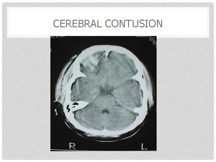
CEREBRAL CONTUSION
MANAGEMENT OF CLOSED HEAD INJURIES • As always ABC’s with C-Spine precautions • IV, O 2, Monitor • Stabilize and resuscitate – – – Sao 2>95% SBP>90 Treat Fever • Head of Bed 30% (once C-Spine cleared) • Stat Head CT with Stat Neurosurgical evaluation • for surgical lesions. Repeat Exams, looking for signs of herniation

SIGNS OF HERNIATION / INCREASED ICP • • Headache, nausea, vomiting Decreasing LOC Sixth nerve paresis (one or both eyes adducted) Decreased respiratory rate Cushing reflex (hypertension/bradycardia/bradynpea) Papilledema Development of signs of herniation – Fixed and dilated pupil – Contralateral hemiparesis – Posturing
HERNIATION SYNDROMES • CPP = MAP – ICP: Must keep CPP >60 mm Hg • Uncal Herniation: – Occurs when unilateral mass pushes the uncus (temporal lobe) through the tentorial incisa, presenting as: • Ipsilateral pupil dilatation • Contralateral hemiparesis • Deepening coma • Decorticate posturing • Apnea and death
HERNIATION SYNDROMES • Cerebellar Herniation – Downward displacement of cerebellar tonsils through the foramen magnum. – Presents as : • Medullary compression • Pinpoint pupils • Flaccid quadriplegia • Apnea and circulatory collapse
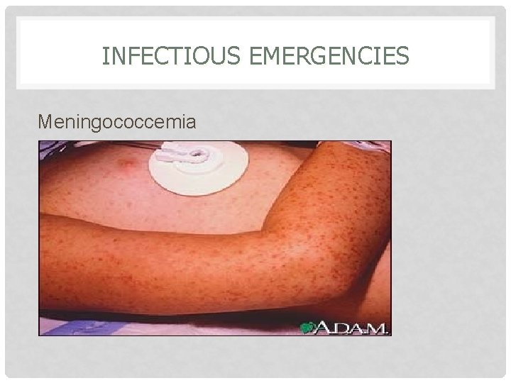
INFECTIOUS EMERGENCIES Meningococcemia
INFECTIOUS NEUROLOGIC EMERGENCIES • Meningitis: inflammation of the meninges • History: – Acute Bacterial Meningitis: • Rapid onset of symptoms <24 hours – Fever, Headache, Photophobia – Stiff neck, Confusion • Etiology By Age: – 0 -4 weeks: E. Coli, Group B Strep, Listeria – 4 -12 weeks: neotatal pathogens, S. pneumo, N. meningitides, H. flu – 3 mos – 18 years: S. pneumo, N. menin. , H. flu – >50/ alcholics: S. pneumo, Listeria, N. menin. , Gram(-) bacilli
MENINGITIS • Physical Exam Pearls – Infants and the elderly lack the usual signs and symptoms, only clue may be AMS. – Look for papilledema, focal neurologic signs, ophthalmoplegia and rashes – As always full exam • Checking for above • Brudzinski’s sign • Kernigs sign – KEY POINT: If you suspect meningococcemia do NOT delay antibiotic therapy, MUST start within 20 minutes of arrival!!!!!
MENINGITIS • Emergent CT Prior to LP – – – Those with profoundly depressed MS Seizure Head Injury Focal Neurologic signs Immunocompromised with CD 4 count <500 • DO NOT DELAY ANTIBIOTIC THERAPY!!
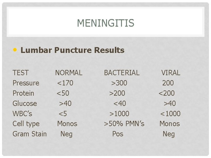
MENINGITIS • Lumbar Puncture Results TEST Pressure Protein Glucose WBC’s Cell type Gram Stain NORMAL <170 <50 >40 <5 Monos Neg BACTERIAL >300 >200 <40 >1000 >50% PMN’s Pos VIRAL 200 <200 >40 <1000 Monos Neg
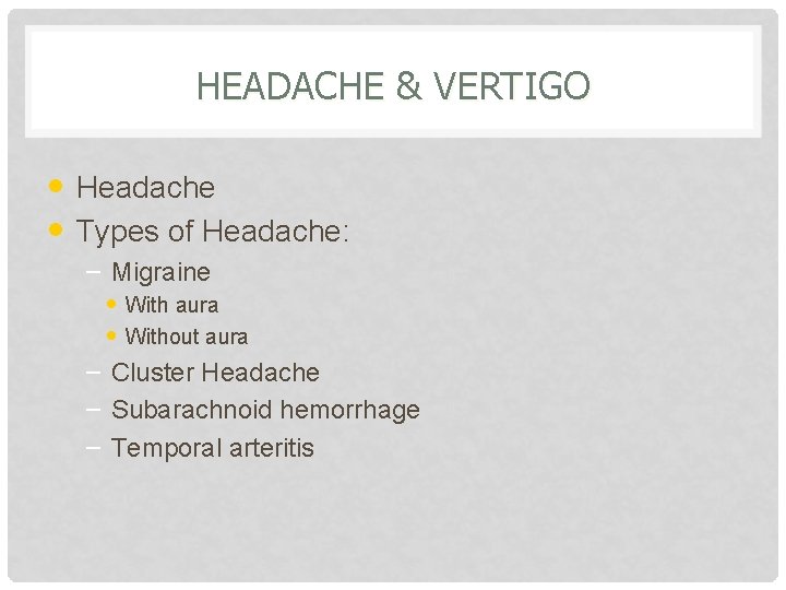
HEADACHE & VERTIGO • Headache • Types of Headache: – Migraine • With aura • Without aura – Cluster Headache – Subarachnoid hemorrhage – Temporal arteritis
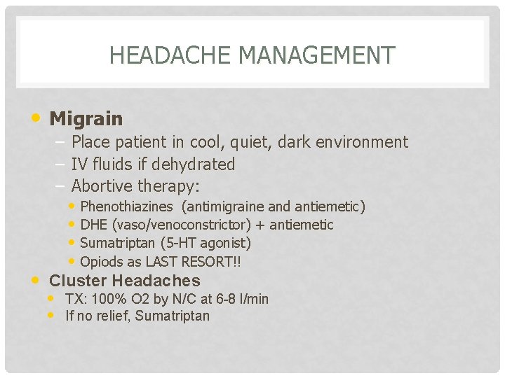
HEADACHE MANAGEMENT • Migrain – – – • Place patient in cool, quiet, dark environment IV fluids if dehydrated Abortive therapy: • Phenothiazines (antimigraine and antiemetic) • DHE (vaso/venoconstrictor) + antiemetic • Sumatriptan (5 -HT agonist) • Opiods as LAST RESORT!! Cluster Headaches • TX: 100% O 2 by N/C at 6 -8 l/min • If no relief, Sumatriptan
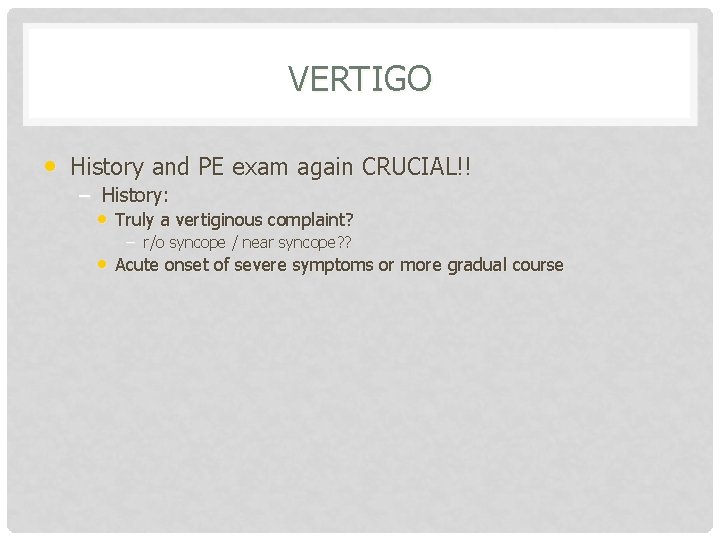
VERTIGO • History and PE exam again CRUCIAL!! – History: • Truly a vertiginous complaint? – r/o syncope / near syncope? ? • Acute onset of severe symptoms or more gradual course
VERTIGO Peripheral Vertigo • History: – Acute onset of severe dizziness, nausea, vomiting. – May be a positional worsening of symptoms – Recent history of URI or similar episodes in past which resolved. • PE Pearls: • Horizontal nystagmus which fatigues • Normal Neuro exam with normal cerebellar function and • gait Reproduction of symptoms with Hallpike maneuver
VERTIGO Central Vertigo • Due to lesions of brainstem or cerebellum • 10 – 15% of cases • Signs & Symptoms: – – Gradual onset of mild disequilibrium Mild nausea and vomiting Nonfatigable nystagmus (any direction) Associated neurological abnormalities: • Ptosis • Facial palsy, dysarthria • Cerebellar findings, ataxia
EMERGENT PERIPHERAL NEUROPATHIES • Acute Toxic Neuropathies – Diptheria (Cornybacterium diptheriae) • Acutely ill patient with fever • Membranous pharyngitis that bleeds • Powerful exotoxin produces widespread organ damage – – – Myocarditis/AV Block, Nephritis, Hepatitis Neuritis with bulbar and peripheral paralysis (ptosis, strabismus, loss of DTR’s) • TX: Parenteral PCN or Erythromycin – Horse Serum antitoxin – Respiratory isolation and admission the rule
EMERGENT PERIPHERAL NEUROPATHIES – Botulism (Clostridium botulinum toxin) • Earliest finding(90%)= Blurred vision, diplopia, ophthalmoplegia, ptosis • Neurologic abnormalities descend and will lastly involve the respiratory musculature and death with 6 hours if not treated! • Mentation and sensation are normal • Remember in infants with FTT (failure to thrive) – Raw honey contains C. botulinum • Tx: Aggressive airway stabilization! • Trivalent serum antitoxin • Lastly, there have been some recently reported cases of hypersensitivity to “Bo-tox”
EMERGENT PERIPHERAL NEUROPATHIES • Tetanus – Symptoms 4 “T”’s • Trismus, Tetany, Twitching, Tightness • Risus sardonicus / opisthotonus • Signs of sympathetic overstimulation – Tachycardia, hyperpyrexia, diaphoresis – Management: • Human Tetanus Immunoglobulin (HTIG) • d. T Toxoid • Metronidazole
EMERGENT PERIPHERAL NEUROPATHIES • Guillain-Barre Syndrome – Most common acute polyneuropathy – 2/3’s of patients will have preceeding URI or gastroenteritis 1 -3 weeks prior to onset – Presents as: paresthesias followed by ascending paralysis starting in legs and moving upwards • Remember Miller-Fischer variant: has minimal weakness and presents with ataxia, arreflexia, and ophthalmoplegia. – DX: LP will show cytochemical dissociation • Normal cells with HIGH protein. – TX: Self limiting, Early and aggressive airway stabilization, Iv. Ig
EMERGENT MYOPATHIES • Myasthenia Gravis – – Most common disorder of neuromuscular transmission An autoimmune disease that destroys acetylcholine receptors (Ach. R) which leads to poor neurotransmission and weakness – Proximal >> Distal muscle weakness – Commonly will present as: • Muscle weakness exacerbated by activity, and is relieved by rest – Clinically: ptosis, diplopia and blurred vision are the most common complaints. Pupil is spared!
EMERGENT MYOPATHIES • Myasthenia Gravis – – Myasthenic crisis = A true emergency!! Occurs in undiagnosed or untreated patients • Due to relative Ach (acetylcholine) deficiency • Patients present with profound weakness and impending respiratory failure – TX: Stabilize and manage airway • Consider edrophonium 1 -2 mg IV (Ach. E inhibitor)
- Slides: 46