
Neurologic Emergencies Acute Neuropathies Amanda Witt MD December
Neurologic Emergencies: Acute Neuropathies Amanda Witt, MD December 18, 2013
Patient #1 28 y/o M admitted for 1 week of leg weakness. He had been unable to walk x 2 days. He had respiratory distress in the ER and was intubated. He did have some flu like symptoms 2 weeks prior to admission. PMH unremarkable, neg tob/Et. OH/drug use Exam: afebrile, awake, following commands on ventilator. FVC about 800 ml. CN significant for mild bifacial weakness, no ptosis or ophthalmoparesis. Increased oral secretions. Strength 3 -/5 in UE and 2+/5 in BLE. Areflexic. Diminished vibratory sense in toes, intact proprioception and pinprick. Downgoing toes. Lumbar puncture: normal opening pressure, protein 85 mg/d. L, 120 cells, 80% lympocytes. Cytology neg. Glucose 58 mg/d. L. SPEP normal
Patient #1: Differential Diagnosis Myelopathy Emergent and treatable – any patient with acute progressive quadriparesis No long tract signs, no sensory level Periodic paralysis Neuromuscular junction disorder Myositis or rhabdomyolysis Unlikely with normal CPK, abnormal spinal tap, and +areflexia Acute Neuropathy AIDP Porphyria Arsenic poisoning Lyme Disease West Nile virus
Patient #1: EMG/NCS Prolonged distal motor latencies, slow conduction velocities, normal CMAP amplitudes. No H reflexes, absent F waves. Low amplitude SNAPs EMG: reduced recruitment only
Patient #1: Continuing Workup MRI C spine normal CMP, TSH, vitamin B 12, vitamin E levels normal Urine test for porphyrins neg, arsenic level neg. RPR neg Hepatitis neg HIV positive Lyme and West Nile not tested
HIV associated AIDP Occurs early during seroconversion and when CD 4 counts are over 500 Similar pathology to AIDP Response to IVIG and plasma exchange is similar HIV associated CIDP can also occur Tip is elevated cell count with elevated protein on LP
Guillain Barre Syndrome Can be associated with infectious cause Takes many forms: Demyelinating sensorimotor “classic” Axonal Motor Sensory Ataxic Autonomic
Peripheral Nerve Complications in HIV Early HIV Disease GBS, mononeuropathies Moderately Advanced HIV disease CIDP, mononeuritis multiplex Diffuse infiltrative lymphocytosis syndrome Syphilitic polyradiculopathy Hepatitis C related neuropathy HTLV type I related neuropathy Motor neuron disease syndrome Advanced HIV Disease Distal symmetric sensory PN Autonomic PN Lumbosacral polyradiculopathy CMV related polyradiculopathy or mononeuritis multiplex Medication-Related Neuropathy Didanosine, stavudine, zalcitabine Others: ethambutol, HMG Co. A reductase inhibitors, isoniazid, Taxol, platins
Other infectious causes of acute weakness and/or paresthesias Lyme: CNs, diffuse burning paresthesias, AIDP-like illness, plexopathy, mononeuritis multiplex (axonal loss) Leprosy: segmental demyelination, then axonal loss Chagas disease: autonomic neuropathy Brucellosis: myositis, AIDP-like, sensory motor PN West Nile Disease: axonal, asymmetric
Patient #2 23 y/o WF was admitted with a 2 day history of increased clumsiness and blurred vision. 2 weeks ago had myalgias and diarrhea. Otherwise PMH and Soc. Hx were unremarkable. Exam: alert and oriented x 4, limitation of gaze in all directions with some movement of right lateral rectus, bilateral ptosis, normal pupils, mild bilateral facial weakness. In 2 days, had complete ophthalmoplegia. Mild proximal leg and foot dorsiflexor weakness, severe limb and truncal ataxia, difficulty walking. Areflexic. Mild decrease vibratory and proprioception in toes. Normal pain and temperature. Downgoing toes.
Patient #2: Differential Diagnosis Myasthenia Gravis Pos: ophthalmoplegia and ptosis Neg: reflexes are usually present, no ataxia Botulism Pos: ophthalmoplegia and areflexia Neg: patients usually very severely weak, not ataxic, and have dilated pupils Mitochondrial Disease: too chronic Brain Stem encephalitis: no UMN signs, normal mental status Wernicke’s encephalopathy: Pos: Acute presentation of ataxia, areflexia, and ophthalmoplegia Neg: Patient’s have altered mental status Multiple sclerosis Pos: ophthalmoplegia and ataxia Neg: not areflexic, usually have other UMN signs
Patient #2: Work up MRI brain negative Lumbar Puncture: protein 60 mg/d. L, 12 Lymph, glucose normal CMP, ANA, TSH, HIV, vitamin E and B 12 normal EMG/NCS with normal motor NCS and F waves, normal rep stim and EMG but evidence of asymmetric axonal sensory NCS abnormalities Edrophonium negative GQ 1 b antibodies abnormally elevated
Diagnosis: Miller Fisher syndrome (variant of GBS) Described in 1956: ophthalmoplegia, ataxia, areflexia 5% of all cases of GBS: affects primarily sensory nerves Ophthalmoplegia presents first, pupils preserved, distal paresthesia, mild weakness, areflexia. Some crossover with brainstem encephalitis (Bickerstaff encephalitis) which also has positive Gq 1 b antibodies Can be associated with campylobacter jejuni infections Treatment with plasmophoresis or IVIG Good prognosis
Acute Cranial Nerve Palsies Miller Fisher variant of GBS Bickerstaff Brainstem Encephalitis Botulism Myasthenia Gravis Vasculitis Paraneoplastic (anti-Hu) – also with sensory findings and ataxia, can be chronic Sarcoidosis Wernicke’s encephalopathy Multiple Sclerosis
Patient #3 34 y/o F was admitted to the hospital with nausea, vomiting, abdominal pain, and weight loss x 3 weeks. 2 days PTA she noticed tingling in her toes, then lost all feeling in the lower extremities by the day of admission. This continued to progress upward to her chest. No bowel/bladder disturbance. PMHx of TBI with bifrontal partial lobectomy and left eye prosthesis. Fam Hx unremarkable. ROS neg for HA, neck pain, dysphagia, dysarthria, fever, chills, cough, chest pain, SOB. Exam: alert, oriented x 4. Frontal cranial deformity. CN intact considering left eye prosthesis. Strength UE 4/5 proximally, 4 -/5 distally. BLE 0/5. Depressed reflexes diffusely. No babinski signs. Loss of pinprick to the waist, loss of vibration and position sense in both legs at feet and ankles.
Patient #3: Initial Hospital Course Cervical and Thoracic MRI: normal IVF and reglan were given. 5 days after admission, she became completely quadriparetic, confused and disoriented. She also developed respiratory failure and was intubated and transferred to the ICU.
Patient #3: Differential Diagnosis Myelopathy First thing to rule out, imaging is negative, and bowel/bladder perserved. Initial “spinal shock” can delay emergence of UMN signs however. Neuromuscular Junction disorder Botulism: pupil normal Myasthenia Gravis: no bulbar findings Organophosphate poisoning: no cholinergic signs Myopathies/Rhabdo: + sensory abnormalities make this less likely Rapidly progressive acute neuropathy GBS, porphyria, arsenic intoxication, acute severe vasculitis
Patient #3: Further testing LP with 45 mg/d. L protein, normal glucose, no cells WBC 18. 7 k Hematocrit 30. 3, hgb 9. 5 gm/d. L, MCV 73 f. L CMP normal except for alk phos of 196 IU/L CK 50 IU/L, LDH 810 IU/L ESR 28 HIV/hepatitis normal Immunofixation with polyclonal Ig. G. Quant Ig normal. She received IVIG over 4 days.
Patient #3: EMG/NCS Predominantly axonal, motor neuropathy. Patchy reduced amplitude CMAPs, diffuse denervation. No demyelinating findings. Differential limits to: Arsenic poisoning Porphyria Acute severe vasculitic neuropathy Axonal GBS
Patient #3: Follow up Testing Cryoglobulins, hepatic profile normal 24 hr urine neg for arsenic Urine normal until exposed to light – became dark tan color. 24 hr urine porphyrin test with aminolevulinic acid of 8. 7 (normal <5), porphobilinogen of 16μG (normal <2 μG), uroporphyrins 458 (normal <45), coproporphyrins 373 (normal <110)
Patient #3: Diagnosis and Treatment She has coproporphyria, a form of hepatic porphyria She was treated with 300 g of glucose/day, IV hydration, and 4 daily treatments of hematin 3 gm/kg. She recovered slowly and transferred to a rehabilitation hospital. Management involves avoiding drugs that might induce attacks (esp if they induce the p 450 system) Hematin produces feedback inhibition to decrease the production of porphyrins MUST monitor for development of SIADH
Take away points for Porphyrias Can be hepatic or erythropoietic in origin depending on which enzyme in the pathway is abnormal Excretion of the porphyrins can be measured in urine and stool Patients with hepatic porphyrias may have attacks of paralysis, psychosis, and abdominal pain (occasionally skin lesions). These are caused by stress, hormones or drugs that induce the P 450 system. Thoracic sensory level is common Neuropathy is axonal with secondary demyelination
Patient #4 76 y/o F was referred for electrodiagnostic testing several days after a sudden onset left foot drop that occurred immediately after a fall. +anorexia, weight loss, malaise, and numbness in both LE x several weeks. PMH + CAD, CEA, aortic valve stenosis s/p replacement, HTN, HLD. Medications include atenolol, HCTZ, amlodipine, synthroid, aspirin Exam: general exam nl; strength intact except for mild weakness in both hands and toe dorsiflexors on the right. Unable to dorsiflex the left foot and toes or evert the left foot. Reflexes absent in ankles, trace at knees, normal in UE. Absent vibration sense in toes and ankles, decreased pinprick, temp, and light touch in both LE in stocking distribution up to calf. Mild decrease of vibratory sense and 2 pt discrimination in fingers.
Patient #4: Differential Diagnosis Peripheral Polyneuropathy, uncertain time frame Acute Left foot drop Metabolic (uremia, diabetes, thyroid, Et. OH) Peroneal mononeuropathy (stretch or compression) Inflammatory L 5 radiculopathy Malignancy: Paraneoplastic or infiltrative Plexopathy Vascular Drugs (amiodorone, colchicine, etc) Toxins (arsenic, lead) Sciatic Nerve Lesion Monomelic presentation of systemic illness Malignancy Motor neuron disease Vasculitis
Patient #4: EMG/NCS 1. Predominantly axonal length dependent sensorimotor peripheral polyneuropathy. 2. Evidence of secondary demyelination affecting mainly the lower extremiteis with more severe involvement of the left peroneal nerve. 3. No evidence of a radiculopathy or lumbosacral plexopathy in either lower extremity.
Patient #4: Initial Laboratory Data Sodium 126 mg/d. L TSH elevated at 47. 4 (nl 0. 35 -5. 50), Glucose 200 mg/d. L Mild hyperlipidemia ESR 85 mm/hr, ANA neg CT chest/abdomen normal
Patient #4: Interim Exam At first, she improved with PT, fluid restriction, and adjustment of her thyroid dose. She was diagnosed with diabetic neuropathy with contribution from hypothyroidism, and a peroneal palsy. She was then re-referred to neurology 2 months later with acute onset right hand weakness> left Exam: +reynaud’s in both hands, strength 2/5 in right wrist and finger extensors, 3/5 in left hand. Hand flexion 4/5, finger flexion 4/5, hand muscles 4 -/5. Left foot drop, mild right dorsiflexion weakness. Reflexes 1 to 2+ in LUE, 0 at right brachioradialis and biceps, and 1+ in triceps. Knee and ankle reflexes absent. Absent vibration in feet, ankles and fingers. Pinprick, touch, and temp sensations were decreased to mild calf, left forearm, and right elbow.
Patient #4: Repeat EMG/NCS Now more severe neuropathy, absent CMAPs in both peroneal motor nerves, evidence of conduction blocks throughout most prominent in left ulnar and median nerves. EMG with diffuse denervation in BLE and right arm. In summary, severe asymmetric polyneuropathy with axonal degeneration and demyelinating features with conduction block.
Patient #4: Repeat Labs Sodium 133, albumin 3 mg/d. L. ESR 105 mm/hr. FANA and HIV neg C-ANCA negative, P-ANCA 4. 6 (nl <0. 8) C 3 and C 4 normal, anti SS-A and SSB normal LP with glucose 96, protein 31, and no cells
Patient #4: Mononeuritis Multiplex due to Vasculitis Quadriceps muscle biopsy: Evidence of neurogenic atrophy with atrophic angular fibers on muscle bx Sural nerve biopsy: small to medium sized vasculitis with infiltration of vessel wall and fibrinoid necrosis Treatment with 60 mg prednisone/day with improvement Aziothioprine added for steroid sparing (worsening diabetes on prednisone alone)
Patient #4: Summary Consider vasculitis with patients with new onset mononeuropathy, mononeuritis multiplex, and even peripheral polyneuropathy Elevated ESR should be pursued aggressively P-ANCA elevated vasculitis, C-ANCA in Wegener’s granulomatosis. Both elevated in polyarteritis nodosa High on the differential should also be malignant causes (POEMS, etc). Immunofixation and skeletal surveys are also needed in the workup.
Other Considerations: POEMS Polyneuropathy, Organomegaly, Edema or Endocrine abnormalities, M protein, Skin disorder Associated with osteosclerotic myeloma (most common), Castleman disease (angiofollicular lymphadenopathy), extramedullary plasmacytomas, Waldenstrom macroglobulinemia, and solitary lytic plasmacytoma. Some have no identifiable malignancy Symptomatic symmetric large fiber polyneuropathy, CN can be affected, papilledema evident in 30 -50%. Usually demyelinating with secondary axonal findings. Ig. G or Ig. Alambda chain monoclonal gammopathy in urine or serum. Immunofixation most sensitive
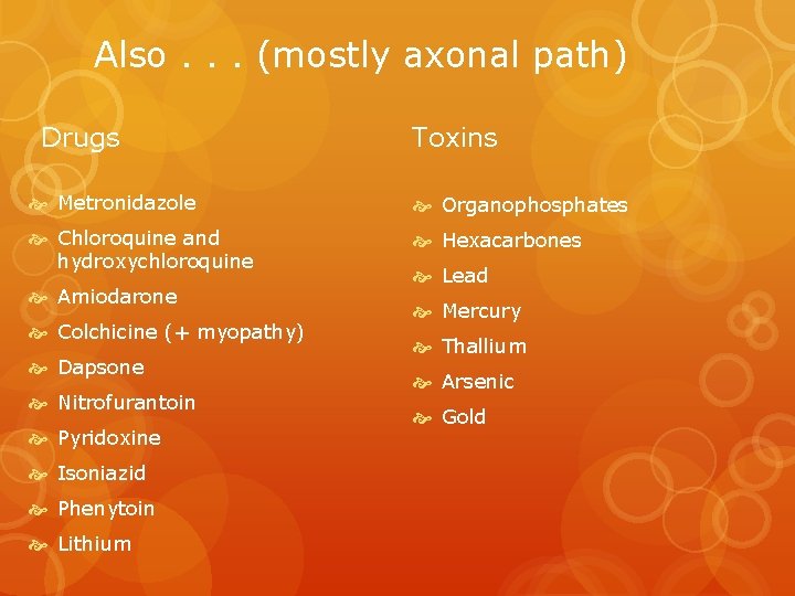
Also. . . (mostly axonal path) Drugs Toxins Metronidazole Organophosphates Chloroquine and hydroxychloroquine Hexacarbones Amiodarone Colchicine (+ myopathy) Dapsone Nitrofurantoin Pyridoxine Isoniazid Phenytoin Lithium Lead Mercury Thallium Arsenic Gold
- Slides: 33