Neurocutaneous Syndromes Neurofibromatosis Tuberous Sclerosis Sturge Weber Neurocutaneous

Neurocutaneous Syndromes ● ● ● Neurofibromatosis Tuberous Sclerosis Sturge Weber

Neurocutaneous Syndromes Neurofibromatosis Tuberous Sclerosis Sturge Weber

Neurofibromatosis ● ● ● Autosomal dominant NF 1 ● 1: 3500 incidence ● Mutation on chromosome 17 NF 2 ● 1: 40, 000 incidence ● Deafness (bilateral) ● CNS tumors ● Mutation on chromosome 22
: ● + FHx")
Neurofibromatosis ● NF 1 criteria (need 2 of the following 9): ● + FHx ( but ~ ½ cases sporadic mutation) ● Skin criteria: ● CAL (need 6+, > 0. 5 cm prepubertal, > 1. 5 cm postpubertal) ● Neurofibromas ● Inguinal / axillary freckling ● Bone criteria: ● Pseudarthrosis (angulation deformity of long bone) ● Scoliosis ● Hypoplasia of sphenoid bone in base of skull ● Eye criteria: ● Lisch nodules (hamartomas in the iris) ● Optic pallor (optic glioma)
 ●")
Neurofibromatosis ● ● Autosomal dominant Two types: ● NF 1 (1: 3500 incidence) ● ● ● chromosome 17 mutation in Neurofibromin NF 2 (1: 40, 000 incidence) chromosome 22 (CNS tumors, deafness) mutation in Merlin NF 1 criteria: ● ● + Family History ( but ~ ½ cases sporadic mutation) Skin ● ● ● Bone ● ● CAL (need 6+, > 0. 5 cm prepubertal, > 1. 5 cm post-pubertal) neurofibromas Pseudarthrosis (angulation deformity of long bone) Scoliosis Absence of sphenoid bone in base of skull Eye ● ● Lisch nodules (hamartomas in the iris) Optic pallor (optic glioma)

CAL Neurofibroma CAL Axillary Freckling

Pseudarthrosis Sphenoid Bone Hypoplasia Scoliosis

Optic Glioma Lisch Nodules

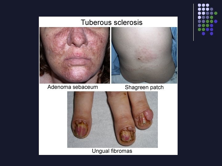
Tuberous Sclerosis ● ● Autosomal dominant Chromosomes 9 and 16 Skin hypopigmentations (“Ash leaf” spots) Benign hamartomas: ● ● ● skin ● adenoma sebaceum on face ● shagreen patch (brown leathery) on forehead or lower back brain, retina, heart, kidney Seizures in 80 -90 %
 and 16 (tuberin) Skin hypopigmentations")
Tuberous Sclerosis ● ● Autosomal dominant Chromosomes 9 (hamartin) and 16 (tuberin) Skin hypopigmentations (“Ash leaf” spots) Benign hamartomas: ● ● ● skin ● adenoma sebaceum on face ● shagreen patch (brown leathery) on forehead or lower back brain, retina, heart, kidney Seizures in 80 -90 %

“Ash Leaf” Spot Shagreen Patch Adenoma Sebaceum

Cortical Tubers Rhabdomyoma
")
Sturge Weber ● ● Unilateral port wine stain over upper face Buphthalmos (infantile glaucoma) �� enlargement of globe, corneal clouding Intracranial leptomeningeal vascular anomaly and calcifications in 90 % Seizures (partial / focal onset)

Port Wine Stain Buphthalmos with enlarged globe, corneal clouding Leptomeningeal Vascular Anomaly


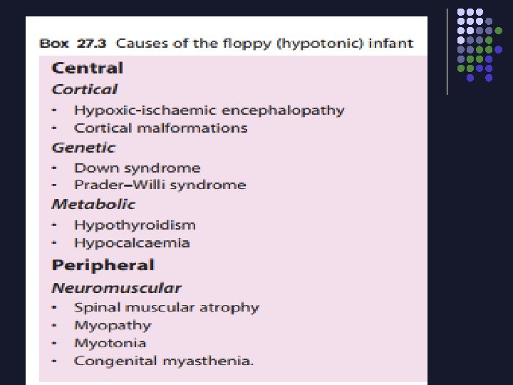
The ‘floppy infant’ ● ● ● picking up the infant, who tends to slip through the fingers hang like a rag doll when suspended prone. There will be marked head lag when the head is lifted by the arms from supine.


INFANTILE HYPOTONIA ● ● ● . Central hypotonia is associated with poor truncal tone but preserved limb tone. Dysmorphic features suggest a genetic cause. Lower motor neurone lesions are suggested by a frog-like posture


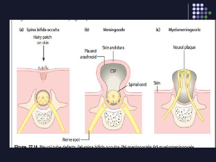
● Neural tube defects and hydrocephalus

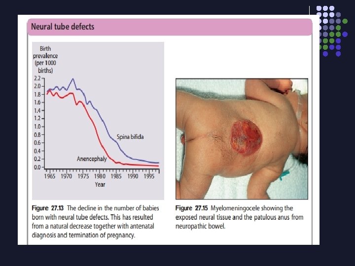
Neural tube defects ● ● failure of normal fusion of the neural plate to form the neural tube during the first 28 days following conception. Mothers of a fetus with a neural tube defect have a 10 -fold increase in risk of having a second affected fetus.

Folic acid supplementation ● ● reduces this risk. High doses are now recommended periconceptually for women with a previously affected infant planning a further pregnancy. Low-dose periconceptual folic acid supplementation is recommended for all pregnancies. In some countries e. g. United States, folic acid is added to flour for bread.

Anencephaly ● This is failure of development of most of the cranium and brain. Affected infants are stillborn or die shortly afterbirth.

Encephalocele ● There is extrusion of brain and meninges through a midline skull defect, which can be corrected surgically. However, there are often underlying associated cerebral malformations

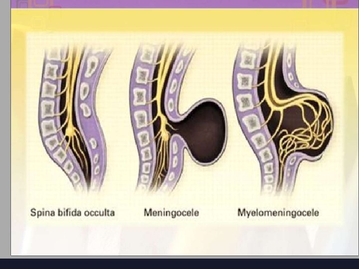
Spina bifida occulta ● ● ● This failure of fusion of the vertebral arch incidental finding on X-ray, +-overlying skin lesion such as a tuft of hair, lipoma, birth mark or small dermal sinus. usually in the lumbar region. There may be underlying tethering of the cord (diastematomyelia). . Neurosurgical relief of tethering is usually indicated.

Meningocele ● Meningoceles usually have a good prognosis following surgical repair.

Myelomeningoceles: ● ● ● Variable paralysis of the legs • Sensory loss • Bladder denervation (neuropathic bladder) • Bowel denervation (neuropathic bowel) • Scoliosis Hydrocephalus from the Chiari malformation (herniation of the cerebellar tonsils and brainstem tissue through the foramen magnum), leading to disruption of CSF flow.

Management – ● ● The back lesion is usually closed soon after birth. physiotherapy helps prevent joint contractures. For sensory loss – skin care is required to avoid the development of skin damage and ulcers. Neuropathic bladder – an indwelling or intermitten catheter may be required for a neurogenic bladder.

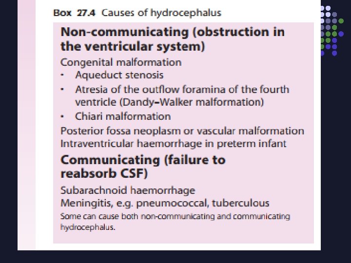
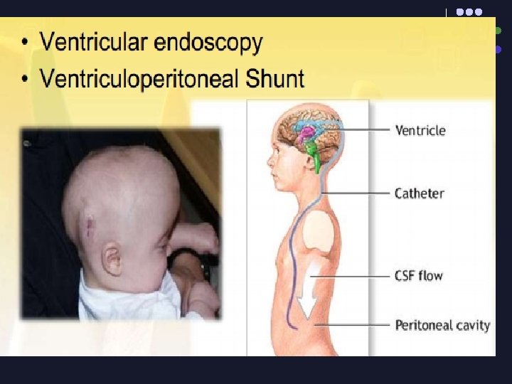
Hydrocephalus ● ● In hydrocephalus, there is obstruction to the flow of cerebrospinal fluid, leading to dilatation of the ventricular system proximal to the site of obstruction. The obstruction may be within the ventricular system or aqueduct (non-communicating or obstructive hydrocephalus), or at the arachnoid villi, the site of absorption of CSF (communicating hydrocephalus)

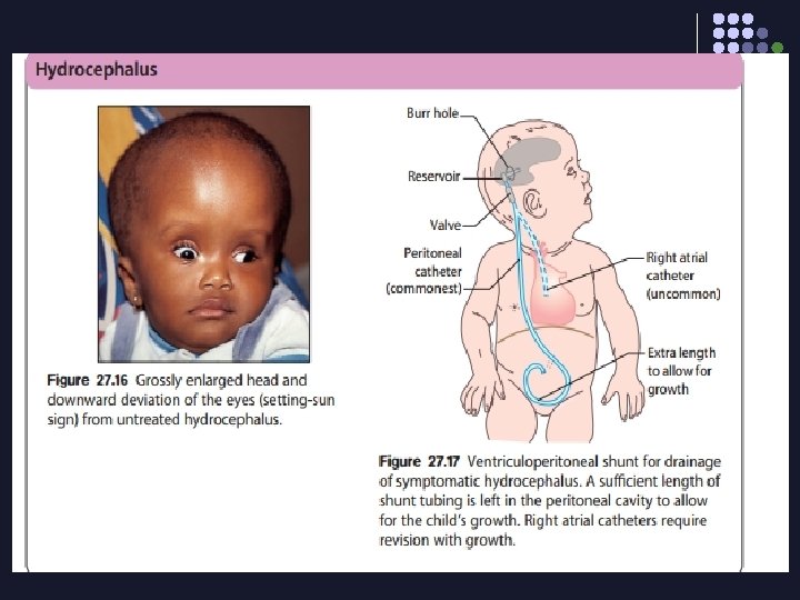
clinical Hydrocephalus In infants, ● increase in head circumference, ● separation of skull sutures, ● bulging of the anterior fontanelle, ● distension of scalp veins and ● sun setting of the eyes • Older children present with raised intracranial pressure •

“Sunsetting Eyes: clinical sign of increased intracranial pressure
- Slides: 39