
Neuro Sarcoid Fred H Hochberg MD SYSTEMIC SARCOID
Neuro. Sarcoid Fred H Hochberg, MD
SYSTEMIC SARCOID • Skin: (20%) as maculopapules, violaceous facial discoloration, erythema noosum, psoriaform changes. • Lofgren syndrome: (nodes, polyarthralgias, fever with HLA-DQB 1) • Eye: iritis, chorioretinitis, conjunctivitis • Polyarthritis (ankles) chronic arthritis • Granulomatous myositis • Salivary swelling 4% • Cardiac conduction changes
Neuro-sarcoid history • 1909 Heerfordt “uveo-parotid” FUO with paralysis of ‘cerebrospinal nerves’ • 1948 Colover. 118 published cases reviewed • 1950’s Sones steroid benefit • 1976 ACE assays (alveolar macroph and giant cells). 60% sensitive, Non-sp: liver, DM, malignancy, Gauchers.
Sarcoid Risk Factors/Staging • African Americans: 4 x with life time risk >2. 4% • I: hilar and R paratracheal nodes: 50% • II: hilar nodes and interstitial: 25% • III: interstitial disease with reduced nodes • IV: fibrotic lung changes
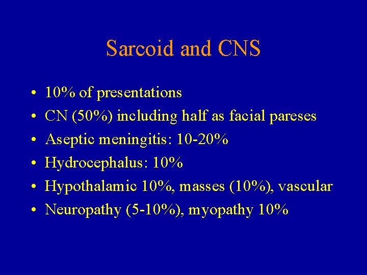
Sarcoid and CNS • • • 10% of presentations CN (50%) including half as facial pareses Aseptic meningitis: 10 -20% Hydrocephalus: 10% Hypothalamic 10%, masses (10%), vascular Neuropathy (5 -10%), myopathy 10%

Table 2. Organ Involvement in Neurosarcoidosis From: Lower: Arch Intern Med, Volume 157(16). September 8, 1997. 1864 -1868
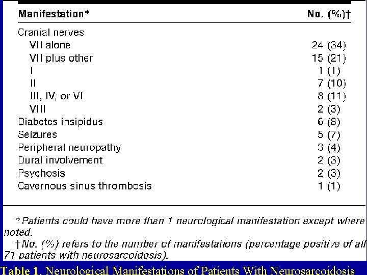
Table 1. Neurological Manifestations of Patients With Neurosarcoidosis
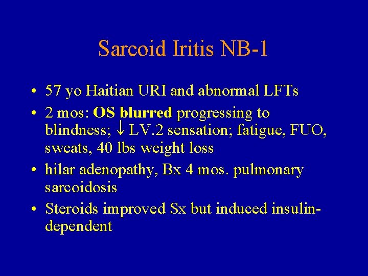
Sarcoid Iritis NB-1 • 57 yo Haitian URI and abnormal LFTs • 2 mos: OS blurred progressing to blindness; LV. 2 sensation; fatigue, FUO, sweats, 40 lbs weight loss • hilar adenopathy, Bx 4 mos. pulmonary sarcoidosis • Steroids improved Sx but induced insulindependent
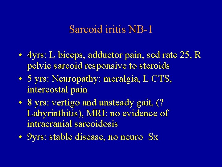
Sarcoid iritis NB-1 • 4 yrs: L biceps, adductor pain, sed rate 25, R pelvic sarcoid responsive to steroids • 5 yrs: Neuropathy: meralgia, L CTS, intercostal pain • 8 yrs: vertigo and unsteady gait, (? Labyrinthitis), MRI: no evidence of intracranial sarcoidosis • 9 yrs: stable disease, no neuro Sx
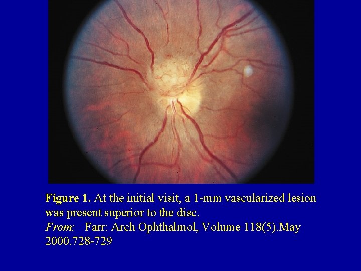
Figure 1. At the initial visit, a 1 -mm vascularized lesion was present superior to the disc. From: Farr: Arch Ophthalmol, Volume 118(5). May 2000. 728 -729
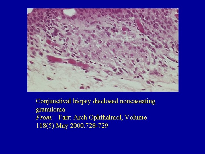
Conjunctival biopsy disclosed noncaseating granuloma From: Farr: Arch Ophthalmol, Volume 118(5). May 2000. 728 -729
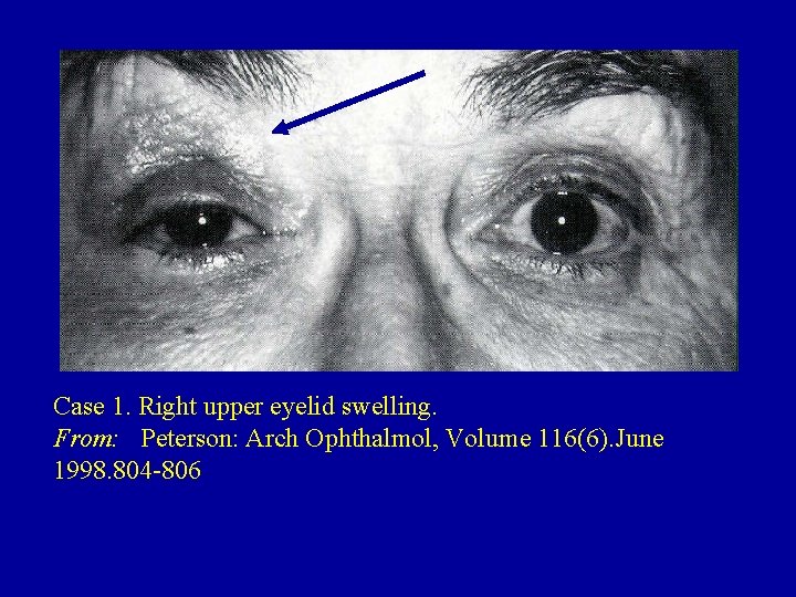
Case 1. Right upper eyelid swelling. From: Peterson: Arch Ophthalmol, Volume 116(6). June 1998. 804 -806
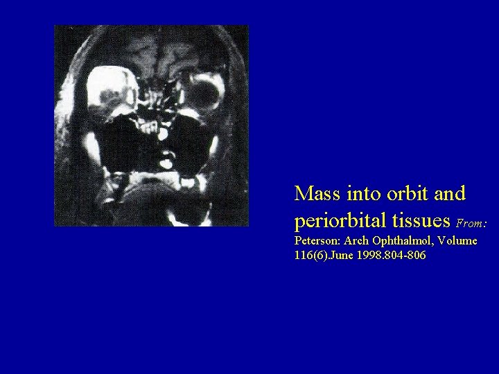
Mass into orbit and periorbital tissues From: Peterson: Arch Ophthalmol, Volume 116(6). June 1998. 804 -806
Cranial Nerves and Sarcoid • Granulomas from meninges into: – II (chiasm), – III (nerve), – VII (peripheral), – VIII (peripheral usually auditory)
Sarcoid and Skull Base • Chronic basilar meningitis-NPH • Endocrine: prolactin high in 1/3 pts, dec. Cortisol, hypogonadism, SIADH or DI • Location is periventricular III and meningeal • Less common is vasculopathy/granuloma into penetrating vessels
An accountant (JP) had been well until six years ago when he had difficulty fixating on the proper line when reading pages with columns of figures. The problem was bilateral. Five years before admission, he noted progressive loss of left vision. Two years later, he experienced anosmia and had urinary frequency and urgency, with nocturia five times nightly.
Sarcoid - Optic chiasm JP • Involves bilateral optic nerve/chiasm, pituitary, infundibulum, hypothalamus, bilateral inferior frontal lobes, mediastinal lymph nodes with secondary panhypopituitarism. ACE: 62 -69. • Plaquenil/Steroids (1 gm medrol-60 mgpred): +/- OU & new hypothalamic lesion. • XRT: 1, 000 c. Gy and C 1 MTX
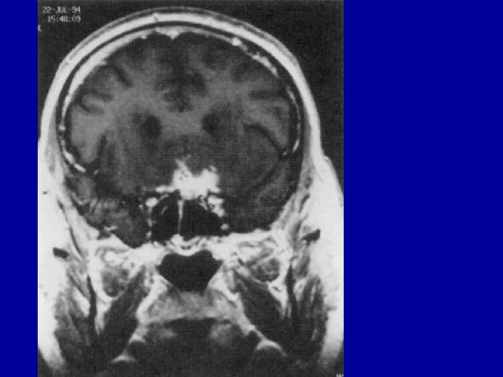
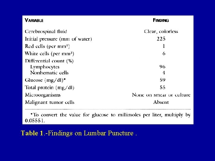
Table 1. -Findings on Lumbar Puncture.
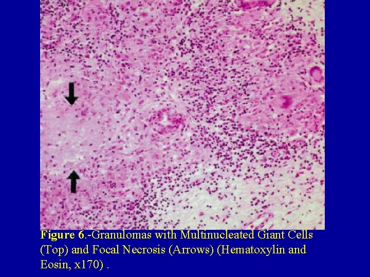
Figure 6. -Granulomas with Multinucleated Giant Cells (Top) and Focal Necrosis (Arrows) (Hematoxylin and Eosin, x 170).
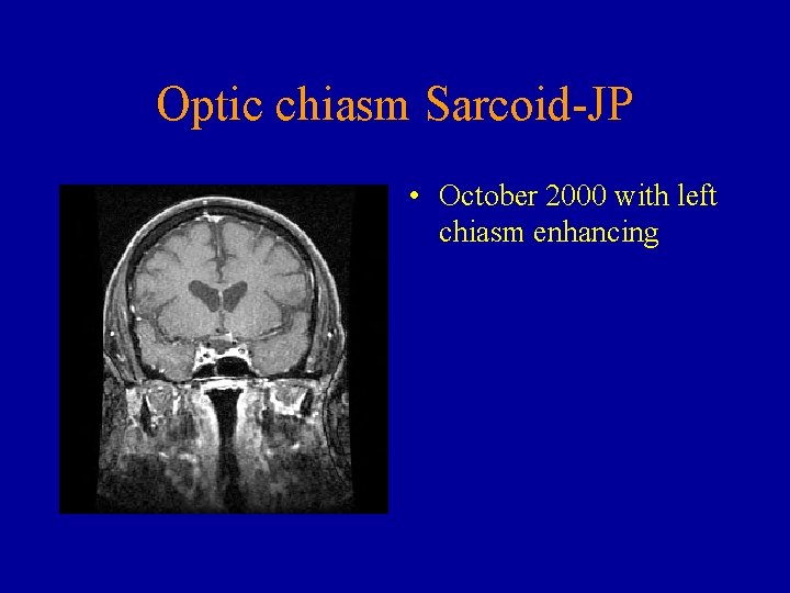
Optic chiasm Sarcoid-JP • October 2000 with left chiasm enhancing
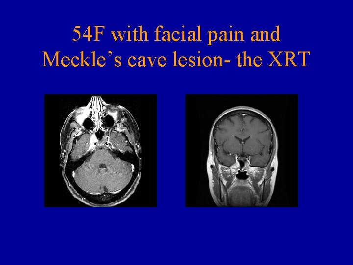
54 F with facial pain and Meckle’s cave lesion- the XRT
Meningeal Sarcoid • Subarachnoid –Ventricular- skull base and SIADH • Sherman and Stern: meningeal disease in 17/17 patients on gadolinium MRI. • Fifteen patients had well-defined leptomeningeal enhancement following contours of the brain
1994: blurry OS from uveitis. Hilar nodes, ACE + 57 Six mos. : vertigo clockwise, fell left and ataxic gait. calcium of 10. 1 CSF 16 white blood cells, 8% polys, 86% lymphocytes Mediastinoscopy: sarcoidosis MRI no lesions. 60 mg prednisone
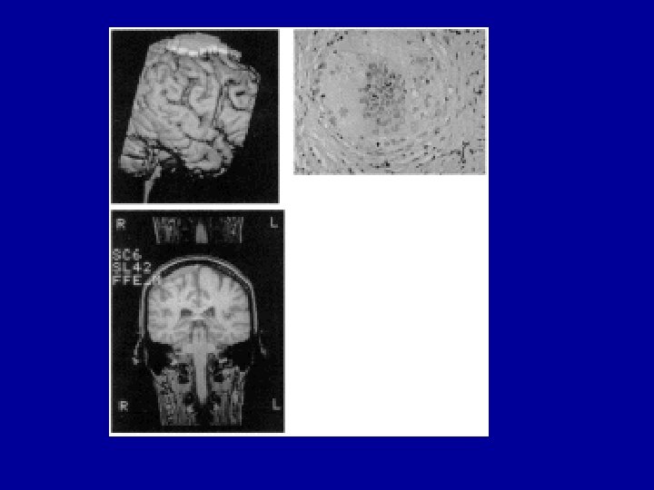
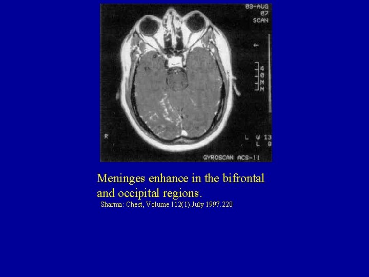
Meninges enhance in the bifrontal and occipital regions. Sharma: Chest, Volume 112(1). July 1997. 220
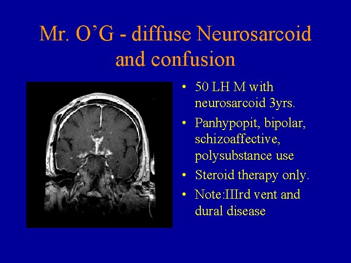
Mr. O’G - diffuse Neurosarcoid and confusion • 50 LH M with neurosarcoid 3 yrs. • Panhypopit, bipolar, schizoaffective, polysubstance use • Steroid therapy only. • Note: IIIrd vent and dural disease
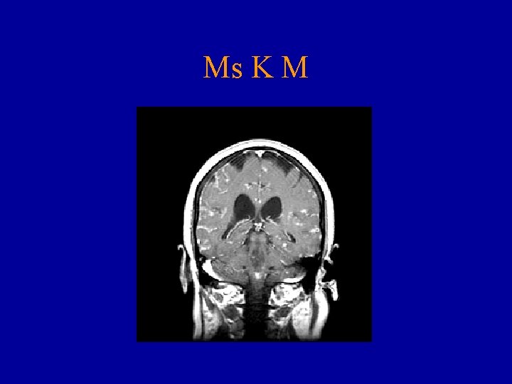
Ms K M
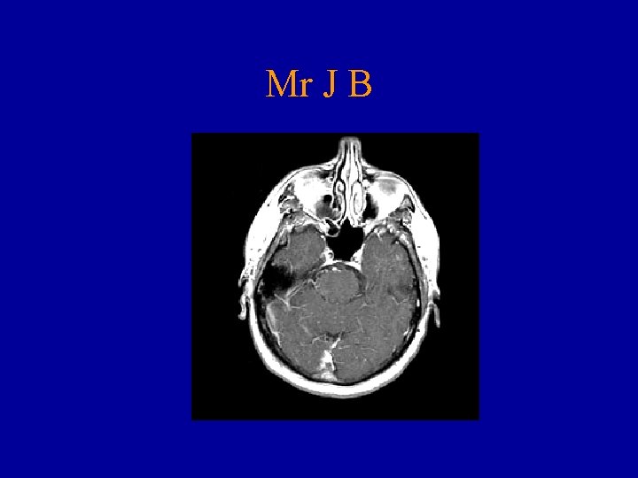
Mr J B
Hypothalamus - Sarcoid • 30 F: L 1/2 P and ICP. Large TP cystic mass • Resected mass and stable 2 years • Then meningoencephalitis, hypothalamic dysfn, hydrocephalus • Sarcoidosis diagnosed and steroids started • Series. Neurochir. 1999
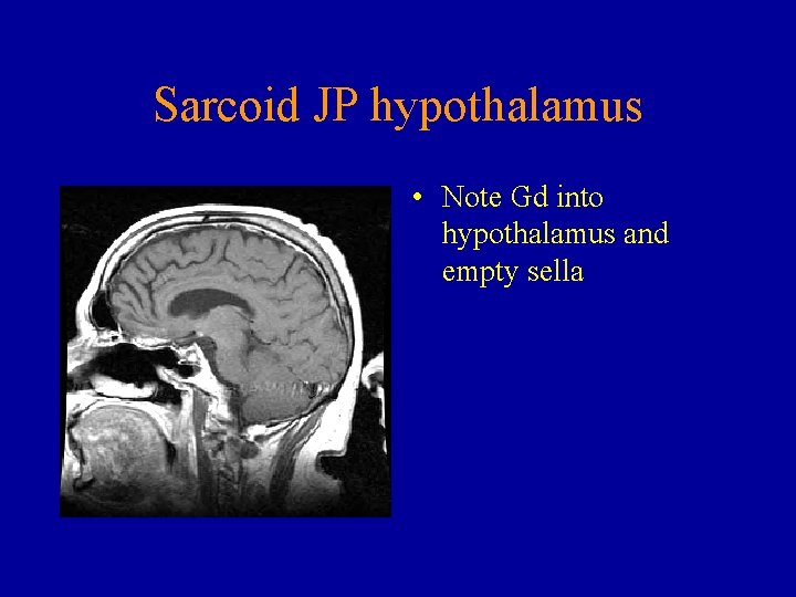
Sarcoid JP hypothalamus • Note Gd into hypothalamus and empty sella
CORD AND ROOT 54 -woman • Pain soles of feet, rectal paresthesias into legs and buttocks. • 8 w: Legs and gait. • Daily frontal and occipital headaches • MRI spine and EMG (mild denervation gluteal muscles. )
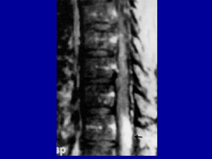
• 50 - African American with numb lateral right foot - 4 -month period. • MRI: cauda equina tumor with negative L 4 -5 hemilaminectomy. Postoperative numb left leg and buttocks. • One month later right-sided facial weakness, diplopia with gaze to the right, right foot sens change and episodic vertigo. • Two months of treatment with dexamethasone (Decadron) did not change his symptoms.
36 -year-old African American • Depression, substance abuse, and "bizarre behavior. " • Six years later auditory and visual hallucinations, disorganized and sexually inappropriate. • Chest: "chronic pulmonary disease pattern" • MRI "diffuse meningeal and perivascular Volume 155(8) August 1998 pp 1106 -1108 enhancement, " The American Journal of Psychiatry • CSF: increased cellularity and protein.
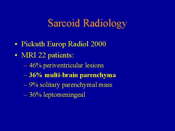
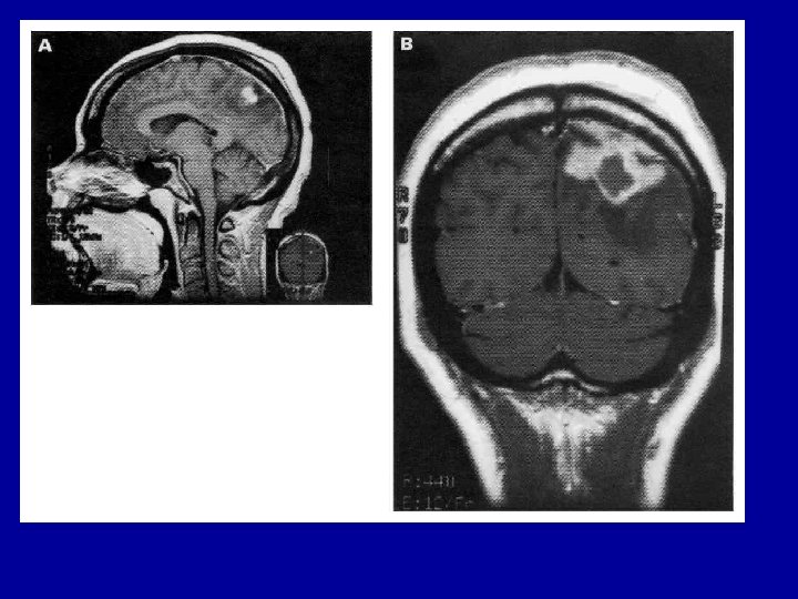
Sarcoid Radiology • Pickuth Europ Radiol 2000 • MRI 22 patients: – 46% periventricular lesions – 36% multi-brain parenchyma – 9% solitary parenchymal mass – 36% leptomeningeal
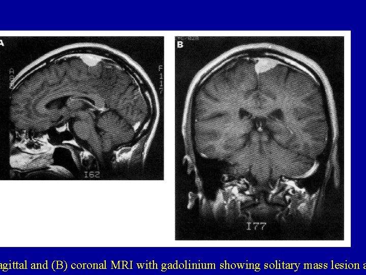
agittal and (B) coronal MRI with gadolinium showing solitary mass lesion a
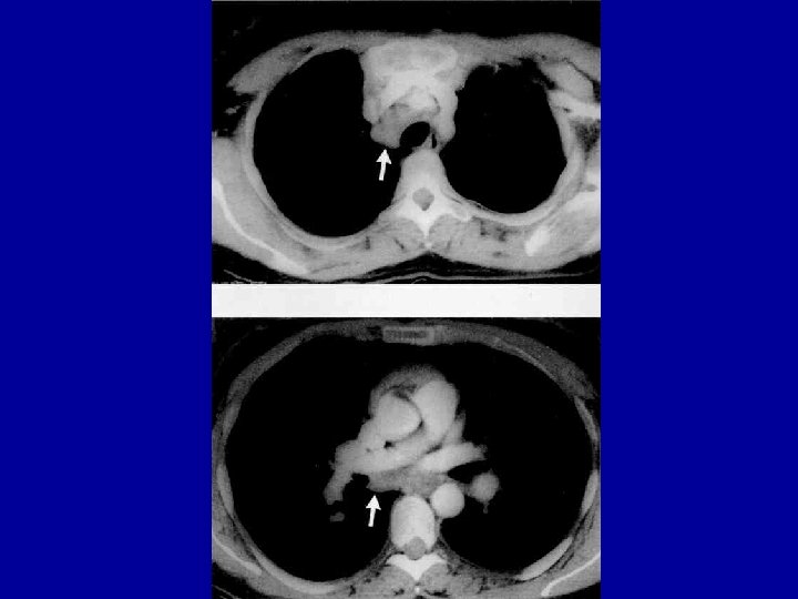
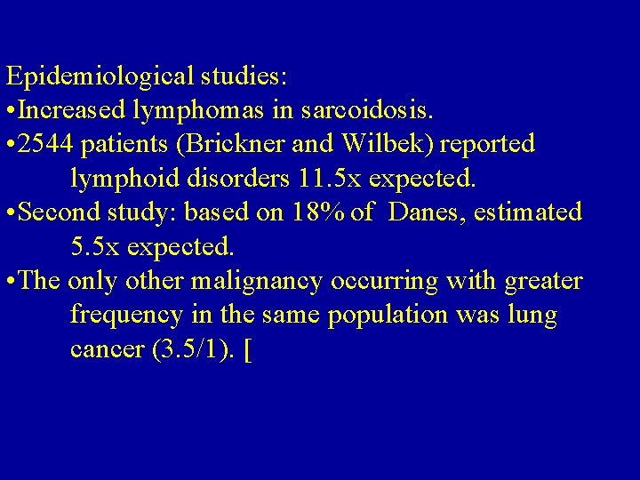
Epidemiological studies: • Increased lymphomas in sarcoidosis. • 2544 patients (Brickner and Wilbek) reported lymphoid disorders 11. 5 x expected. • Second study: based on 18% of Danes, estimated 5. 5 x expected. • The only other malignancy occurring with greater frequency in the same population was lung cancer (3. 5/1). [
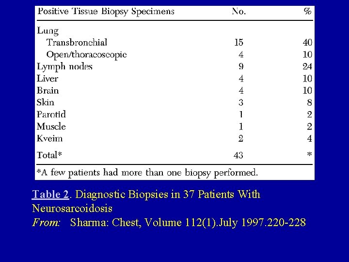
Table 2. Diagnostic Biopsies in 37 Patients With Neurosarcoidosis From: Sharma: Chest, Volume 112(1). July 1997. 220 -228
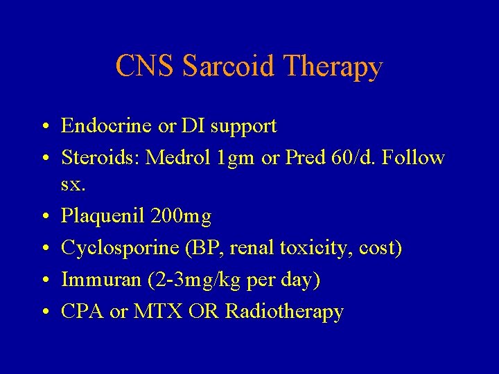
CNS Sarcoid Therapy • Endocrine or DI support • Steroids: Medrol 1 gm or Pred 60/d. Follow sx. • Plaquenil 200 mg • Cyclosporine (BP, renal toxicity, cost) • Immuran (2 -3 mg/kg per day) • CPA or MTX OR Radiotherapy
Patients were treated with cyclophosphamide Cyclophosphamide was administered intravenously every 2 weeks. The initial dose was 500 to 700 mg and doses were escalated until either neutropenia or other side effects were noted.
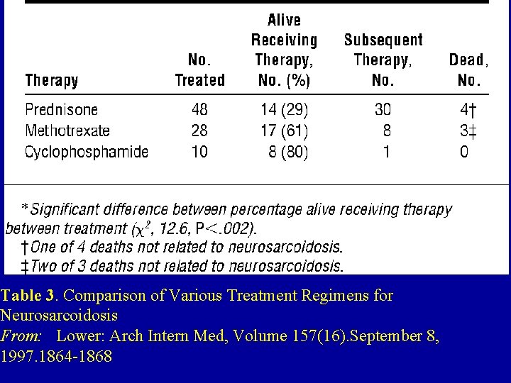
Table 3. Comparison of Various Treatment Regimens for Neurosarcoidosis From: Lower: Arch Intern Med, Volume 157(16). September 8, 1997. 1864 -1868
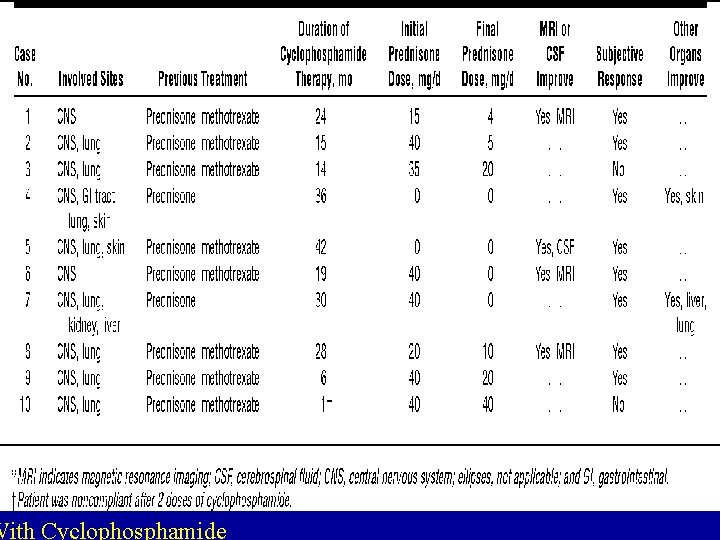
With Cyclophosphamide
NEUROSARCOID 71 of 554 patients with sarcoidosis. Seventh (facial) paralysis in 39 patients. 24 patients as only manifestation recovery in all but 1 patient. Forty-eight patients with disease other than facial nerve palsy received corticosteroids Corticosteroids benefited only 14 patients (29%). Methotrexate treated 17 (61%) of 28 patients cyclophosphamide controlled 9 (90%) of 10 patients
Methotrexate protocol: initially 10 mg oral per week as a single weekly dose. GI effects avoided by taking two 5 -mg doses 12 hours apart once a week. CBC, monthly renal and hepatic function. Methotrexate adjusted if decreased white blood cell count etc. In some patients who did not respond, the dosage was increased <15 mg/week.
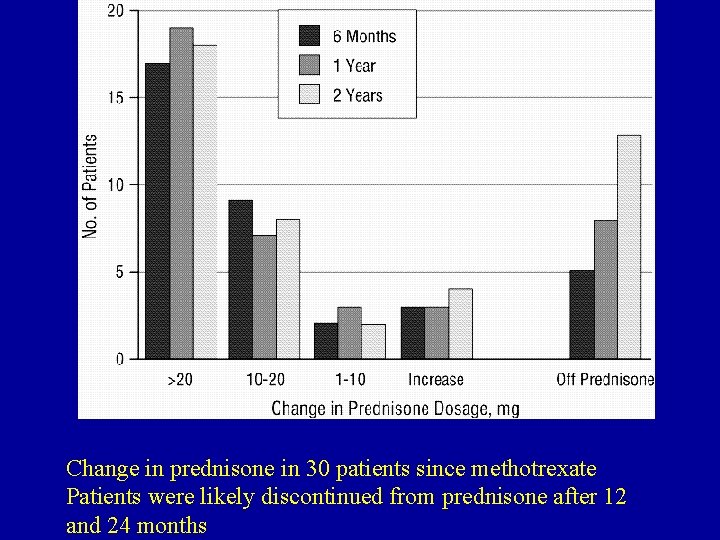
Change in prednisone in 30 patients since methotrexate Patients were likely discontinued from prednisone after 12 and 24 months
- Slides: 48