NEURAL TUBE DEFECTS Done by mohamad naghawi 3


NEURAL TUBE DEFECTS Done by: mohamad naghawi 3
, (0. 6")
Neural tube defects • Account for most congenital anomalies of the (CNS), (0. 6 -1. 3 cases per 1000 live births) in the United States. • Result from failure of the neural tube to close spontaneously between the 3 rd and 4 th wk of development in utero. 4

Neural tube defects • Second most common disability in childhood following cerebral palsy. • Second most common congenital anomaly after cardiac defects. 5

Epidemiology: • Celtic ethnic group, as Welsh, Irish & Scotch. • Female predominance, 60 - 70% of cases. • Geographical varitation, with England & china having higher rates. • The incidence has declined signifacntly in the past 3 decades due to: 1 - screening techniques, as US & AFP. 2 - termination of preg. 3 - administration of folic acid. 6

Folic Acid • Prevents up to 70% of NTDs. § U. S. Public Health Service recommends that all women capable of becoming pregnant consume 400 micrograms (0. 4 milligrams) folic acid daily. § All pregnant give 0. 4 milligrams folic acid daily as prophylactic dose § But if there are risk factor for NTD as previous baby , , the prophylactic dose should be 4 milligram) 7

Etiology: • the precise cause is still unknown • 1 - malnutrition. • 2 - medications; as antiepileptics (valproate, carbamazepine). • 3 - previous NTD child; increases the risk to 3%. • 4 - radiation. • 5 - certain medical conditions; as IDDM. • 6 - alcohol & smoking; alcohol increases the risk while smoking reduces it. 8

Embryology • The primary defect is a failure of the neural folds to fuse in the midline and form the neural tube, which is neuroectoderm. • The subsequent defect is the maldevelopment of the mesoderm, which, in turn, forms the skeletal and muscular structures that cover the underlying neural structures. 9

• The human embryo passes through 23 stages of development after conception, each occupying approximately 2 -3 days. • Two different processes form the CNS. The first is primary neurulation, which refers to the formation of the neural structures into a tube, thereby forming the brain and spinal cord. • Secondary neurulation refers to the formation of the lower spinal cord, which gives rise to the lumbar and sacral elements. • Any disruption during stages 8 -10 can cause a neural tube defect (NTD). The neural plate is formed at stage 8 (days 17 -19), the neural fold occurs at stage 9 (days 19 -21), and the fusion of the neural folds occurs at stage 10 (days 22 -23). 10

Detection: Prenatal screening: • 1 - maternal AFP in the 15 - 20 wk. • 2 - amniotic AFP. • 3 -US is 95% specific. 11

• AFP is the major serum protein in early embryonic life and is 90% of the total serum globulin in a fetus. • it’s first made in the yolk sac and later in the GI system and liver of the fetus. • Serum maternal AFP measurement of more than 1000 ng/ ml is abnormal. & could be : • if AFP IS more than 1500 ng/ml there will be : Anencephaly , Spina bifida cystica Encephalocele (leaking), Conjoined twins, Omphalocele, Turner syndrome, Gastroschisis , Exstrophy of the cloaca , Oligohydramnios & fetal death. 12

Pathology: • neural tube defects can be open (neural structures that communicate with the atmosphere) or closed (skin covered). They can be ventral or dorsal midline defects. 13
 Spina bifida , • Occulta • Aperta •")
The major neural tube defects 1) Spina bifida , • Occulta • Aperta • Cystica; meningocele & myelomeningocele. or 2) bifid cranium; • Bifid cranium occultum. • Encephalocele. • Anencephaly. 14

Bifid cranium occultum: • The most benign type of cranium bifidum occultum is the persistent parietal foramina or persistent wide fontanelle. • The parietal foramina can be transmitted as an autosomal dominant trait via a gene located on the short arm of chromosome 11. • mostly asymptomatic, & most of these skull defects close over time • Diagnosis by occlusion. • Note : the anterior fontanel is closer between 7 to 18 month , , if the closer before the 7 month this called early closer of anterior fontanel • And if the closer after 18 month , , this cold delay closer of anterior fontanel 15

Delay closer of anterior fontanel DD • • 1 - hypothyroidism 2 - hydrocephalus 3 - rickets 4 -Bifid cranium occultum

Spina Bifida Occulta • The post. Vertebral arch has a closure defect within it, but there is no herniation of the neural tube. • It’s found in up to 10% of the population. • Most individuals are asymptomatic and lack neurological signs. • No hydrocephalus or Chiari II malformation. • Cutaneous stigmata as a hairy patch, dermal sinus tract, dimple, hemangioma, or lipoma may exist and point to the underlying spina bifida, • Note; For differentiation, dimples below the gluteal fold are benign & non neurological, as PNS. Dimples above should be evaluated further. 17

Other signs that should elucidate your suspicion: • Radiologic signs ; Hemivertebrae , Scoliosis • Orthopedic findings; as asymmetry or foot deformities • Neurological problems: as weakness, atrophy or asymmetry, loss of sensation, painless sores, hyperreflexia , unusual back pain, radiculopathy • Urologic problems ; as Neurogenic bladder or incontinence 18

• A spine X-ray shows a defect in closure of the posterior vertebral arches and laminae, typically involving L 5 and S 1. • There is no abnormality of the meninges, spinal cord, or nerve roots. • occasionally associated with more significant developmental abnormalities of the spinal cord, including syringomyelia, diastematomyelia, and a tethered cord. • The cord is occasionally tethered by a shortened thickened filum terminale (hypertrophic). ﺍﻛﺜﺮ ﺍﻟﻤﺮﺍﺕ ﻣﻨﻌﻤﻞ mri 19

Impairments associated with Spina Bifida Physiological changes below the level of the lesion generally include: abnormal nerve conduction, resulting in: Þ somatosensory losses Þ motor paralysis, including loss of bowel and bladder control 20

Impairments associated with Spina Bifida Anatomical changes below the level of lesion: Þ musculoskeletal deformities (scoliosis) Þ joint and extremity deformities (joint contractures, club foot, hip subluxations, diminished growth of non-weight bearing limbs) Þ osteoporosis. 21

meningocele & myelomeningocele Majd suaifan 22

Meningocele A meningocele is formed when the meninges herniates through a defect in posterior vertebral arches, or anterior sacrum. 23

• The spinal cord is usually normal and assumes a normal position in the spinal canal, although there may be tethering of the cord, othe congenital anomally associated syringomyelia, or diastematomyelia. • A fluctuant midline mass that may transilluminate occurs along the vertebral column, usually in the lower back. Most meningoceles are well covered with skin and pose no immediate threat to the patient. • In asymptomatic children with normal neurolgic findings and full thickness skin covering the meningocele, surgery maybe delayed or sometimes not performed. 24

• The least common form of spina bifida. • In a posterior meningocele, the outer faces of some vertebrae are open (unfused) and the meninges are damaged and pushed out through the opening, appearing as a sac or cyst which contains cerebrospinal fluid. • In an anterior meningocele, the inner faces of vertebrae are affected and the cyst protrudes into the retroperitoneum or the presacral space. 25
 constipation. • 2)")
• An anterior meningocele comes with Symptoms of: • 1) constipation. • 2) bladder dysfunction. • 3) Female patients may have associated anomalies of the genital tract, including a rectovaginal fistula and vaginal septa. • Plain X-ray demonstrate a defect in the sacrum. • And CT scanning or MRI outlines the extent of the meningocele. 26

MYELOMENINGOCEL E Characterized by protrusion of the neural elements through a vertebral defect into a meningeal lined sac. Typically, the cord at this level is not fused. but is in it’s flattened embryological state , with the nerve roots arising from the ventral surface & the open central canal lying dorsally. Remnants of neural tissue are visible beneath the membrane, which may occasionally rupture and leak CSF. 27

Myelomeningocele 28

Myelomeningocele • Myelomeningocele represents the most severe form of dysraphism involving the vertebral column and occurs with an incidence of approximately 1/2, 000 live births. • A myelomeningocele may be located anywhere along the neuraxis, but the lumbosacral region accounts for at least 75% of the cases. 29

Etiology • The cause of myelomeningocele is unknown. • a genetic predisposition exists; the risk of recurrence after one affected child increases to 3– 4% and increases to approximately 10% with two previous abnormal pregnancies. • There is strong evidence that maternal periconceptional use of folic acid supplementation reduces the incidence of neural tube defects in pregnancies at risk by at least 50%. • To be effective, folic acid supplementation should be initiated before conception and continued until at least the 12 th wk of gestation when neurulation is complete. • In normal risk pregnancies, women should take 0. 4 mg of folic acid daily. And in high pregnancy (previously affected child), supplementation should be started 4 mg daily, beginning 1 month prior to planned conception. 30

Cont. • Certain drugs—including drugs that antagonize folic acid such as trimethoprim and the anticonvulsants: Valproic acid, carbamazepine, phenytoin, and phenobarbital increase the risk of myelomeningocele. • Valproic acid causes NTDs in approximately 1 -2% of pregnancies whe administered during pregnancy. 31

Clinical Manifestations The condition produces dysfunction of many organs and structures, including the skeleton, skin, and genitourinary tract, in addition to the peripheral nervous system and the CNS. ● lesion in the low sacral region causes bowel and bladder incontinence associated with anesthesia in the perineal area but with no impairment of motor function. ● ﻛﻞ ﻣﺎ ﻛﺎﻧﺖ ﺍﻋﻠﻰ ﻛﻞ ﻣﺎ ﻛﺎﻥ ﺍﻟﺘﺎﺛﺮ ﻋﻠﻰ ﻣﻮﺗﻮﺭ ﻓﻨﻜﺸﻦ ﺍﻛﺜﺮ ● ﻛﻞ ﻣﺎ ﻛﺎﻧﺖ ﺍﻭﻃﻰ ﻛﻞ ﻣﺎﻛﺎﻥ ﺍﻟﺘﺎﺛﻴﺮ ﻋﻠﻰ ﺍﻟﺴﻔﻨﻜﺘﻴﺮ ﺍﻛﺜﻴﺮ ● 32

• Newborns with a defect in the midlumbar region typically have a saclike cystic structure covered by a thin layer of partially epithelialized tissue • Examination of the infant shows • a flaccid paralysis of the lower extremities, an absence of deep tendon reflexes, • a lack of response to touch and pain, • and a high incidence of postural abnormalities of the lower extremities (including clubfeet and subluxation of the hips). • Constant urinary dribbling and a relaxed anal sphincter may be evident. 33

• Hydrocephalus in association with a type II Chiari defect develops in at least 80% of patients with myelomeningocele. • Generally, the lower the deformity in the neuraxis (e. g. , sacrum), the less likely is the risk of hydrocephalus. *Congenital downward displacemet of cerebellar vermis and tonsils through the foramen magnum. 34

• Ventricular enlargement may be indolent and slow growing or may be rapid, causing a bulging anterior fontanel, dilated scalp veins, setting-sun appearance of the eyes, irritability, and vomiting associated with an increased head circumference. • Not infrequently, infants with hydrocephalus and Chiari II malformation develop symptoms of hindbrain dysfunction, including difficulty feeding, choking, stridor, apnea, vocal cord paralysis, pooling of secretions( involvement of cranial nerve) and spasticity of the upper extremities, which, if untreated, can lead to death. 35

MYLEOMENINGOCEL E Management Multidisciplinary team approach 1. Assessment of the sac & it’s coverings. 2. Site , extension , there are lack CSF 3. Neurological examination of lower limb ( power , tone , deep tendon reflex , coordination , sensation , examine the sphincter ) 4. Examination for other associated conditions. 5. Counseling & careful discussions with the parents. 6. Surgical procedures. 36

Treatment • Surgery can be delayed for several days (except when there is a CSF leak or not cover with skin) • most infants require a shunting procedure for hydrocephalus. 37

Prognosis • For a child who is born with a myelomeningocele and who is treated aggressively, the mortality rate is approximately 10– 15%, and most deaths occur before age 4 yr. • At least 70% of survivors have normal intelligence, but learning problems and seizure disorders are more common than in the general population. • Because myelomeningocele is a chronic handicapping condition, periodic multidisciplinary follow-up is required for life. 38

Microcephaly Encephalocele Anencephaly By: Muthanna Al-Abbadi

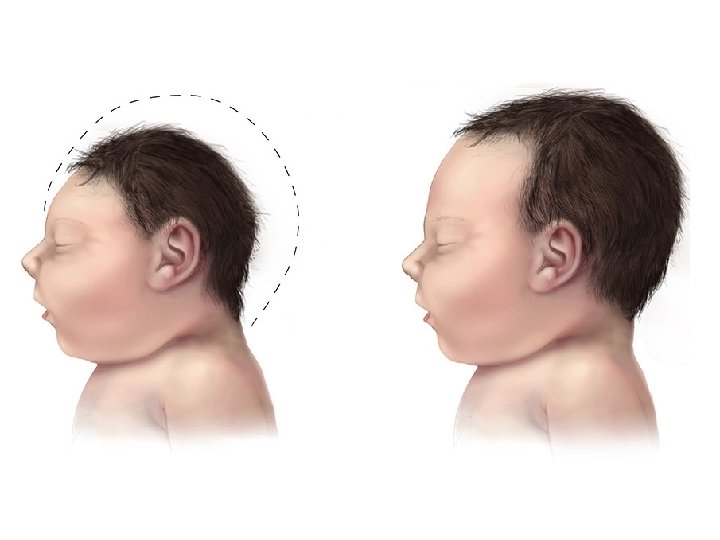
Microcephaly • Microcephaly is defined as a head circumference that measures more than 3 SD below the mean for age and sex • This condition is relatively common, particularly among developmentally delayed children • Subdivided into 2 main groups: 1. Primary (genetic) 2. Secondary (non-genetic)

Primary Microcephaly • Refers to a group of condition that usually have no associated malformations or are associated with a specific genetic syndrome • Affected children are usually identified at birth
 • Autosomal Dominant • Syndromes: Down,")
Causes of Primary Microcephaly • Familial (Autosomal Recessive) • Autosomal Dominant • Syndromes: Down, Edward, Cri-du-Chat, Cornelia de Lange, Rubinstein-Taybi, and Smith-Lemli-Opitz • X-Linked

Secondary Microcephaly • Results from a large number of agents that can affect a fetus in utero or an infant during periods of rapid brain growth, particularly the first 2 years of life • May be identified at birth or later

Causes of Secondary Microcephaly • Congenital Infections: CMV, Rubella, Toxoplasmosis • Drugs: Alcohol, (fetal alcohol syndrome main cause in eurob area, Hydantoin • Radiation • Meningitis/Encephalitis • Malnutrition • Hyperthermia • HIE • Syndromes: Rett, Seckel, Angelman

Clinical Manifestations • Small Head Circumference, Sloping Forehead, and Small Anterior Fontanelle • Developmental Impairments • Organ Malformation • Visual Impairment • Hearing Loss • Learning Difficulties • Seizures • Movement Disorders • No any them with familial

Diagnosis • Investigations are determined by the history and physical examination. If the cause is unknown: • Mother’s serum phenylalanine level (brain damage) • Karyotype (chromosomal syndromes) • MRI (structural abnormalities of the brain) • CT (intracerebral calcifications) • Fasting plasma and urine amino acid analysis • Serum ammonia • TORCH titers, HIV testing, and urine culture (CMV)

Treatment • No specific treatment to fix microcephaly • Supportive genetic and family counseling • Placement in an appropriate program that will provide for maximal development of the child • Controlling seizures and neuromuscular symptoms • Adequate nutrition • Note • HC : 35 CM AT BIRTH < and in first three month increase 2 cm every month and in second three month increase 1 cm every month <<and in the last 6 month increase 3 cm as 0, 5 cm every month <and in the least of life increase 12 cm

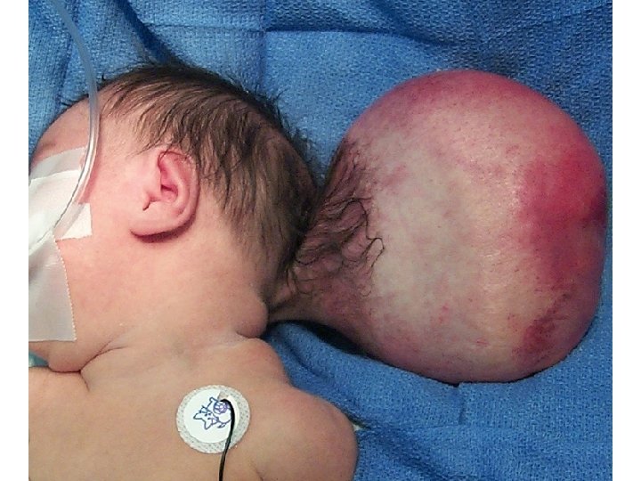
Encephalocele • An encephalocele is a protrusion of the brain through a defect in the skull (cranium bifidum) that is "closed" or covered with skin • Cranial encephalocele contains the sac plus cerebral cortex, cerebellum, or portions of the brainstem • It could be Primary (congenital) that present at birth, its major types are: sincipital, basal, occipital. It could be Secondary (acquired) due to trauma or post-surgical defects

Encephalocele • The cranial defect occurs most commonly in the occipital region at or below the inion. • Frontal or nasofrontal encephaloceles are also common in certain parts of the world. Some frontal lesions are associated with cleft lip/palate • Patients are at increased risk for developing hydrocephalus, because of: aqueductal stenosis, Chiari malformation, or the Dandy-Walker syndrome

Meckel-Gruber Syndrome • Cranial encephalocele is most commonly part of a syndrome called Meckel-Gruber syndrome. • It is a rare autosomal recessive condition that is characterized by: • Occipital encephalocele • Cleft lip/palate • Microcephaly • Microphthalmia • Abnormal genitalia • Polycystic kidneys • Polydactyly ﻻﺯﻡ ﻧﺪﻭﺭ ﻋﻠﻴﻬﻢ ﻣﻊ encephalocel

Clinical Manifestations • The clinical features of encephalocele are variable depending upon the type (location) and severity • Sincipital encephaloceles may be occult lesions that are noticeable or may present with marked craniofacial deformities • Basal encephaloceles may or may not be apparent on external inspection. Affected patients may present as: a nasal or epipharyngeal mass, difficulty breathing, recurrent upper tract infections, nasal discharges, recurrent meningitis, or CSF leaks

Clinical Manifestations • Occipital encephaloceles usually are obvious at the time of birth • Those of relatively large size may be associated with cranial nerve deficits, poor sucking and feeding, spasticity, blindness, seizures, or developmental delay • Neurologic deficits may progress after birth if hydrocephalus develops • Occipital encephalocele also may be associated with hind-brain anomaly (Chiari III malformation)

MRI demonstrating a Chiari III malformation with cervico -occipital encephalocele

Diagnosis • The diagnosis is apparent at birth in most of the occipital encephaloceles • But shold be antinatal • Basal encephaloceles may present as a midline mass in the nose or may not be visible. They also tend to present with meningitis • An encephalocele may be mistaken for a nasal polyp if it is located within the nose or for a soft tissue tumor if it is covered with skin and anterior to the nose.

Diagnosis • Transillumination of the sac can indicate the presence of neural tissue. • X-ray of the skull and cervical spine • Ultrasonography is most helpful to identify the contents of the sac • MRI and CT • Diagnosis in utero is possible by: maternal serum AFP levels, U/S, MRI

Treatment • If diagnosed prenatally, vaginal delivery is safe if the lesion is small. C/S is required if the lesion is large • Surgical treatment is appropriate in most cases (removing the overlying sac and closing the defect)

Prognosis • Patients are at risk for vision problems, microcephaly, intellectual disability, and seizures • Patients with neural tissue within the sac and associated hydrocephalus have the poorest prognosis

Anencephaly • It is an open defect in the calvaria, meninges, and skin, such that the cranial neural tube is exposed • It results from failure of the rostral neuropore to close around postovulatory day 25 • It is a severe defect and is not compatible with survival. Infants that are alive at birth generally die within hours, but occasionally survive for a few days or weeks • Incidence is 1 in 1, 000 (most end in miscarriage). Recurrence is common

Clinical Features • In the most common type of anencephaly, the forebrain and variable amounts of upper brainstem are involved • Exposure in utero results in destruction of neural tissue, which appears as a hemorrhagic, fibrotic, nonfunctioning mass • The hypothalamus is typically missing. The cerebellum, brainstem, optic nerves, and spinal cord may be malformed. Underdevelopment or absence of the pituitary leading to adrenal hypoplasia is always present

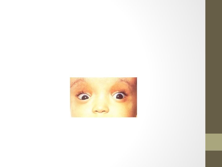
Clinical Features • The frontal, parietal, and portions of the occipital bones are most often affected • The absent cranial vault results in the characteristic appearance of bulging eyes and absent neck • The defect in the skull sometimes extends through the level of the foramen magnum and involves the cervical spine (holoacrania) • Neonates typically have brainstem function, with spontaneous breathing and often with suck, root, and gag responses. However, they are permanently unconscious

Additional Anomalies • Folding of the ears • Cleft lip/palate • Congenital heart defects • Omphalocele • Pulmonary, renal, and skeletal malformations • Aganglionosis

Diagnosis • Maternal serum AFP level • Fetal ultrasonography

Management & Prevention • Prenatal detection is usually followed by termination of the pregnancy • There are no neurosurgical management options • Prevention is the most important aspect of management of anencephaly • Periconceptional folic acid supplementation is recommended, higher doses are recommended for women taking anticonvulsants or has previous history of anencephaly • Don’t do Resuscitation

Thank You

hydrocephalous Marah Masadeh Supervised by dr. omar nafi

Definition • Hydrocephalus is not a specific disease; it represents a diverse group of conditions that result from impaired circulation and/or absorption of CSF or, in rare circumstances, from increased production of CSF by a choroid plexus papilloma • There is abnormal enlargement of cerebral ventricles and/or subarachnoid space as a result of excess cerebrospinal fluid (CSF) accumulation.

• The incidence of congenital hydrocephalus is 3 per 1, 000 live births in US. • The incidence of acquired hydrocephalus is not known exactly due to the variety of disorders that may cause it.
 choroid plexus in the ventricular system(75%).")
physiology • Production of csf by : 1) choroid plexus in the ventricular system(75%). 2) extrachoroidal sources, including the capillary endothelium within the brain parenchyma(25%). q control of production: a) Adrenergic nerves : diminishes CSF production b) cholinergic nerves: may double the normal CSF production rate
 In a normal child, approximately 20 m.")
• Production of CSF : 1) In a normal child, approximately 20 m. L/hr of CSF. 2) The total volume of CSF approximates 50 m. L in an infant and 150 m. L in an adult. q Most of the CSF is extraventricular.

CSF FLOW

CSF absorbtion • CSF is absorbed : A. primarily by the arachnoid villi through tight junctions of their endothelium by the pressure forces B. lymphatic channels directed to the paranasal sinuses, along nerve root sleeves C. the choroid plexus itself.
: obstruction within the ventricular")
Types of hydrocephalous • obstructive or non communicating hydrocephalus(most commonly): obstruction within the ventricular system • Non obstructive or communicating hydrocephalus: resulting from obliteration of the subarachnoid cisterns or malfunction of the arachnoid villi.

obstructive or non communicating hydrocephalus • an abnormality of the aqueduct of Sylvius or a lesion in the fourth ventricle. • aqueductal stenosis results from: all ventrical dilated except four ventricle 1) an abnormally narrow aqueduct of Sylvius that is often associated with branching or forking. 2) In asmall percentage : inherited as a sex-linked recessive trait. q association with aqueductal stenosis : a) neural tube closure defects, including spina bifida occulta b) neurofibromatosis.

obstructive or non communicating hydrocephalus • Aqueductal gliosis • neonatal meningitis or a subarachnoid hemorrhage in a premature infant • Intrauterine viral infections • mumps meningoencephalitis • A vein of Galen malformation. • Lesions or malformations of the posterior fossa : 1) posterior fossa brain tumors 2) Chiari malformation 3) the Dandy-Walker syndrome

Nonobstructive or communicating hydrocephalus • a subarachnoid hemorrhage • Pneumococcal and tuberculous meningitis • Leukemic infiltrates.

Clinical manifestations q. Depends on : • age at onset • the nature of the lesion causing obstruction • duration and rate of increase of the intracranial pressure (ICP).

• • • Enlargement of the head …. HC(for children less 3 years ) the anterior fontanel is wide open and bulging the scalp veins are dilated. forehead is broad eyes might deviate downward because of impingement of the dilated suprapineal recess on the brainstem tectum, producing the setting-sun eye sign. q. Long-tract signs as CP 1) Brisk tendon reflexes 2) Spasticity 3) clonus (particularly in the lower extremities), 4) Babinski sign

• In an older child, the cranial sutures are less accommodating so that the signs of hydrocephalus may be subtler q common to both age groups: • Irritability, • Lethargy • poor appetite • Vomiting Ø in older patients: headache is a prominent symptoms. ØHeadache and vomiting and dementia think about hydrocephalus

• A gradual change in personality and deterioration in academic productivity suggest a slowly progressive form of hydrocephalus ﻋﻠﻰ ﺍﻟﺴﺮﻳﻊ ﺑﻌﻤﻠﻪ ct • head circumference often indicate an increased velocity of growth.

examination • Percussion of the skull: a cracked pot sound or Macewen sign, indicating separation of the sutures. • A foreshortened occiput suggests Chiari malformation, • a prominent occiput suggests the Dandy-Walker malformation. • Papilledema • abducens nerve palsies • Pyramidal tract signs, which are most evident in the lower extremities. • Diagnosis is CT

Diagnosis and differential diagnosis • Familial cases suggest X-linked or autosomal hydrocephalus secondary to aqueductal stenosis. ﺛﺎﻧﻲ ﺑﻴﺒﻲ • A past history of prematurity with intracranial hemorrhage, meningitis, or mumps encephalitis. • café-au-lait spots/megalencephaly…NF • inspection, palpation, and auscultation of the skull and spine. • head circumference • Dealy closer of anterior frontanel

• size and configuration of the anterior fontanel • Inspection of the back (tufts of hair, lipoma, or angioma)…spinal dysraphism • a prominent forehead or abnormalities in the shape of the occiput • A cranial bruit…vein of Galen arteriovenous malformation( behind ear ) • Transillumination of the skull. . massive dilation of the ventricular system or in the Dandy-Walker syndrome

• Inspection of the eyegrounds: 1. chorioretinitis suggests an intrauterine infection, such as toxoplasmosis. 2. Papilledema(older children). q The head might appear enlarged secondary to: 1) a thickened cranium resulting from chronic anemia ( thalssemia) 2) Rickets 3) osteogenesis imperfecta 4) epiphyseal dysplasia.

• bilateral parietal bone prominence…Chronic subdural collections • Measurement of parents’ head circumferences…Familial megalencephaly is inherited as an autosomal dominant trait and is characterized by delayed motor milestones and hypotonia but normal or near-normal intelligence.

• Plain skull films: 1. separation of the sutures 2. erosion of the posterior clinoids in an older child 3. an increase in convolutional markings (beaten-silver appearance) on the inside of the skull with long-standing increased ICP • The CT scan and/or MRI along with ultrasonography for cause and severity.

beaten-silver appearance increase. ICP

Chiari malformation • Type I : displacement of the cerebellar tonsils into the cervical canal • produces symptoms during adolescence or adult life • usually not associated with hydrocephalus. q. Patients complain : • recurrent headache • neck pain • urinary frequency • progressive lower-extremity spasticity. • Syringomyelia : Syrinx of the spinal cord especially the cervical region should be looked for on MRI imaging.

type I Chiari malformation

type II Chiari malformation • a failure of pontine flexure development during embryogenesis, and results in elongation of the fourth ventricle and kinking of the brainstem, with displacement of the inferior vermis, pons, and medulla into the cervical canal • Progressive hydrocephalus with a myelomeningocele.
 produce • Stridor • weak cry • apnea, • which may be")
qduring infancy(10%) produce • Stridor • weak cry • apnea, • which may be relieved by shunting or by decompression of the posterior fossa. qduring childhood: • abnormalities of gait • Spasticity • increasing incoordination (including the arms and hands)

small, slitlike fourth ventricle, which has been pulled into a vertical position Cerebellar tonsil foramen magnum

treatment • surgical decompression • If asymptomatic or mildly symptomatic patients may be managed conservatively

Dandy-Walker malformation • a cystic expansion of the fourth ventricle in the posterior fossa • midline cerebellar hypoplasia, which results from a developmental failure of the roof of the fourth ventricle during embryogenesis

Dandy-Walker malformation • 90% of patients have hydrocephalusa. q associated anomalies, including • agenesis of the posterior cerebellar vermis and corpus callosum. q Infants present with : • a rapid increase in head size • a prominent occiput. • Transillumination of the skull may be positive. q Most children have evidence of ( on neurological examination ) • long-tract signs • cerebellar ataxia • delayed motor and cognitive milestones q Management : shunting the cystic cavity (and on occasion the ventricles as well) in the presence of hydrocephalus

• Serial measurement of the head circumference discover the Chiari malformation , Dandy-Walker malformation and hydrocephalous

All ventricle dilated in Dandy. Walker malformation dilated lateral ventricles large posterior fossa cyst

splaying of the cerebellar hemispheres by the dilated fourth ventricle

The incomplete vermis now becomes recognizable. decreased size temporal horns
medical management: use of acetazolamide and furosemide(temporarily). 2)extracranial")
Therapy • depends on the cause. 1)medical management: use of acetazolamide and furosemide(temporarily). 2)extracranial shunts, particularly a ventriculoperitoneal shunt 3)Endoscopic third ventriculostomy has evolved as a viable approach and procedure might need to be repeated to be effective. Ventricular shunting may be avoided with this approach. q The major complications of shunting are : • Occlusion (characterized by headache, papilledema, emesis, mental status changes) • bacterial infection (fever, headache, meningismus), Staphylococcus epidermidis. . • intrauterine surgical management of fetal hydrocephalus (high rate of associated cerebral malformations)

Prognosis qdepends on the cause of the dilated ventricles. qincreased risk for various developmental disabilities. q. The mean intelligence quotient is reduced particularly for performance tasks as compared with verbal abilities. • abnormalities in memory function. q. Vision problems are common: because the occipital cortex affected • Strabismus • visuospatial abnormalities • visual field defects • optic atrophy with decreased acuity secondary to increased ICP. • The visual evoked potential latencies are delayed and take some time to recover after correction of the hydrocephalus. • some children show aggressive and delinquent behavior. • Accelerated pubertal development in patients with shunted hydrocephalus or myelomeningocele.
- Slides: 103