Neural Anatomy and Function NERVOUS SYSTEMS Central nervous


 Nerves – Motor (Alpha) Nerves •")


 • Synapse")
 1.")


 1. Motor cortex 2. Motor neurons 3. Muscles")


- Slides: 43
Neural Anatomy and Function
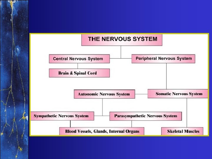
NERVOUS SYSTEMS • Central nervous system • Peripheral nervous system
CENTRAL NERVOUS SYSTEM p. 33 • Brain – Cerebral Cortex/Cerebrum • Motor cortex – Basal Ganglia/Diencephalon – sensory input – Cerebellum – motor control – Brain stem – sensory input • Spinal Cord
PERIPHERAL NERVOUS SYSTEM • Somatic – Sensory (Gamma) Nerves – Motor (Alpha) Nerves • Autonomic [FYI] – Parasympathetic – Sympathetic
Spinal Nerves p. 35
NERVE ANATOMY • A single nerve cell is called a neuron • A bundle or group of neurons make up a nerve • A nerve contains both afferent and efferent nerves
Afferent Neuron • Carry impulse towards the CNS (e. g. sensory nerves) • Synapse Towards CNS
Efferent Neuron • Carry impulse away from the CNS (e. g. motor nerves) 1. Stimulatory 2. Inhibitory
NEURON ANATOMY • Dendrite • Cell Body • Axon
AXON 1. 2. 3. 4. Conduction Myelin sheath Synapse Neurotransmitter
CONDUCTION Resting State: Na outside, K inside
CONDUCTION 4. Na K Pump 3. Repolarization 1. Depolarization 2. Propagation
MYELIN SHEATH
SYNAPSE
SYNAPSE
SYNAPSE • Quick Time Movie
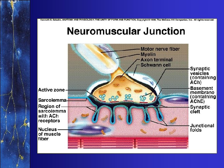
NEUROMUSCULAR JUNCTION
NEUROMUSCULAR JUNCTION
MOTOR UNIT • Motor unit = one motor nerve + all the muscle fibers it innervates. • Types – Fast (alpha -1) – Slow (alpha -2)
3 1 2
MOTOR UNIT Fast Slow
MUSCLE TENSION or FORCE or STRENGTH 1. Number of MU stimulated 2. Frequency of stimulation to each MU 3. Type of MU stimulated
MUSCLE STIMULATION: How does it begin?
1. Volitional Control (Motor Cortex) 1. Motor cortex 2. Motor neurons 3. Muscles
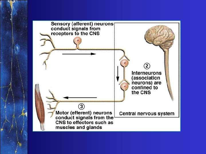
2. Reflex Control 1. Afferent neuron – Sensory neuron 2. Efferent neuron – Motor neuron
PROPRIOCEPTION & KINESTHESIS p. 37 • Proprioception – The ability to sense the position and location and orientation and movement of the body and its parts • Kinesthesis – The ability to feel movements of the limbs and body
PROPRIOCEPTION • Proprioceptors of the joints and skin – Meissner’s corpuscles – Ruffini’s corpuscles – Pacinian corpuscles – Krause’s end-bulbs
PROPRIOCEPTION • Proprioceptors of the muscles – Muscle spindles – Golgi tendon organs
Muscle Spindles
muscle spindle
Muscle Spindles • Provide proprioception • Sense the amount of stretch and the rate of stretch • Reflexes involving the muscle spindles is how we ‘feel’ a movement was done correctly • Spindles are a part of learning; we develop such reflexes as we practice skills and movements
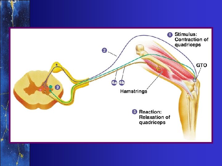
Golgi Tendon Organs
Golgi Tendon Organs • Sensitive to muscle tension and active contraction • Protect muscle from excess contraction force • Stimulation of GTO an afferent impulse is sent to the central nervous system • In turn, efferent impulses are sent to the… – Agonist muscle causing it to relax – Antagonist muscle causing it to contract
Neuromuscular Summary • An example from baseball. • A pitcher throws a curve ball to Sammy Sosa • Sammy’s eyes see the ball coming towards him and is able to identify the pitch as a curve ball • Sensory nerves (afferent) send that information to the CNS • In the CNS the sensory nerve synapses with motor nerves • The motor nerves in turn stimulates the muscles (deltoid and pectoralis major) required to swing the bat in the proper position to hit the ball
Neuromuscular Summary • Inside the fibers of the deltoid and pectoralis major, calcium is released • The calcium allows myosin heads to attach to actin • When the heads swivel the fibers of the deltoid muscle will shorten • The shortening of the fibers will pull on the humerus causing Sammy to swing the bat • The muscle spindles “tell” the CNS when the arm is in the correct position • If all goes as planned, the deltoid and pectoralis major will move his arms into a position to hit the ball
Neuromuscular Summary • An example from weight training. • A man is having his muscular strength tested on the bench press • For his first lift, 50 pounds is put on the bar • He will be using his pectoralis major muscle with has 500 motor units (300 slow twitch and 200 fast twitch) and his triceps muscle • His CNS stimulates 280 motor units leading to his pectoralis major muscle (180 slow twitch and 100 fast twitch) • End nerve ending stimulates a separate muscle fiber
Neuromuscular Summary • Calcium is released, myosin attaches to actin and swivels. • The pectoralis major and triceps muscles shortens and his arm extends outward raising the bar with 50 pounds on it. • After a few minutes of rest, 100 pounds is place on the bar • This time he stimulates 380 motor units (260 slow twitch and 120 fast twitch) • The weight is successfully lifted • After a few minutes of rest, 150 pounds is place on the bar
Neuromuscular Summary • This time he stimulates 460 motor units (280 slow twitch and 180 fast twitch) • As the pectoralis major muscle contracts the GTO in the pectoralis major are stimulated • They stimulate a sensory nerve leading to the CNS • In the CNS, the sensory nerve synapses with a motor nerve that will inhibit (relax) the pectoralis major muscle • The man is unable to successfully lift the 150 pounds.