Nervous System Tissues Dr Anderson GCIT Functions of

 – deliver information from the")

 Division – deliver information to the brain via")

 • Characteristics – VERY long lived (100+ years) – Do not")

 – secrete protein to provide • A protective")


")
 – Take messages from")

 conc. of cations (net positive) conc. of cations (net negative)")

 – Inside the cell is")


 total reversal of membrane potential (from -70")


")


 to happen anywhere")
- Slides: 42
Nervous System Tissues Dr. Anderson GCIT
Functions of the Nervous System • Sensation (sensory neurons) – deliver information from the outside environment to the brain • Integration (associative neurons) – process gathered information, generate thoughts and retain memories • Motor Output (motor neurons) – delivers a message from the brain (or as a reflex) to the muscles or glands
Major Divisions • Central Nervous System – Brain and Spinal Cord • Peripheral Nervous System – innervate different parts of the body to connect to the brain or spinal cord – Spinal nerves – Cranial Nerves
Peripheral Nervous System • Afferent (Sensory) Division – deliver information to the brain via impulses from specific nerves (heat, cold, pain, stretch, etc. ) • Efferent (Motor) Division – Carry impulses from the brain to muscles and glands – Somatic nervous system – controls voluntary muscles – Autonomic nervous system – controls involuntary actions
Autonomic Nervous System • Sympathetic Branch – generally stimulates activity in body tissues not under conscious control (e. g. increasing secretion of adrenaline in a dangerous situation) • Parasympathetic Branch – generally inhibits activity in tissues not under conscious control (e. g. preventing the bladder from flexing when holding urine)
Neurons (Nerve Cells) • Characteristics – VERY long lived (100+ years) – Do not divide once they differentiate and their role is set (? ) – Very high metabolic rate (i. e. high demands for O 2)
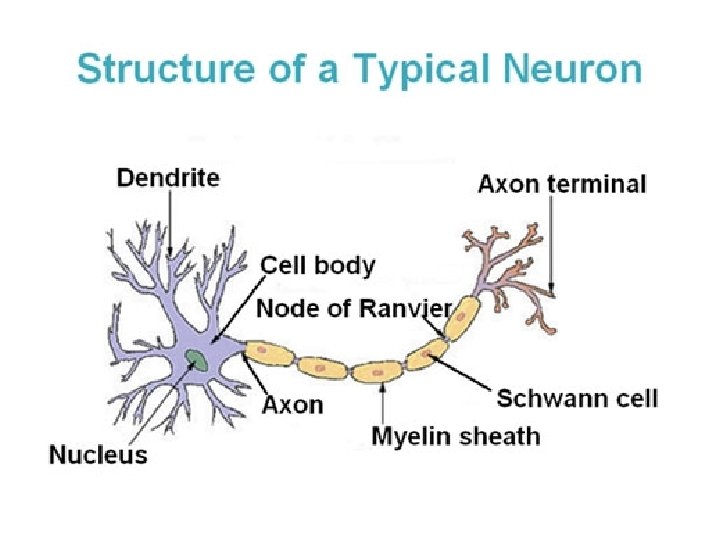
Neural Processes • Dendrites – receptive region of the neuron – Send/Receive information to/from other nerves • Axon – area between the cell body and the nerve terminus – Vary greatly in length from a few micrometers up to over a meter!
Nervous Tissue - Neuroglia (supporting cells) – secrete protein to provide • A protective scaffolding for neurons (matrix) • Insulation around long nerve cells (axons) to speed up action potential propagation
Neuroglia in the CNS • Microglia – serve as dormant immune cells – Upon sensing damage or disease of nervous tissue, microglia transform into a special type of macrophage that “eats” the pathogens or damaged nerve cells – This is critical, as immune cells are prevented from entering the CNS!!
Neuroglia in the CNS • Ependymal Cells – epithelial cells of the CNS, they line the cavities of the brain and spinal cord allowing diffusion • Ciliated to circulate cerebro-spinal fluid throughout the CNS
Neuroglia in the CNS • Oligodendrocytes – Add insulation to nerve fibers in the CNS • Myelin Sheaths
Neuroglia in the PNS • Schwann Cells – surround and form myelin sheaths around the nerve fibers in the PNS – also facilitate the regeneration of damaged peripheral nerves
Myelin Wrapping
Axons • Axons are the conducting region of the cells, generating and conducting action potentials along the nerve cell membrane (axolemma) • Generally one branch, but may have collaterals • Axon ends at its terminals (buttons) – up to 10, 000 branches or more – Function?
Myelin Sheath • Wraps around nerve axons – Protects the axolemma – Electrically insulates the axon while increasing the speed of action potential transmission • In the PNS, Schwann cells wrap around the nerve, making concentric rings • Eventually the cytoplasm of the cells are removed from the cell, leaving the Schwann cell membrane (the myelin sheath)
Myelin Sheath • As Schwann cells wrap around the nerve axon at regular intervals (about 1 mm apart) there are gaps in the myelin sheath called the Nodes of Ranvier
Cell Insulation Myelinated Unmyelinated Faster action potential Slower action potential transmission No loss of signal between cells Potential loss of signal between cells “White matter” in CNS “Grey matter” in CNS
Neuron Classification • Multipolar – 3 or more processes (1 axon and multiple dendrites) • Bipolar – 1 axon and 1 dendrite • Unipolar – 1 process splits (double-ended axon)
Functional Classification • Sensory neurons – (generally unipolar, some bipolar) – Take messages from sensory receptors to the CNS • Motor neurons – (multipolar) – Deliver impulses to effector organs (glands/muscles) • Associative neurons (most are multipolar) – In CNS - integrate sensory and motor
Membrane Potentials • Review! • Voltage – the electric “pressure” between two points (unit - Volts) • Current – flow of electrical charge (unit – amps) • Resistance – the hindrance to charge flow (unit – Ohms)
Electrochemical Potential (Neurolemma) conc. of cations (net positive) conc. of cations (net negative)
Channel Proteins • Let ions in or out What do these channels respond to? • Chemically gated proteins – • Voltage-gated proteins – • Mechanically-gated proteins-
The Electrochemical Gradient • Resting potential – (polarized state) – Inside the cell is more negative than outside the cell due to ion concentrations – Range from -40 to -90 m. V, depending on neuron type – How is this potential maintained?
Ion Dynamics and Transport Proteins • Proteins are specific in shape to allow only certain ions through (usually in one direction) • Leakage channels – ions move passively according to electrochemical gradients – Both Na+ and K+ • Sodium-potassium pump maintains the gradient (using ATP in the process)
How do Nerves Work? • Depolarization – ions are suddenly transported across the neurolemma (nerve cell membrane), causing the interior of the cell to become less negative • Graded potentials – slow, local depolarization – can initiate action potentials • Action potentials – fast, long distance depolarization
Graded Potentials • Local depolarizations in a single cell membrane • Voltage declines with distance from the stimulus • Response depends on magnitude of stimulus – Examples? • Slow, short-distance signals, but can initiate long-distance signals
Action Potentials • A very brief (milliseconds) total reversal of membrane potential (from -70 m. V to +30 m. V) • http: //www. youtube. com/watch? v=7 Eyhs. O ewn. H 4
What does this graph show?
AP Propagation • As voltage gated proteins are stimulated to open and shut, the nerve impulse moves away from the initial site of stimulation
Threshold and “All or Nothing” • Not all nerve stimulations start action potentials • However, if a value is reached (15 -20 m. V difference from resting) the nerve initiates an action potential • Because of this, nerves either ‘fire’ at this minimal level of stimulation, or not at all
Degree of Stimuli • All action potential are the same, either the nerve fires or it does not • How does the CNS know whethere is a strong or weak stimulus? • Answer:
Synapses • How do neurons communicate with other cells? • Synapses are microscopic gaps between nerve cells and the cells with which they send/receive information
Synapses • Electrical - Cells are basically “connected” via gap junctions that allow ions to flow directly between cells • Chemical – release and/or receive messages from adjacent cells via neurotransmitters
Electrical vs. Chemical Synapses
Neurotransmitters - Mechanism 1. AP at the axon terminal opens Ca channels, letting Ca into the cell 2. Ca causes vesicles containing neurotransmitters to be released 3. Neurotransmitters travel across synaptic cleft to receiving cell
Neurotransmitters - Mechanism • Neurotransmitter binds to receptors on the receiving cell • Chemically sensitive channel proteins open and allow ions to flow into receiving cell • Enzymes break down neurotransmitter to release them from receptors and degrade them
Excitatory vs. Inhibitory • Some neurotransmitters increase the rate at which neurons fire (excitatory) • Some neurotransmitters decrease the rate of neuron firing (inhibitory) • Given what you know about nerve physiology, HOW does this happen? ? ?
Nerve Conduction Velocities Unmyelinated Slow conduction velocity Myelinated Fast Conduction Velocity
Saltatory Conduction • “Jumping” depolarization on myelinated nerves • Only happens where nerve cell membranes are exposed to the extracellular environment • Speeds up nerve impulses and preserves signal strength
Saltatory Conduction
What’s the Difference? • Unmyelinated cells allow voltage changes (ion flow) to happen anywhere along the neurolemma • Myelinated cells only allow ion flow to happen at the Node of Ranvier – Increases electrical “pressure” at the node – Prevents the “leaking” of ions, keeping their concentration high