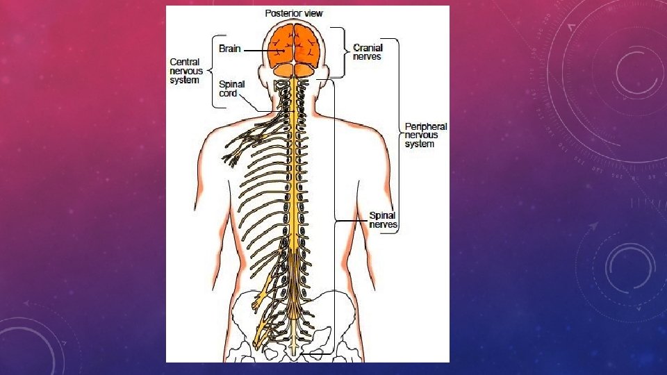
NERVOUS SYSTEM THE SPINAL CORD AND SPINAL NERVES


 -It is a nice, sunny day. . .")


, NOREPHINEPHRINE (NORADRENALINE) üACTEYLCHOLINE is the neurotransmitter that sends the")


 DIENCEPHALON – Area between the cerebral hemispheres and the brain stem. üIt")
 üPROTECTIVE STRUCTURES")

 ü A clear liquid that circulates in and around the brain")
 is divided into 4 visible")


 FRONTAL LOBE – Human’s have the")
 PARIETAL LOBE – Middle top (SUPERIOR) part of the brain. ü The")
 OCCIPITAL LOBE – Most posterior part of the brain. • Controls")


 MEDULLA OBLONGATA üBottom of the brain that connects the brain stem to the")


 – a tool used to measure the electric currents running through the")
 - a needle between the lower bony vertebrae")

 – When the myelin sheath is")

 - A short period of insufficient blood supply to the")


")


")

- Slides: 80
NERVOUS SYSTEM THE SPINAL CORD AND SPINAL NERVES CHAPTER 9
üThe NERVOUS SYSTEM acts as the CHIEF COORDINATOR for all the other systems. üThe nervous system is responsible for detecting as well as responding to all the changes (STIMULI) that the body faces. üTHERE ARE 2 PARTS TO THE NERVOUS SYSTEM: ü 1) CENTRAL NERVOUS SYSTEM – includes the brain and spinal ü 2) PERIPHERAL NERVOUS SYSTEM – includes all those nerves NOT included in the Central Nervous System üThe PNS consists of the following: üCRANIAL NERVES – they carry impulses to and from the brain üSPINAL NERVES – they carry messages to and from the spinal cord.
üEFFECTOR – Any tissue or organ that carries out a command from the nervous system. All muscles and glands fall into this category. üSOMATIC NERVOUS SYSTEM – Controlled voluntarily and all the effectors are skeletal muscle. üAUTONOMIC NERVOUS SYSTEM - (VENTRAL NERVOUS SYSTEM) – they control all the smooth muscle, cardiac muscle and glands.
• SYMPATHETIC NERVOUS SYSTEM – It is a nice, sunny day. . . you are taking a nice walk in the park. Suddenly, an angry bear appears in your path. üDo you stay and fight OR do you turn and run away? üThese are "Fight or Flight" responses. üIn these types of situations, your sympathetic nervous system is called into action - it uses energy - your blood pressure increases, your heart beats faster, and digestion slows down ühttps: //www. youtube. com/watch? v=O 16 k. GZ-Gm 74 – 16 sec
PARASYMPATHETIC NERVOUS SYSTEM (VISCERAL NERVOUS SYSTEM) -It is a nice, sunny day. . . you are taking a nice walk in the park. ü This time, however, you decide to relax in comfortable chair that you have brought along. ü This calls for "Rest and Digest" responses. ü Now is the time for the parasympathetic nervous to work to save energy. ü This is when blood pressure can decrease, pulse rate can slow, and digestion can start.
https: //ww w. youtube. c om/watch? v=0 IDg. Bl. CH Vs. A - 11
NEURONS AND THEIR JOBS üNEURONS –highly specialized cells of the nervous system üPARTS OF A NEURON: ü 1. CELL BODY – contains the nucleus and all other normal eukaryotic organelles ü 2. DENDRITES – fibers that conduct impulses to the cell body. ü Highly branched. The name dendrite in Greek means “tree” ü They are the receptors, meaning they RECEIVE the message first. ü 3. AXONS – Fibers that send messages AWAY from the cell body to either other neurons, muscles, or glands. ü It is made up of a single fiber which branches at the very end.
ü 4. MYELIN SHEATH – Made up of a fatty material called MYELIN that covers some axons. ü Job is to protect and insulate the fibers of the axon. ü In the PNS, this covering is produced by special connective tissues called SCHWANN CELLS that wrap around the axon like a jelly roll. ü When this is complete there are tiny spaces in between called NODES OF RANVIER. These tiny gaps are important for allowing the nerve impulses to travel much faster. ü 5. NEURILEMMA – Thin outermost coating of the Schwann cells. ü This is a tool to help repair peripheral nerves when injured. ü This repair is often very slow and uncertain
üNerves of the CNS are myelinated but DO NOT HAVE SCHWANN CELLS, therefore they have no Neurilemma. üBecause of this lack of neurilemma, CNS cells CAN NOT REPAIR THEMSELVES. üThis is why spinal cord injuries are often lifelong. üMYELINATED AXONS are often called WHITE FIBERS and are found in the WHITE MATTER of the brain and spinal cord. üThose axons that are not covered by a myelin sheath are called GRAY MATTER.
PNS NEURON
TYPES OF NEURONS üNerves that send messages TO the Spinal cord or BRAIN are called SENSORY NEURONS (UNIPOLAR or AFFERENT NEURONS). üThose that send the messages FROM the brain to the body are called MOTOR NEURONS ( MULTIPOLAR or EFFERENT) üNerves that send messages within the brain and spinal cord (CNS) are called INTERNEURONS (CENTRAL or BIPOLAR NEURONS)
ü A bundle of neurons in the PNS is called a called TRACTS. NERVE. In the CNS they are ü NEUR/O – means nerves ü The connective tissue around an individual fiber is called ENDONEURIUM ü The connective tissue around a fascicle is called a PERINEURIUM ü The connective tissue around the whole nerve is called the EPINEURIUM ü The Cranial nerves that contain only sensory fibers are called (AFFERENT NERVES) SENSORY ü Whereas, the cranial nerves that contain motor fibers are called (EFFERENT NERVES) MOTOR ü However most of the cranial as well as the spinal nerves contain both types of fibers
üThe nervous system also contains cells that support and protect. All these cells together are called NEUROGLIA or GLIAL cells. üSchwann cells are examples of neuroglia üDifferent type of Neuroglia: üThose that protect nervous tissue üThose that support nervous tissue üThose that aid in repair of other cells üThose that act as phagocytes to remove pathogens üThose that regulate fluids around and between cells
üASTROCYTES – Have a starlike appearance. They are found in the brain where they attach to capillaries and help protect the brain from harmful substances. üNeuroglia continue to reproduce their entire life, so most tumors of the nervous system are neuroglia.
üElectric charges carried by neurons is known as POTENTIAL üA NERVE IMPULSE starts with a organ then travels to the brain. üThis movement back to the brian is called the ACTION POTENTIAL. üOnce the impulse travels from the dendrite down the axon, it will jump over a space called the SYNAPSE with the help of neurotransmitters. üThe first neuron is called the PRESYNAPTIC CELL and the second one is called the POSTSYNAPTIC CELL.
üOther neurotransmitters are EPHINEPHRINE (ADRENALINE), NOREPHINEPHRINE (NORADRENALINE) üACTEYLCHOLINE is the neurotransmitter that sends the signal across the last neuron that connects to the muscles. • https: //www. youtube. com/watch? v=p 5 z. Fg. T 4 aof. A – neurotransmitter video (2)
• SPINAL CORD ü Links the PNS and the brain. ü Protected by VERTEBRAE, which continue from the occipital bone to coccyx. ü All the gray matter (UNMYELINATED) and white matter (MYELINATED CELLS) are arranged in columns running dorsally, one on top of the other. ü There are 2 pairs of columns ü DORSAL HORNS ü VENTRAL HORNS
REFLEXES üSometimes reactions are needed to happen so quickly that the signal does not travel from body part to brain and back. üSome travel from body part to Spinal cord then back. These are called REFLEXES. üREFLEXES – rapid uncomplicated automatic responses involving few neurons. ü 2 types of reflexes” ü SPINAL – these are the quick ones that are normally because of a sudden unexpected stimuli ü STRETCH – These happen when a muscle is stretched and responds by contracting. ( These are often used in physical exams to test the condition of your nervous system)
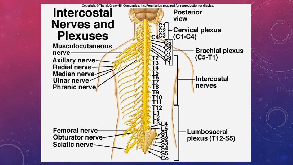
SPINAL NERVES ü 31 pairs of spinal nerves: each pair numbered based on the section of spinal cord it arises. üEach nerve is attached to the spinal cord by 2 roots: DORSAL ROOT and VENTRAL ROOT üGANGLION – collections of nerve cell bodies located outside the CNS.
BRANCHES OF SPINAL NERVES üNetworks of nerves fall into 3 groups: üCERVICAL PLEXUS – deliver muscles to the neck and receives impulses from the neck and back of head. The nerve that triggers the diaphragm is found here. üBRACHIAL PLEXUS – sends impulses from the shoulder, arm, forearm, wrist and hand üLUMBOSACRAL PLEXUS – Supplies the PELVIS AND LEGS üThe largest branch of the lumbosacral plexus is the SCIATIC NERVE – Passes through the dorsal side of pelvis, beneath the gluteus maximus, down the back of the thigh and down the dorsal lower leg to the foot. . At the beginning it is near 1 inch thick
• All skin sections are supplied by regions of nerves called DERMATOMES. • GANGLIA -a structure containing a number of nerve cell bodies, typically linked by synapses, and often forming a swelling on a nerve fiber.
AUTONOMIC NERVOUS SYSTEM • Regulates all the hollow organs and glands • Automatically • CELIAC GANGLION - Control the digestive organs • SUPERIOR MESENTRIC GANGLION – Controls the large and small intestines • INFERIOR MESENTRIC GANGLION – Controls the lower large intestine, urinary and reproductive systems
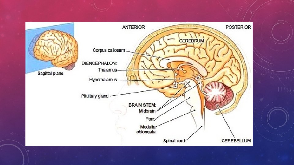
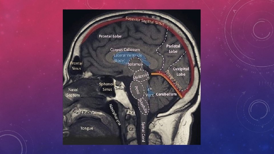
CHAPTER 10 – NERVOUS SYSTEM - BRAIN üThe Brain is protected by membranes, fluid and bones of https: //vm. tiktok. com/4 ub. Nkv/ - brain facts the skull üThe brain is divided into 4 major sections: ü 1) CEREBRUM – The largest part of the brain üDivided into a right side and a left side. üThese are called the RIGHT AND LEFT CEREBRAL HEMISPHERE üThese are separated by a large groove called the LONGITUDINAL FISSURE
ü 2) DIENCEPHALON – Area between the cerebral hemispheres and the brain stem. üIt includes the thalamus and hypothalamus ü 3) BRAIN STEM – connect the cerebrum and diencephalon with the spinal cord. üSuperior (TOP) part of the brain stem is the MIDBRAIN. üInferior (BELOW) the midbrain is the PONS followed by the MEDULLA OBLONGATA üRight below the MEDULLA OBLONGATA is where the spinal cord connects to the brain. This is where the foramen Magnum (hole in the skull) allows the spinal cord to exit the brain. ü https: //vm. tiktok. com/4 upvko/
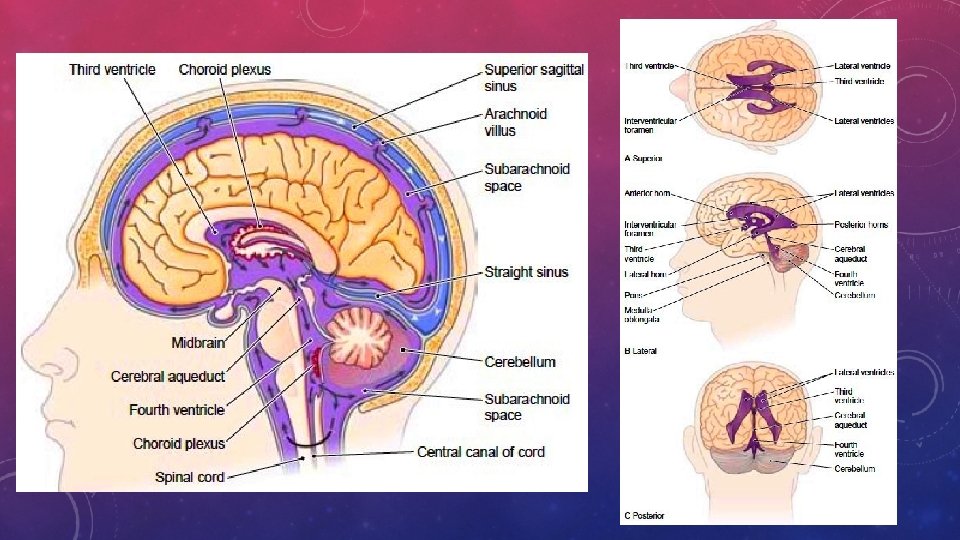
üCEREBELLUM – Posterior to the cerebrum ( Means little brain in greek) üPROTECTIVE STRUCTURES OF THE BRAIN AND SPINAL CORD üMENINGES – 3 layers of connective tissue that surrounds both the brain and the spinal cord to form a complete enclosure. üOUTER MOST IS CALLED: DURA MATER – thickest and toughest. There also areas of space called DURAL SINUSES which help drain blood from brain tissue
üMiddle layer of Meninges is called the ARACHNOID. This layer allows space for the movement of cerebrospinal fluid between all membranes üInner most layer is the PIA MATER. This holds blood vessels that supply nutrients and oxygen to the brain and spinal cord.
CEREBROSPINAL FLUID (CSF) ü A clear liquid that circulates in and around the brain and spinal cord. ü It’s job is to support nervous tissue, cushion shocks that would otherwise damage delicate structures as well as carry nutrients to cells and take wastes from cells of the brain/spinal cord. ü The CSF forms 4 spaces called ventricles in the brain. ü Each of these ventricles have a vascular network (Blood Vessels) called the CHOROID PLEXUS ü The largest ventricles are found in the 2 cerebral hemispheres. ü Extensions of these ventricles in the cerebrum are called HORNS. ü All ventricles communicate with each other through the FORAMINA (holes) ü 3 rd ventricles is surrounded by the diencephalon. ü The fluid flows from the 3 rd to the 4 th by the cerebral aquaduct
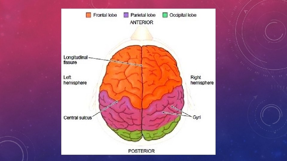
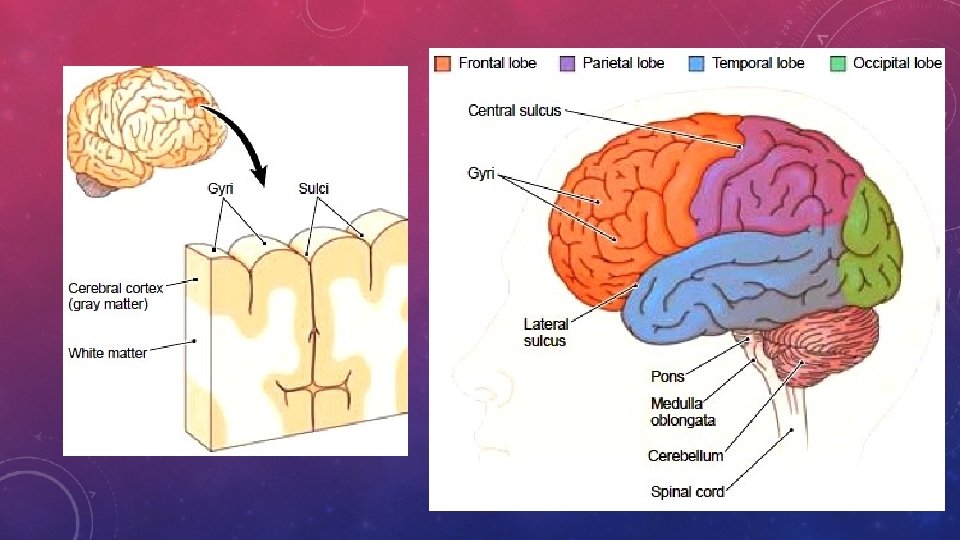
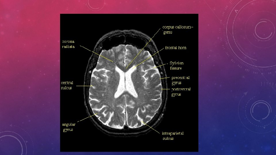
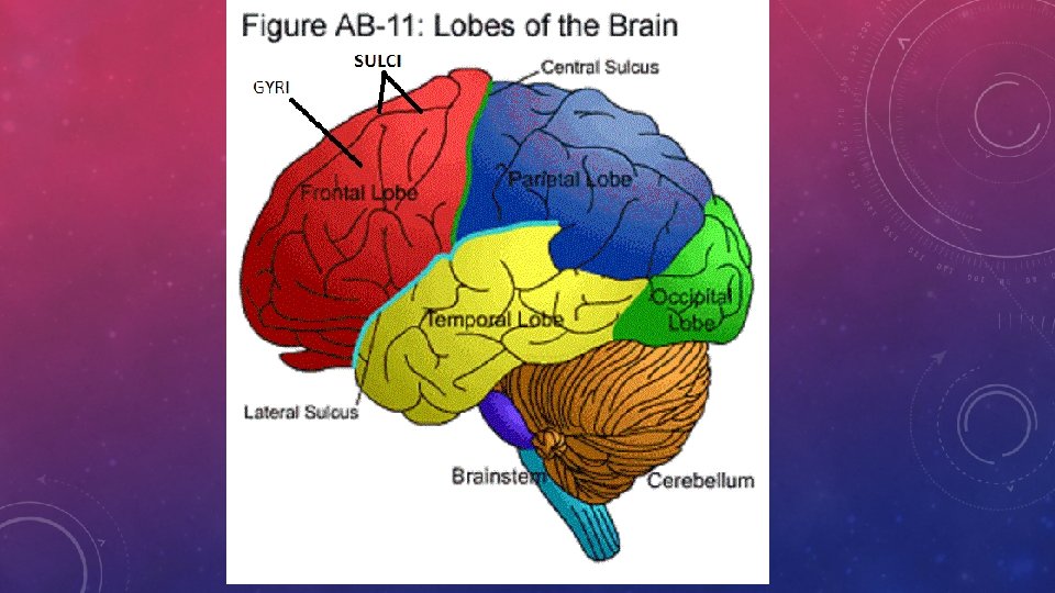
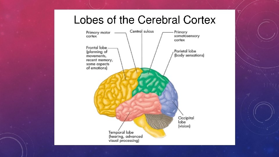
CEREBRAL HEMISPHERES ü Each side of the brain (HEMIPSHERE) is divided into 4 visible lobes. ü FRONTAL, PARIETAL, TEMPORAL and OCCIPITAL LOBES ü There is a 5 th lobe called the INSULA that is under the surface. ü The outer most tissue of the cerebral hemispheres is gray matter called CEREBRAL CORTEX. This is thin and is responsible for conscious thought, reasoning, abstract mental functions. ü The raised folds of the cortex called GYRI (Ji-Ri) ü The grooves in between the Gyri are called SULCI. There are many Sulci but 2 are very important: üCENTRAL SULCUS – between the frontal and parietal lobes üLATERAL SULCUS – Found between the frontal, parietal lobe and the temporal lobe
üInside the cerebral hemispheres, it is mainly white matter with a couple sections of gray. üThese gray matter areas are called BASAL NUCLEI or BASAL GANGLIA. üThese help regulate body movement and the muscles of facial expression. üThe neurotransmitter in this area is called DOPAMINE. üThe white matter found at the bottom of the longitudinal fissure is called the CORPUS CALLOSUM. üThis is the area that allows the left and right hemisphere to communicate with each other.
• https: //www. youtube. com/ watch? v=k. MKc 8 nf. PATI – Bozeman (13) • How the brain works (7) https: //www. youtube. com/ watch? v=9 Uukcd. U 258 A • Corpus Callosum tiktok https: //vm. tiktok. com/48 Mt. Fs/
FUNCTIONS OF THE CEREBRAL CORTEX üThis is where all impulses are received analyzed. üThe brain stores information and that is what forms our memories in the cortex. üThe cortex also controls things like discrimination. Judgment and all the associations we make with things. All our conscious an voluntary actions also arise from this area.
4 MAIN PARTS OF THE CEREBRUM ü 1) FRONTAL LOBE – Human’s have the largest frontal lobe of all animals. Anterior most part. üContains the PRIMARY MOTOR AREA which controls the following; ü all conscious control of the skeletal muscles üWritten and Motor Speech üProblem solving üBehavior, Personality and Mood üConcentrating and thinking
ü 2) PARIETAL LOBE – Middle top (SUPERIOR) part of the brain. ü The GYRUS in this area contain the PRIMARY SENSORY AREA – skin impulses such as touch, pain, and temperature are interpreted here. ü This area also is responsible for estimating distances, sizes and shapes, some language, body awareness and attention ü 3) TEMPORAL LOBE – Found on both sides of the brain. üAUDITORY AREA is found here. This is where all hearing impulses are interpreted. üLANGUAGE and MEMORY üOLFACTORY AREA is also here. Sense of smell from the nose
• 4) OCCIPITAL LOBE – Most posterior part of the brain. • Controls the vision area for interpreting all impulses from the retina in the eye.
3 R
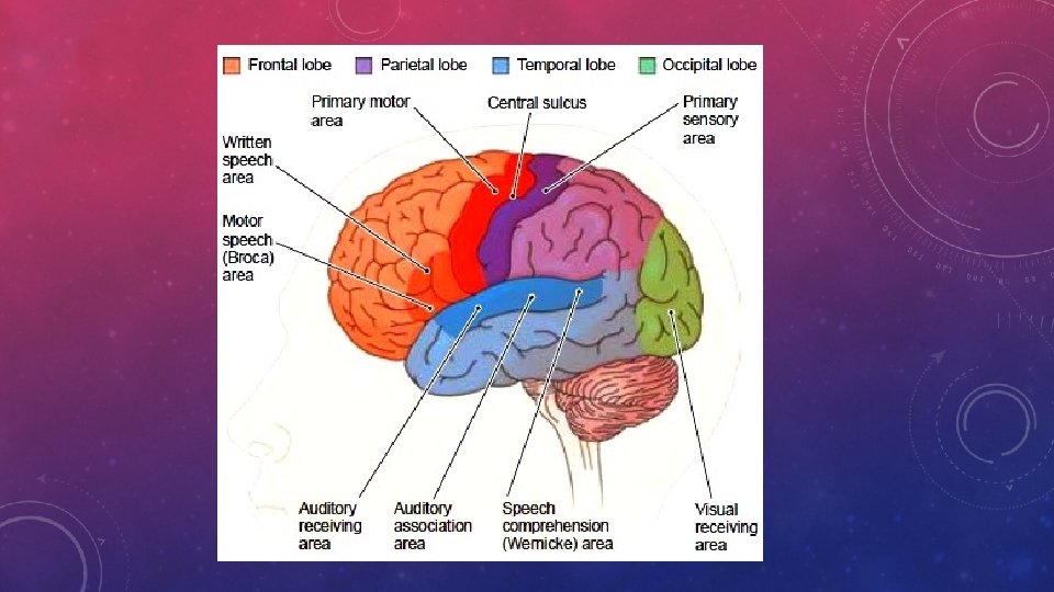
üIn the TEMPORAL LOBE, there are 3 auditory areas; üAUDITORY RECEIVING AREA – detects sound impulses transmitted from the environment üAUDITORY ASSOCIATION AREA – Interprets those sounds üSPEECH COMPREHENSION AREA or WERNICKE (VE-nihke) – recognizes speech and the meaning of words ü If someone suffers a stroke, this area is often affected. üIN the FRONTAL LOBE there are MOTOR AREAS for spoken and written communication üMOTOR SPEECH AREA (BROCA AREA) – controls the tongue, soft palate and larynx üIN THE OCCIPTIAL LOBE – the visual areas are found. There is only 1 area without a specific name
MEMORY üSHORT TERM MEMORY – retention of bits of information for a few seconds or perhaps a few minutes after which the information is lost unless reinforced. üLONG-TERM MEMORY – storage of information that can be recalled at a later time. These tend to be more fixed the more often a person repeats the remembered experience. ü So Short term memories can become long term if the person repeats it often enough.
THE DIENCEPHALON üAlso known as the INTERBRAIN. It is located between the cerebral hemispheres and the brain stem. üIncludes the THALAMUS and the HYPOTHALAMUS. üTHALAMUS – Controls all sensory impulses and directs them to the particular area of the cerebral cortex. üHYPOTHALAMUS – Lower in the DIENCEPHALON. ü Helps maintain Homeostasis by controlling body temperature, water balance, sleep, appetite and some emotions suc has fear and pleasure. ü Also controls both the Sympathetic and Parasympathetic systems. ü So it also influences the heart rate, blood pressure, hormone secretion and other vital body functions.
• https: //www. youtube. com/watch? v=j. Hxy. P-n. Uh. UY – actual brain (6)
üBetween the cerebrum and the diencephalon is a region called the Limbic System. This area is involved in emotional states and behavior. üHIPPOCAMPUS is included here and is involved in learning and the formation of Long term memories. üIt also is responsible for wakefulness and sleep.
THE BRAIN STEM üMade of 3 parts: Midbrain, Pons and the Medulla Oblongata ü 1. MID BRAIN üMost superior part üCenters for reflexes involving the eyes and ears üSome Cranial nerves originate here
2. THE PONS üLies in between the midbrain and the medulla oblongata. üConnects the 2 hemispheres of the cerebrum to the brain stem üResponsible for allowing impulses to travel to and from both parts üAlso responsible for certain involuntary reflexes such as controlling respiration and more of the cranial nerves found here
3) MEDULLA OBLONGATA üBottom of the brain that connects the brain stem to the spinal cord. üControls the muscles of respiration üRegulates the rate and force of your heartbeats üRegulates the contraction of smooth muscle in the Blood vessels so it controls the blood flow and blood pressure. üThe last of the cranial nerves are found here https: //www. youtube. com/watch? v=cu 7 A 8 LIz. L 1 o – 1 ½ min.
THE CEREBELLUM üMade of 3 parts: Vermis and the 2 hemispheres lateral üHelps coordinate voluntary muscles. Diseases of this area normally involve muscle jerkiness or tremors üHelps maintain balance üHelps maintain muscle tone
BRAIN STUDIES üNeurologist - a physician specializing in diseases of the brain, spinal cord and nerves. üCT SCAN ( COMPUTED TOMOGRAPHY) – Provides pictures of bone, soft tissue and cavities of the brain. üMRI – (MAGNETIC RESONANCE IMAGING) – More detailed views of the brain than the CT Scan. Can see brain bleeds (Hemorrhaging) that the CT scan not. üPET – (POSITRON EMISSION TOMOGRAPHY) – visualizes the brain in motion. ühttps: //www. youtube. com/watch? v=GHLBc. Cv 4 rqk – How PET SCANS WORK. (2)
üELECTROENCEPHALOGRAPH (EEG) – a tool used to measure the electric currents running through the billions of neurons in the brain. üElectrodes are placed on the head and pick up electrical signals produced as the brain functions. üThese signals are then amplified and recorded to produce brain waves seen on the machine. üUsed to study sleep patterns, detect disease such as epilepsy, strokes, encephalitis, dementia, etc. , located tumors, study the effects of drugs and to help determine brain death. üCheck out the scans on page 212 ühttps: //www. youtube. com/watch? v=C 0 OUex 2 pi. Gc – (6)
üLumbar puncture or spinal tap (LP) - a needle between the lower bony vertebrae of our spinal column allows a physician to sample the fluid, cerebrospinal fluid (CSF), surrounding the brain and spinal cord. üLab tests on the fluid are used for diagnostic purposes such as presence of bacteria in meningitis, special proteins in multiple sclerosis, or blood cells
• How does the brain work – excellent video by universal channel. 54 min. https: //www. youtube. com/watch? v=UTc Oc 80 Z 2 n 0 -
DISEASES OF THE NERVOUS SYSTEM üMS (MULTIPLE SCLEROSIS) – When the myelin sheath is randomly disintegrated. Causing short circuits. ühttps: //www. youtube. com/watch? v=M 7 O 78 Lvr. NSQ – 5 min üCerebrovascular accident (CVA) - the fancy name for a “stroke”. üA blood vessel in the brain may burst causing internal bleeding. üOr, a clot may arise in a brain blood vessel (a thrombus), or arise elsewhere (embolus) and travel to get stuck in a brain vessel which then deprives brain tissue of oxygen. üDepending upon the area of the brain involved, the patient may suffer paralysis, loss of speech or loss of vision. https: //www. youtube. com/watch? v=f. Kr. XCly 1 k. K 0 (3)
üISCHEMIC STROKES – When a blood vessel becomes blocked and restricts blood flow to a specific area. ü These can start in the brain or start somewhere else and travel to the brain. üHEMORRHAGIC STROKES – When a blood vessel breaks open and bleeding on the brain occurs. High Blood Pressure is a common cause. üSUBARACHNOID HEMORRHAGE – When blood spills into the CSF. ü APHASIA – a common aftereffect of a stroke. Loss or difficulty speaking ü https: //www. youtube. com/watch? v=SJYZ-As. K 18 k – medicine for strokes (3) üTPA – clot dissolving medication https: //www. youtube. com/watch? v=d. Clu 6 n. C 2 q. Ns
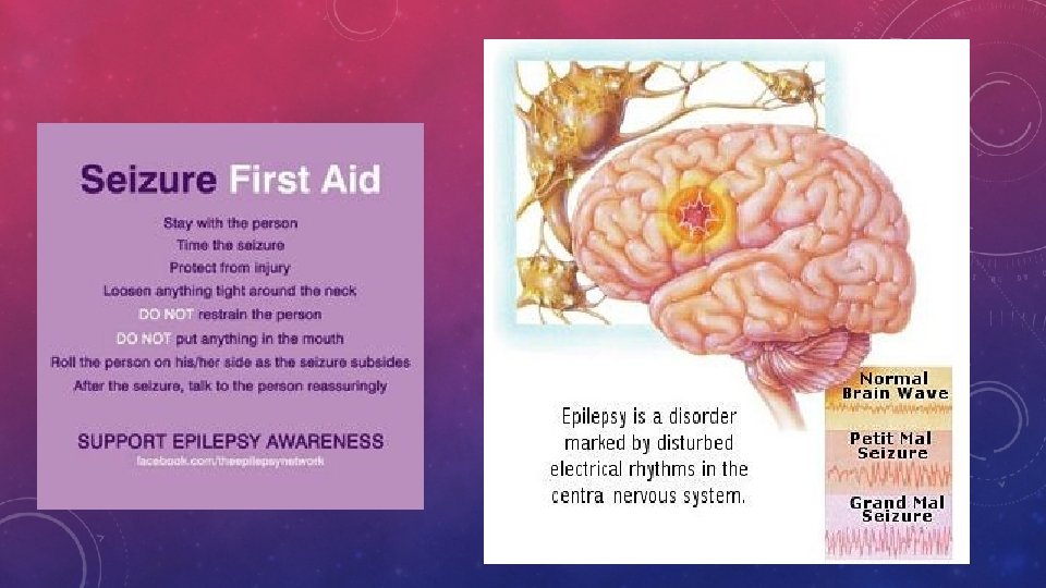
üTRANSIENT ISCHEMIC ATTACK (TIA) - A short period of insufficient blood supply to the brain can have the same signs and symptoms as a stroke such as weakness in an arm, a partial loss of vision, but the problem lasts less than 24 hours. üPeople who get TIA’s are at increased risk of having a stroke in the future. üEpilepsy - a Greek word for “seizure. ” Convulsions is another term used. üSeizures may have many causes and not all seizures are epilepsy. High fevers in young children may trigger seizures which are short in duration, easily controlled and, typically, have no permanent aftereffects. üEpilepsy is a specific condition which may occur at any age, seizures are more intense, longer lasting in duration, and recur with some frequency. The condition may be controlled with medication, or if unresponsive to drugs, may require surgery. üTik tok - https: //vm. tiktok. com/X 3 ns. SV/
üMENINGITIS – Inflammation of the brain and the meninges. üUsually caused by bacteria that enter the ear, nose and throat. üCan occur often where people live in close quarters. (College Dorms are a great place) üHeadache, stiff neck, nausea and vomiting are symptoms üDiagnosed by a lumbar puncture. üBacterial meningitis is treated with antibiotics but left untreated can lead to death. üViral Meningitis – more rare and must heal itself
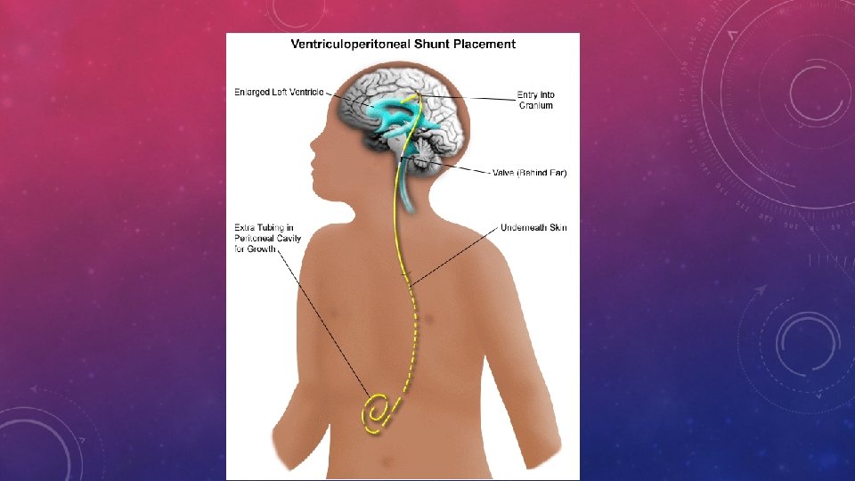
üENCEPHALITIS – inflammation of the brain. üCaused by viruses such as HIV, Rabies, Polio, West Nile, etc. üSymptoms, VERY HIGH FEVER, vomiting, coma üHYDROCEPHALUS –abnormal accumulation of CSF within the brain. üCan be caused by overproduction of it or lack of proper drainage. üThis puts extreme pressure on the brain against the skull and cam lead to brain tissue becoming destroyed. üCaused by congenital malformations, tumors, inflammation or hemorrhage. ü More common in infants and results in the cranium itself enlarging. ü Treated by a shunt. (bypass drainage tube)
üCEREBRAL PALSY – caused by brain damage occurring before or during childbirth. üTUMORS – Can occur at any age üMost originate from the NEUROGLIAS and are called Gliomas. üThese are unique in the fact that the do NOT metastasize to other body organs. üA common treatment for gliomas in the radiation seeds. The capsules implanted in the brain that release drugs on a schedule.
üEPIDURAL HEMATOMA – A bleed between the dura mater and the skull. Can put pressure on the blood vessels and interput blood flow to the brain. Headaches, partial paralysis, dilated pupils and coma. Pressure will need to be relieved of death can result. üSUBDURAL HEMATOMA – a bleed that results from a tear in a wall of a dural sinus. This often results from a blow to the front or back of the head. üINTRACEREBRAL HEMATOMA – A bleed into the brain tissue üCRANIOTOMY – When a portion of the skull is removed to reduce swelling.
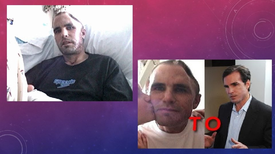
https: //www. youtube. com/watch? v=rx 3 nf. UKvr. Z 8 – 7 brain injury http: //www. cbsnews. com/videos/gabby-giffords-speaks-four-yearsinto-her-recovery/ - (9) Gabby today
üCEREBRAL CONCUSSION – results from a blow to the head causing the brain to slam against the skull. üSymptoms can include – loss of consciousness, headache, dizziness, vomiting, impaired brain function, uneven and unresponsible pupil reactions. üAny injury to the brain can result in TRAUMATIC BRAIN INJURY (TBI). üTraumatic brain injury (TBI), also known as intracranial injury, occurs when an external force traumatically injures the brain üEach year an estimated 1. 5 million Americans üMost commonly caused by motor vehicle accidents, gunshot wounds, falls, shaken baby syndrome and second impact syndrome ( a second head injury before the first one is fully healed. ) ü https: //www. youtube. com/watch? v=F 4 fo. Y 1 Etm. Ko – 11 Preston’s story
üALZHEIMERS – results from an unexplained degeneration of the cerebral cortex and hippocampus. ü Gradual and eventually causes severe intellectual impairment with mood changes and confusion. ü There are some medicines that will delay the progression of the disease, but there is currently no cure. üMULTI INFARCT DEMENTIA – accumulation of brain damage resulting from chronic ischemia ( lack of blood supply) Symptoms are similar to Alzheimer s. üPARKINSON’ S DISEASE – progressive neurologic condition characterized by tremors, rigidity of limbs and joints, slow movement and impaired balance. ü Normally a result of the part of the brain that produces dopamine. This results in lost of voluntary movement. Muhammad Ali ü To little dopamine ü Treated by a drug called L DOPA – which mimics the effects of dopamine ü ** Parkinson’s treatment - https: //www. youtube. com/watch? v=w. ZZ 4 Vf 3 Hin. A – 57 sec
• Gilles de la Tourette syndrome is a condition that causes a person to make repeated, quick movements or sounds that they cannot control. • These movements or sounds are called tics. Caused by too much dopamine • The condition is commonly called TOURETTE SYNDROME. • The early symptoms of Tourette syndrome are almost always noticed first in childhood, with the average onset between the ages of 7 and 10 years. • males are affected about three to four times more often than females. It is estimated that 200, 000 Americans have the most severe form of Tourette syndrome, • Simple motor tics are sudden, brief, repetitive movements that involve a limited number of muscle groups. • Some of the more common simple tics include eye blinking and other vision irregularities, facial grimacing, shoulder shrugging, and head or shoulder jerking. Simple vocalizations might include repetitive throat-clearing, sniffing, or
ühttps: //www. youtube. com/watch? v=Xn 5 D 44 uto. BI – Muhammad Ali (3) üThere are 12 pair of Cranial nerves. Numbered using roman numerals Beginning anteriorly. Pg. 217 ü The first two pairs are the only 2 NOT ARISING FROM THE BRAIN STEM. üOLFACTORY NERVE – Smell üOPTIC NERVE – vision üOCULOMOTOR NERVE – contraction of eye muscles üTROCHLEAR NERVE – supplies one eyeball muscle üTRIGEMINAL NERVE – great sensory nerve of the face. This is the nerve that the dentist anesthetizes to work on your teeth.
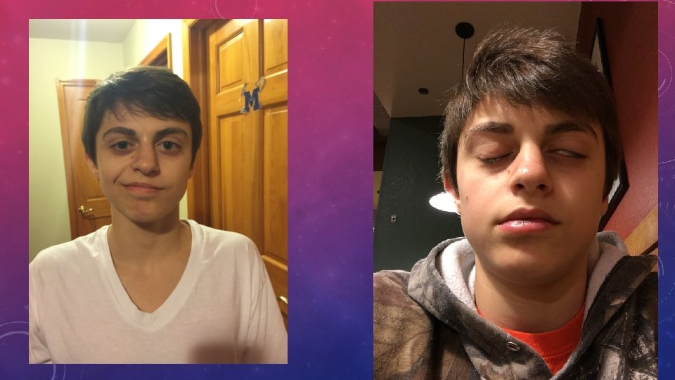
üABDUCENS NERVE – another eyeball muscle nerve üFACIAL NERVE – Facial expression as well as taste, salivary glands and lacrimal (tear duct). This is the one damaged by BELL’S PALSY üVESTIBULOCOCHLEAR NERVE – hearing and equilibrium üGLOSSOPHARYNGEAL NERVE – Back of tongue and the swallowing muscles in the pharynx (Throat) üVAGUS NERVE – LONGEST supplies most of the organs of the thoracic and abdominal cavities as well as the voice box (LARYNX). üACCESSORY NERVE – Controls the neck muscles , trapezius and sternocleidomastoids üHYPOGLOSSAL NERVE – controls the muscles of the tongue.
üBELL’S PALSY – partial facial paralysis caused by damage to the facial nerve. Usually on one side of the face. Normally caused by some type of infection. üNEURALGIA – Nerve Pain üNEUROPATHY - disease or dysfunction of one or more peripheral nerves ( general name for all nerves that bring messages to and from the brain), typically causing numbness or weakness. üNervous system – one of the first to develop as an embryo. (by the end of 3 rd week)
DAY 12 Day 1
• https: //www. youtube. com/watch? v=BR 2 EKZB_qz 4 – parkinsons treatment (2)
Documentary on Tourettes https: //www. youtube. com/watch ? v=dt. CUYUBOnzk – 7 min http: //www. youtube. com/watch ? v=v. ZQhp 3 y. Eg. YM - Michael J Fox – 14 Review of how the brain works. https: //www. youtube. com/watch ? v=9 Uukcd. U 258 A – 7 http: //bobwoodrufffoundation. or g/woodruff/ - 7 -