Nervous System Textbook Reference chapter 7 Objectives Unit

Nervous System Textbook Reference: chapter 7

Objectives Unit 7 7. 1 Name the organs of the nervous system and their functions within each classification of the nervous system 7. 2 Describe the anatomy of a neuron and each part’s function 7. 3 Differentiate between the 3 types of neurons and their functions 7. 4 Describe the changes in a neuron when it is polarized, depolarized & re-polarized 7. 5 Name the 4 main regions of the brain and their subdivisions 7. 6 Name the purpose and structure of the spinal cord 7. 7 Describe a reflex arc, its purpose and the anatomy of the process 7. 8 Compare how different neuroglial cells work to support neurons

Objectives Unit 7 7. 9 Explain how an action potential is transmitted 7. 10 Describe the unique functions of the anatomy of a brain 7. 11 Identify the parts of a sheep brain 7. 12 Identify the homeostatic mechanisms of the nervous system

The Function n To act as the master controlling and communicating system for the body http: //www. agen. ufl. edu/~chyn/age 2062/lect_20/nervsys. gif
 n n Includes brain and spinal cord")
Structural Classifications n Central Nervous System (CNS) n n Includes brain and spinal cord Acts as integrating and command center n Peripheral Nervous System (PNS) n Includes nerves outside of CNS • Spinal nerves • Cranial nerves n Links all parts of body by carrying impulses
 LOOK THEM UP!! You are")
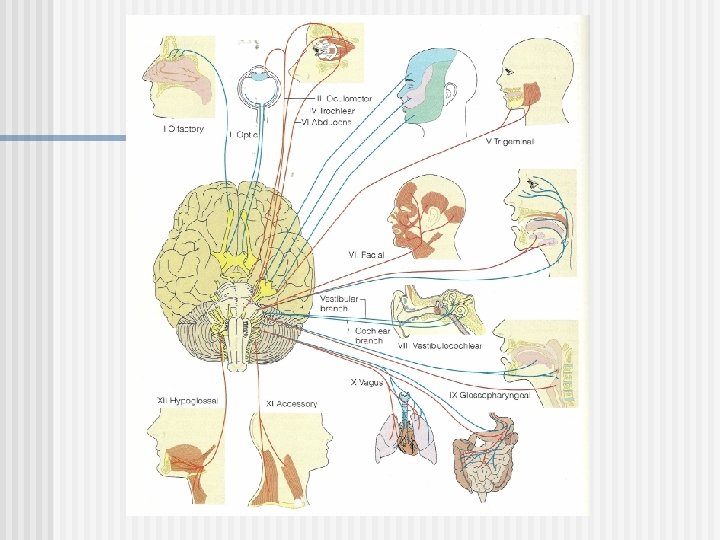
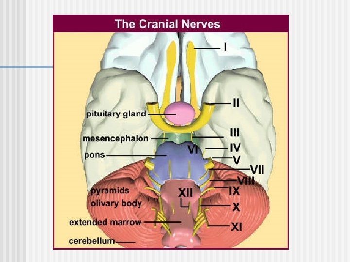
PNS Assignment There are 12 cranial nerves (pgs. 231232) LOOK THEM UP!! You are responsible for knowing their number, name and function. For example: Cranial nerve I: olfactory, sense of smell n Quiz to be announced!!

PNS: Cranial Nerves 12 pairs n n n I. Olfactory II. Optic III. Oculomotor IV. Trochlear V. Trigeminal VI. Abducens n n n VII. Facial VIII. Vestibulocochlear IX. Glossopharyngeal X. Vagus XI. Accessory XII Hypoglossal “Oh, oh, to touch and feel very good velvet, ah!”
 Somatic system: voluntary nervous system Contains sensory and")
Subdivisions of PNS n 1. ) Somatic system: voluntary nervous system Contains sensory and motor nerves n Makes you aware of the world around you & allows you conscious control n
 Autonomic System: involuntary nervous system Consists of motor")
Subdivisions of PNS n 2. ) Autonomic System: involuntary nervous system Consists of motor nerves that carry impulses to organs, blood vessels and glands n The unconscious or “automatic control” n

Subdivisions of Autonomic System n A. Sympathetic Division n n Active during stress heart rate, respiratory rate and rate of ATP breakdown n B. Parasympathetic Division n Active during conditions of normal organ functioning

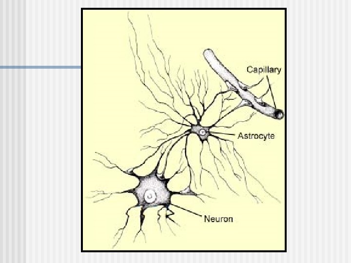
Cells of the Nervous System n 1. Neuroglia Acts to support neurons n Makes up ~ 90% of brain and spinal cord (s. c. ) n There are 5 different types of neuroglial cells n

Assignment n n n Spilt your paper into 4 grids-1 grid for each cell type Using pages 204 -207 in your text, draw the different types of neuroglia and list their location and function within the nervous system below each drawing DO NOT draw Schwann cells but do list its location and function

Cells of the Nervous System n 2. Neurons The conducting nerve cell n Senses changes in environment, integrates information, carries out motor responses n Structure: each neuron varies in shape and size, but all have 3 main parts (dendrite, axon and cell body) n

Neuron Anatomy A. Dendrite: a process off of the cell body; receives impulses from adjacent neurons & conducts electrical impulses TOWARD the cell body; ~1000/neuron

Neuron Anatomy B. Axon: aka nerve fiber; acts to transmit impulses AWAY from the cell body; 1 axon/neuron C. Cell Body: contains the nucleus and metabolic center of the cell

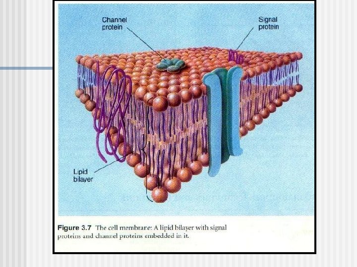
Neuron Anatomy D. Myelin sheath: Lipid covering on some neurons. It encloses the axon & speeds up the rate of nervous conduction i. Gray matter: non-myelinated nerve fiber ii. White matter: myelinated nerve fiber

Myelinated Neuron Non myelinated Neuron

Multiple Sclerosis

Neuron Anatomy E. Nodes of Ranvier: gaps in myelin sheath @ regular intervals; aids in increasing speed of nervous conduction See separate notes for how to conduct nervous impulses

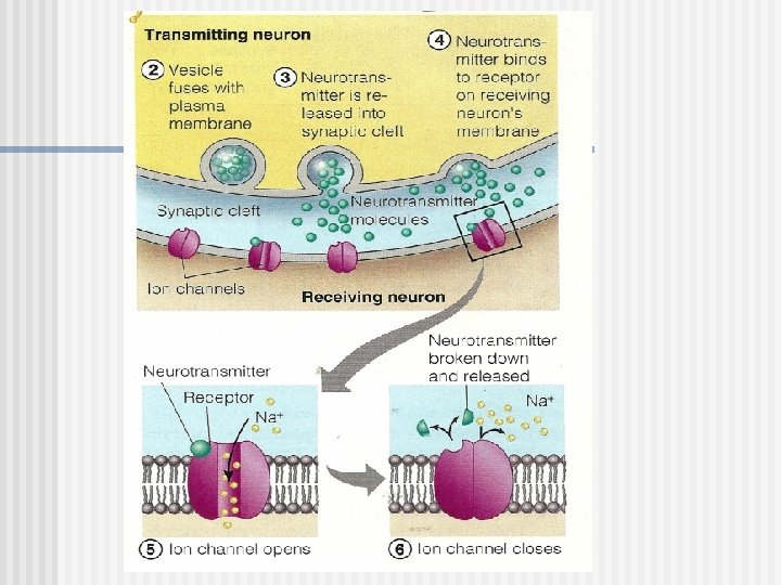
How neurons communicate at synapses

Nerve Conduction Polarized – Nerve at Rest

Nerve Conduction Stimulus initiates neurotransmitter

NERVE CONDUCTION Depolarization & generation of the action potential

NERVE CONDUCTION Propagation of the action potential

Nerve Conduction Repolarization

Once impulse stops, the Na – K pump restores the neuron to its resting state.

Impulse Conduction Unmyelinated neurons slower than myelinated n Impulses on myelinated neurons seem to jump from one node of Ranvier to the next n
 Neurons: transmit impulses from PNS to CNS")
3 Types of Neurons 1. Afferent (sensory) Neurons: transmit impulses from PNS to CNS i. e. pain receptors and proprioceptors 2. Efferent (motor) Neurons: transmit impulses from CNS to muscles or glands
: located within CNS and conducts impulses")
3 Types of Neurons 3. Interneurons (association neurons): located within CNS and conducts impulses between sensory and motor neurons

Neurons Classified by Function

Reflex Arc

Reflex Center n n n Reflex: an extremely rapid response to an emergency situation Occurs in spinal cord or lower brain; does not require thinking The Reflex Arc: n n n Receptor: at end of sensory neuron; can generate an action potential to CNS Association neurons: receive info. from sensory neuron and routes response to motor neuron Motor neurons: conduct impulses to effectors

Think About This n n n A finger comes in contact with a hot stove. Draw and explain the reflex arc, involved in this emergency situation. Label the 3 different neurons in your picture Include the stove, finger, arm and spinal column in your drawing/explanation

5 basic elements of all reflex arcs

Central Nervous System Left Brain Right Brain § Language § Visual/spatial info. y r o § Logic § Imagination e h t s i h t ? § Analytical thought § Insight s e e u o r t d l § 3 -D forms o Bu Skill § Scientific h l l i t s § Numbers § Musical Awareness § R hand control § L hand control

'Right Brain' or 'Left Brain' Myth Or Reality? By John Mc. Crone n n n The 2 sides are actually more complementary…. Left: more detail oriented Right: concentrates on the broad background picture Example: when listening to speech, the left brain concentrates on the vocabulary and grammar, while the right brain picks up the intonation and emotion Example: right side strokes

Boy Brain vs. Girl Brain? ? Structural Differences n Males: more white matter n larger hypothalamus n n Girls: more gray matter n larger corpus callosum n

Boy Brain vs. Girl Brain? ? Functional Differences n Females n n n Better verbally Memory fluency Color id Written tests Un-timed tests Better grades More nurturing More accurate/detail oriented Navigation by landmarks More subdued/passive Interpreting emotions & hidden meanings More intuitive n Males n n n n Visual-spatial activities Aiming at moving or stationary objects Throwing and intercepting projectiles Navigation by distance, time or spatial clues Better at multiple choice tests More aggressive-enjoys rough-housing Excel in higher levels of math

Central Nervous System Brain and Spinal Cord n Brain will be learned in inquiry assignment n

CNS: The Brain n 4 major portions: Cerebrum: largest; sensory & motor function, higher thought, memory & reasoning n Cerebellum: coordination of skeletal muscles n Diencephalon: sensory information n Brain stem: connects parts of nervous system; controls visceral activities n

CNS: Cerebral Lobes n Frontal Lobe: anterior portion n n Primary motor area Concentration, planning, problem solving Broca’s Area: motor speech Temporal Lobe: lateral lobe n n n Interpretation of sensory impulses, memory, visual & auditory patterns Auditory area Olfactory area

External Brain Diagram

CNS: Cerebral Lobes n Parietal Lobe: superior, lateral lobe Sensory area n Understanding speech; using words n n Occipital Lobe: posterior lobe Vision n Combining visual images; visual recognition n

Internal Brain Structures

Internal Brain Structures
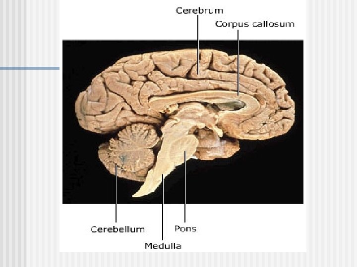

Connecting the Cerebral Hemisphers Corpus Callosum: white matter fiber tract for communication between right and left halves n Example: n left side = sense of others n right side = sense of self n Becoming an adult means connecting these n

CNS: Cerebellum n n Located inferior to occipital lobes and posterior to brain stem Functions in: positioning of limbs, coordinating skeletal movements, maintenance of posture, coordinating cognitive processes

CNS: Diencephalon n Located between cerebral hemispheres; superior to midbrain n Thalamus: central relay center for sensory impulses; allows awareness of pain, touch & temperature Hypothalamus: regulates visceral activities; links nervous and endocrine systems Also holds: optic chiasma, posterior pituitary gland, pineal gland
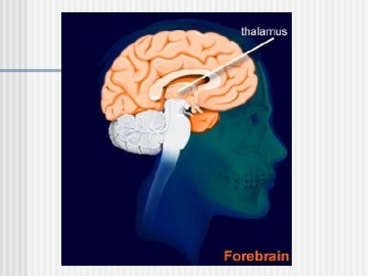

CNS: Diencephalon Amygdala: the emotional center n Hippocampus: short term memory n Doesn’t develop until about 3 years of age n Teens let this part of the brain “run things” while adults use their frontal lobe n

CNS: Brainstem Connects cerebrum to spinal cord n Midbrain: inferior diencephalon and superior pons n n n Visual, auditory reflex centers; head movement for hearing Pons: rounded bulge inferior to midbrain and superior to medulla n Relays sensory impulses; helps regulate breathing

CNS: Brainstem n Medulla Oblongata: from pons to spinal cord n Cardiac center, vasomotor center, respiratory center

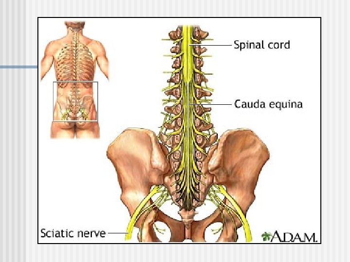
CNS: Spinal Cord A continuation of the brain running from base of brain to 1 st or 2 nd lumbar vertebrae n Is enclosed within spinal column n Is ~ size of thumb except at enlarged cervical and lumbar plexuses (spinal nerve distribution to upper and lower limbs) n

PNS: Spinal Nerves 31 pairs formed from ventral and dorsal roots of spinal cord n Plexuses: complex network of spinal nerves n Serves motor and sensory needs of limbs n Cervical, brachial, lumbar, sacral n

Nerve Plexes

PNS: Plexuses n n n Cervical Plexuses: C 1 -C 4; feeds muscles and skin of neck Brachial Plexuses: C 5 -T 1; deep in shoulders b/n neck & armpits Lumbosacral Plexuses: T 12 -coccyx n n n Extends from lumbar region into pelvic cavity Feeds muscles & skin of lower abdominal wall, external genitalia, legs and feet Feeds muscles & skin of arms

CNS: Spinal Cord n n Provides a 2 -way pathway to and from the brain Serves as a reflex center Has 31 linear segments giving way to 31 spinal nerves (relay info. from PNS to CNS and vice versa) Cauda Equina: collection of spinal nerves at the inferior end of the vertebral column

http: //anatomy. med. umich. edu/modules/spinal_cord_module/Files/entire%20 spinal%20 cord. jpg

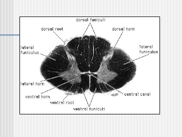
Spinal Cord Cross-Section n Gray Matter: unmyelinated n Located in center of cord forming an “H” • Posterior Horn: upper “arms” of H; contains terminal ends of sensory neurons • Anterior Horn: lower “legs” of H; contain cell bodies of motor neurons • Lateral Horn: between post. & ant. horns; contain cell bodies of motor neurons found within autonomic nervous system

Cross Section Spinal Cord

Spinal Cord Cross-Section Gray Commissure: horizontal bar connecting right and left sides of the “H” n Central Canal: in center of gray commissure; carries cerebrospinal fluid throughout canal n

Spinal Cord Cross-Section n White Matter: myelinated fibers Surrounds central gray matter n Divided into anterior, posterior and lateral columns n Each column holds longitudinal nerve tracts n • Ascending tracts: carry sensory info to brain • Descending tracts: carry motor info. away from brain
: n")
Protections of CNS n Bone: skull and vertebral column n Cerebrospinal Fluid (CSF): n n n Made from the blood Forms a watery cushion that protects tissue from trauma Changes in composition indicates brain pathology

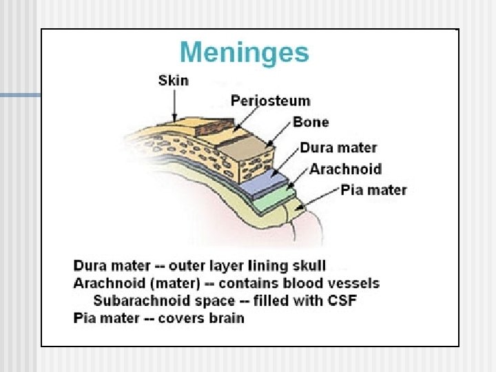
Protections of CNS n Meninges: connective tissue membranes n n n A. Dura mater: tough outer covering of brain and s. c. B. Arachnoid mater: middle layer C. Pia mater: inner surface which clings tightly to surface of brain and s. c. • Subarachnoid space: separates the arachnoid and pia maters; filled with CSF

Protections of CNS n Blood-Brain Barrier n n Composed of least permeable capillaries in body Brain depends on a very constant internal environment • Allows: water, glucose, and essential amino acids • Disallows: urea, protein and most drugs • Cannot block: fats, respiratory gases and fat soluble molecules (alcohol, nicotine and anesthetics)

Nervous System Ongoing Assignment 7. How is the high sodium ion concentration maintained on the outside of the plasma membrane? 8. The rapid movement of which ions into the cell leads to depolarization? 9. What two changes in the plasma membrane lead to repolarization? 10. What is the chemical that carries an impulse between the synaptic cleft between neurons?

Nervous System Ongoing Assignment 11. What is the outermost meningeal layer? 12. How does the posterior gray horn differ functionally from the anterior gray horn in the spinal column? 13. What are the conduction tracts in the spinal column? 14. What are the components of a reflex arc?
- Slides: 76