Nervous System Sensory Mechanisms and Motor Mechanisms CVHS

 – brain and spinal")
 • PNS – sense stimuli inside and outside the body,")

 nodes")


 Skeletal Muscle • two kinds • fast-twitch (white meat)")


- Slides: 29
Nervous System, Sensory Mechanisms and Motor Mechanisms • CVHS • Chapters 37 -39
Organization of the Nervous System • Central Nervous System (CNS) – brain and spinal cord • Peripheral Nervous System (PNS) – nerves outside of CNS – cranial nerves • connect brain w/ upper body – spinal nerves • Connect spine w/ regions of the body below the head
Peripheral Nervous System (PNS) • PNS – sense stimuli inside and outside the body, helps to control internal environment • Sensory/Afferent Division – send impulses to the CNS • Motor/Efferent Division – send impulses away from the CNS – effectors: control voluntary and involuntary muscles
Autonomic Nervous System • Involuntary: smooth and cardiac muscle – Sympathetic • increases energy consumption and prepare for action • Fight or Flight response – Parasympathetic • Enhance activity to gain and conserve energy
Neuron cell body myelin sheath synaptic terminal dendrites Schwann cells synapse axon (hillock) nodes of Ranvier terminal branches
Neuron Structure • Cell body: – performs cellular functions • Dendrites: – Receive information • Axon (hillcock): – Point @ which impulse starts when threshold is exceeded • myelin sheath: – lipid layer surrounding nerve cell • Nodes of Ranvier: – Holes in myelin where Na can move into the cell • Schwann Cell: – produce myelin in the PNS • synaptic terminal: – End of axon • Synapse: – communication junction between 2 nerve cells, where neurotransmitters move
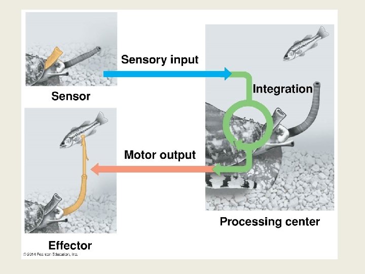
Functional Organization of Neurons 3 Classes of Neurons • sensory neurons convey impulse from sensory receptors to CNS • interneurons integrate sensory input and motor output • motor neurons convey impulses from CNS to effector cells • Function as a reflex arc: • Sensory neuron, to interneuron, to motor neuron
The Knee-jerk Reflex 1. Tap patellar tendon 2. Sensory receptors sense stretch in quadriceps 3. Sensory neurons convey info. to spinal cord 4. Synapses with motor neuron in spinal cord 5. Motor neuron conveys signal to quadriceps 6. Synapse with interneuron in spinal cord 7. Interneurons inhibit other motor neurons (hamstring) 8. Prevents the hamstring from contracting (no resistance to quads contracting).
The Nature of Neural Signals Membrane Potential • the difference in voltage across the plasma membrane + arises from differences in ionic composition (Na+/K+ pump) - normal: positive outside; negative inside (-70 m. V)
Action Potential Excitable Cells • cells that have the ability to change their membrane potentials • neurons and muscle cells • Resting potential (unexcited) • Change from resting potential can result in active electrical impulse • Gated ion channels open or close in response to stimuli • Hyperpolarization • increase in the electrical gradient opens K+ channel; • increase outflow of K+; more negative, no impulse • Depolarization • reduction in the electrical gradient • opens Na+ channel • increase inflow of Na+; less negative, can cause nerve impulse • action potential: a brief reversal of membrane polarity
Graded Potentials and the Action Potential in a Neuron
Propagation of the Action Potential • Membrane becomes depolarized by reacting to a stimulus. – Must cross a threshold of -55 m. V – Action potential – all or none response – Increase frequency = increased stimulus • Na+ rushes into the axon causing the charge reversal from – to + inside the axon • K+ leaves the axon after the action potential is finished – Refractory period – another action potential can not occur yet – Resting potential is restored
Action Potential
Saltatory Conduction • speeds the propagation of action potential + nodes of Ranvier: gaps between myelinated regions - action potentials “jump” from node to node
Conversion of Signal: Electrical to Chemical • Depolarization causes influx of Ca 2+ • Release of synaptic vesicle contents • Neurotransmitter released into cleft • Molecules bind to receptors • Opens ion channels
Diversity of Nervous Systems
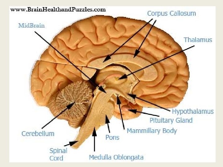
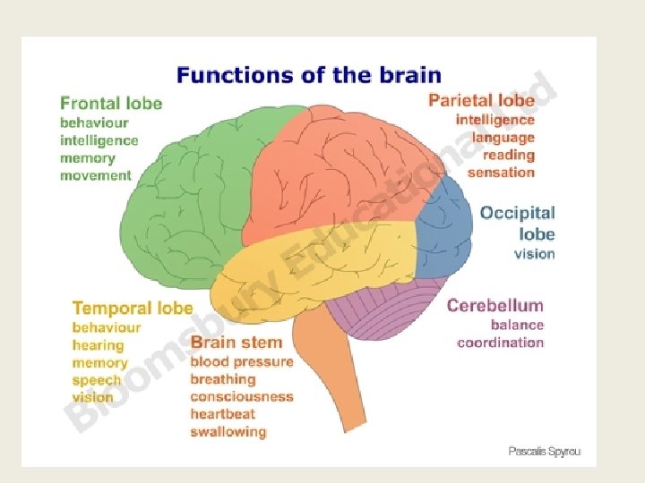
The Brain Structures • Cerebrum – Cerebral cortex – outer portion of the cerebrum – gray matter – Cerebral hemispheres – left and right sides – Corpus callosum – allows communication between the left and right hemispheres • Brainstem – Medulla oblongata – controls autonomic and homeostatic functions – Pons – regulates breathing – Midbrain – large scale body movements - walking • Cerebellum – coordination, learning, and decision making • Diencephalon – Thalamus – input center for sensory info and output center for motor info – Hypothalamus – homeostasis – links to the endocrine system
Cerebral Hemispheres • • Language • Math • Logical operations Visual and auditory details • • Left • • • Right Pattern recognition Face recognition Spatial relations Nonverbal thinking Emotional processing Understanding and reacting to stress Music If you’ve always thought of yourself as a “numbers person” or a creative sort, this research doesn’t change anything. But it’s probably inaccurate to link these traits to one side of your brain. We still don’t know a lot about what determines individual personality; but it seems unlikely that it’s the dominance of one side of the brain or the other that matters. From: Right brain/left brain, right?
Cerebral Cortex • Frontal lobe – Speech • Temporal lobe – Smell – Hearing • Occipital lobe – Vision • Parietal lobe – Speech – Taste – Reading
Muscle Contraction (Ch 39. 1) Skeletal Muscle • two kinds • fast-twitch (white meat) • Tend to go anaerobic • slow-twitch (dark meat) • myoglobin-rich • “twitch” • contraction of protein filaments causes muscles to shorten - thin (actin) and thick (myosin) bands - interleaved with each other myosin grabs actin and pulls - sliding filament theory of muscle contraction
Muscle Contraction Sliding Filament Theory • relaxed muscle + length of each sarcomere is greater - Z-line to Z-line • Contracting Muscle + actin/myosin slide past each other - shortening the sarcomere • Contracted Muscle (maximum) + actin filaments overlap each other - sarcomere is very short
Myosin & Actin Interactions
Regulation of Muscle Contraction • ACh released @ synaptic terminal, diffuse across cleft & bond to muscle cell receptors • Action potential moves down PM along T-tubule • Action Potential triggers Ca 2+ release from sarcoplasmic reticulum • Ca 2+ bond to troponin in thin filament • Myosin cross bridges alternative attach to actin pulling thin filament toward sarcomere • Cystolic Ca 2+ removed • Tropomyosin blocks myosin binding sites, contraction ends and muscle relaxes