Nervous System Sensory Input Sensory organs detect stimuli


 that travel through blood to affect other sites")
 •")


- Slides: 18
Nervous System • Sensory Input – Sensory organs detect stimuli, and transmit messages via sensory neurons • Integration – Interneurons of the brain and spinal cord receive information from sensory neurons and relay appropriate signals to motor neurons • Motor Output – Response to sensory input occurs when motor neurons conduct message of interneurons to effector cells such as muscles or secretory glands • The Peripheral Nervous System (PNS) is made up of sensory and motor neurons found throughout the body • The Central Nervous System (CNS) consists of interneurons of the spinal cord and brain
Axon Single long fiber that branches into synaptic terminals Transmits signals to interneurons or effector cells • Cell Body – Houses the nucleus and other typical organelles • Dendrites – Short numerous fibers projecting from the cell body – Receive incoming nerve impulses from sensory organs or interneurons • Myelin Sheath/Schwann Cells – Resembles a chain of beads – Insulating sheath surrounding the axon of neurons – Made up of supporting cells known as Schwann Cells • Nodes of Ranvier – Spaces between Schwann cells – The only points along the axon where signal transmission occurs
• Membranes are selectively permeable – Proteins and other large organic molecules are mostly negatively charged and do not easily cross membranes – Voltage-gated channels in the membrane move Na+ and K+ ions across the membrane • Facilitated diffusion but transport proteins are open or closed depending on voltage across the cell membrane – Sodium-potassium pumps actively transport Na+ out in exchange for bringing in K+ • Results in more negative charged molecules in the cytoplasm than in the fluid immediately outside the cell when the neuron is at rest • Because negative and positive ions attract the charge difference results in stored, potential energy – This energy can be measured in millivolts – The resting potential of a neuron is -70 millivolts (m. V)
• Stimuli trigger the reversal of the resting potential resulting in the transmission of a nerve signal (action potential)
Action Potentials propagate along axons • Action potentials are localized events • As the action potential passes in one region of the axon it triggers action potentials further down the axon – Opening of K+ channels at the end of an action potential in one region of the axon trigger the opening of Na+ channels in the next region of the axon – Opening of Na+ channels begins the cycle of a new action potential in the next region of the axon • Action potentials are all or none events, frequency of action potentials determine the intensity of the sensation
Neurons Communicate at Synapses • Synapses are the junctions between two neurons or between a motor neuron and an effector cell. • Electrical synapses involved continued propagation of the action potential into subsequent neurons • Chemical synapses allow for more complex and varied signals – Chemical synapses exist where gaps (synaptic clefts) between neurons occur – Chemicals called Neurotransmitters are released by exocytosis by the synaptic terminal of the sending neuron • Neurotransmitters bind to receptor proteins on the surface of receiving cells triggering new action potentials in those cells
The hypothalamus connects the nervous and endocrine system • Hypothalamus – part of the brain, master control center of endocrine system – Receives information from the nervous system about internal and external conditions – Will send out nervous or endocrine signals • Endocrine signals directly control the pituitary gland which in turn secretes hormones that regulate other endocrine organs/glands
Endocrine glands secrete regulatory chemicals (Hormones) that travel through blood to affect other sites in the body (target cells) • Thyroid – regulates development and metabolism – Maintain normal blood pressure, heart rate, muscle tone, digestion ad reproductive functions • Pancreas – manages cellular fuel (Insulin and Glucagon) • Control balance of the amount of glucose available in the blood and the amount stored in body cells as glycogen • Adrenal glands – response to stress – Epinephrine (adrenaline) and norepinephrine (noradrenaline) • “fight or flight” • Gonads – sex hormones, stimulate development and maintenance of reproductive systems, respectively – Females (Estrogen and Progestins) – Males (androgens, i. e. , testosterone)
Hormones Target Cells by Two Signaling Mechanisms Amine, Peptide, and Protein Hormones (proteins) • Bind to cell surface receptor proteins and act via signal transduction pathways • Small, nonpolar molecules that diffuse into cells Steroid Hormones (lipids) • Bind to receptors in the cytoplasm or on the nuclear envelope and trigger the transcription of specific genes
The Endocrine System Maintains Homeostasis Through Negative Feedback
Nonspecific Defenses Against Infection Nonspecific defenses don’t distinguish between infectious microbes • Skin creates a physical barrier – most bacteria and viruses cannot penetrate intact skin – Secretions (tears, sweat, saliva) are acidic and inhibit microbial growth • Mucous Membranes guard exterior openings of organ systems – The acidic environment of the digestive tract kills most microbes swallowed – Nasal hair filters incoming air and mucous traps most microbes and cilia in the trachea sweeps mucous upward out of the respiratory tract • Phagocytic Cells – Neutrophils, Monocytes, and Macrophages are nonspecific, phagocytic cells in blood vessels and interstitial fluid that engulf bacteria and viruses – Natural Killer Cells attack cancer cells and virus infected body cells • Antimicrobial Proteins – Interferons are proteins produced by infected cells that help protect other uninfected cells – Complement proteins amplify the actions of other immune cells
The Inflammatory Response 1. Damaged cells release alarm signals such as histamines 2. Blood vessels dilate and become “leakier” due to histamine 3. Blood flow to damaged area increases and delivers phagocytic white blood cells to engulf bacteria and dead cells. 4. Clotting proteins and platelets in blood increase and help heal the injury 5. Results in redness , heat, swelling, and pus at the site of inflammatory response
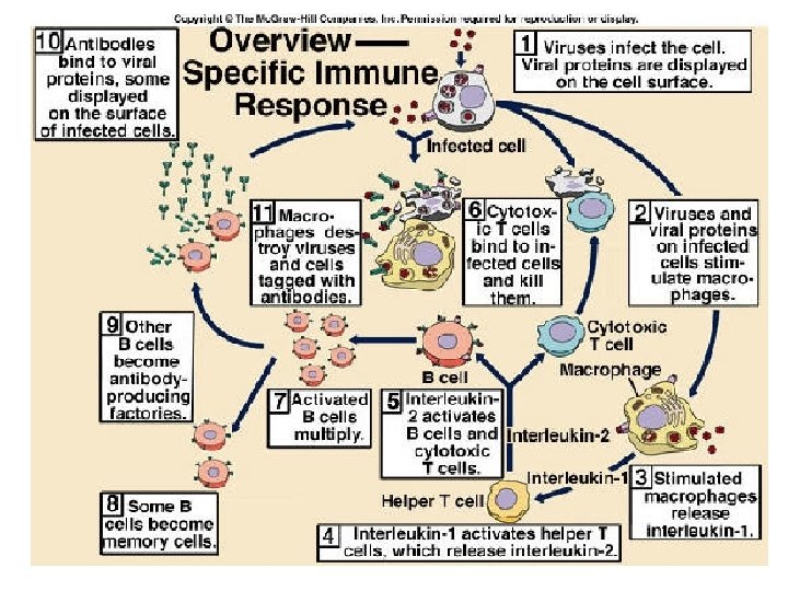
Immune Response • Specific Immunity involves production of antibodies – Antibodies are proteins that bind to specific antigens and which are produced by immune cells – Antigens are molecules that can generate an immune response (antibodygenerating molecules) • Antibodies help inactivate pathogens
Lymphocytes Produce the Immune Response • Lymphocytes are white blood cells that spend most of their time in the organs and tissues of the lymphatic system • Lymphocytes are produced by stem cells in the bone marrow – B-lymphocytes, B-cells, mature in the bone marrow – T-lymphocytes, T-cells, leave the bone marrow to mature in the thymus gland. • Both B and T cells eventually end up in the lymph nodes and other lymphatic organs
B-Cells and Humoral Immunity • When stimulated by antigens, B-cells rapidly divide – Some become inactive memory Bcells that help the immune system respond faster then next time it encounters the same antigen – Others become plasma cells that actively produce antibodies to combat the current infection
Helper T-Cells and Cell-mediated Immunity • T-cells attack antigen-presenting cells (infected body cells that are now displaying foreign antigens) • Helper T-Cells bind to antigen presenting cells and triggers the production of 2 proteins, interleukin-1 and 2 – stimulates growth and division of the Helper T-cell, which creates more Helper T-Cells, both memory T-cells and active helper T-cells – Stimulates B-cells to divide and produce more plasma cells which secrete more antibodies – Stimulates cytotoxic T-cells, the only cells that actually kill other cells
Cytotoxic T-Cells and Cell-Mediated Immunity • Cytotoxic T-cells bind to antigen presenting cells in the same way as helper T-cells • Cytotoxic T-cells then synthesize and secrete several proteins – Perforin, a protein that makes holes in the infected cells membrane and lyses it – A protein that triggers apoptosis (synthesis of a lethal protein that causes programmed cell death)