NERVOUS SYSTEM Organization of the Nervous System Structural


 � Brain � Spinal chord � Act")
")


 of the CNS � Astrocytes � Anchor neurons to their nutrient")


 of the PNS � Schwann cells � Form � the myelin")


 Gyri")


- Slides: 53
NERVOUS SYSTEM
Organization of the Nervous System � � Structural Classification Functional Classification
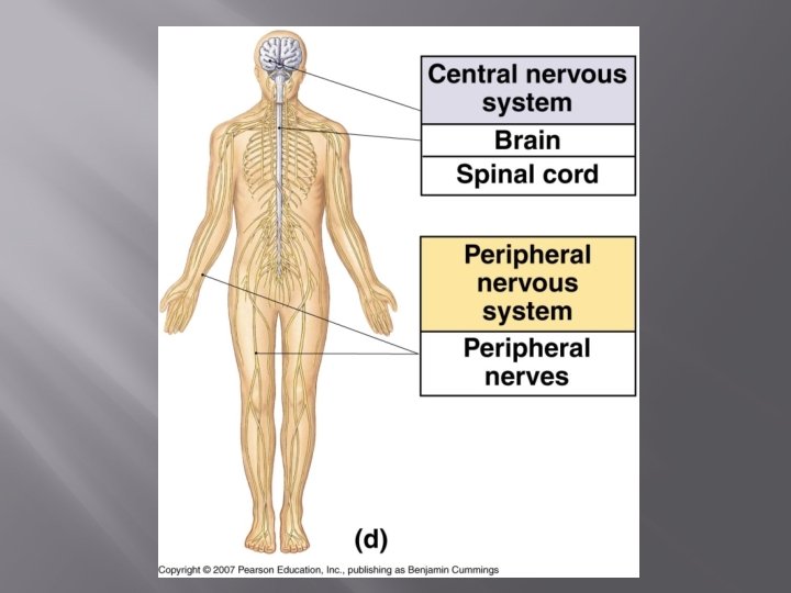
Structural Classification � Central Nervous System (CNS) � Brain � Spinal chord � Act as the integrating and command center of the nervous system � Peripheral Nervous System (PNS) � The part of the nervous system outside of the CNS � Spinal nerves Carry impulses to and from the spinal chord � Cranial nerves Carry impulses to and from the brain
Functional Classification � � Concerned only with PNS structure Two subdivisions � Sensory (afferent) division � Motor (efferent) division
Sensory Division � Consists of nerves that convey impulses TO the central nervous system
Motor Division � � Carries impulses FROM the CNS to the effector organs, muscles, and glands Two subdivisions � Somatic nervous system Voluntary control Most skeletal muscles actions � Autonomic nervous system Involuntary control Cardiac and smooth muscle control Two subdivisions �Sympathetic �Parasympathetic
Nervous Tissue: Structure and Function � Supporting Cells � Do not transmit nerve impulses � Do not lose their ability to divide � Neurons � Transmit nerve impulses � CNS Occurs in clusters called nuclei Bundles of fibers are called tracts � PNS Clusters are called ganglia Bundles of fibers are called nerves
Supporting Cells (neuroglia) of the CNS � Astrocytes � Anchor neurons to their nutrient supply � Make exchanges between the blood and neurons Protect the neurons from harmful substances � Mop � up leaked potassium and neurotransmitters Microglia � Phagocytes � Ependymal � Cilia � circulate the cerebrospinal fluid Oligodendrocytes � Produce the myelin sheaths
Astrocytes
Microglia Cell
Ependymal Cells
Oligodendrocyte
Supporting Cells (neuroglia) of the PNS � Schwann cells � Form � the myelin sheaths Satellite cells � Protective, cushioning cells
Schwann Cell
Satellite Cell
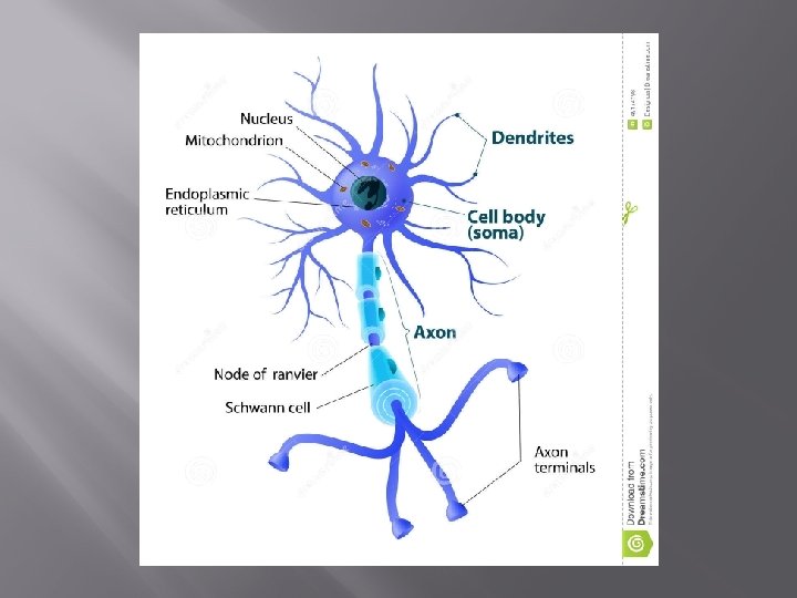
Neurons � Anatomy � Cell body � Processes � Myelin sheaths � Classification � Functional classification � Structural classification � Physiology � Nerve impulses � Reflexes
Anatomy � Cell Body � Metabolic � Amitotic � center of the neuron Processes � Vary from � Dendrites microscopic to 4 feet long convey messages toward the cell body May have many � Axons convey messages away from the cell body Only one per neuron Have many axon terminals per axon �Terminals contain neurotransmitters � Synaptic cleft Gap between two neurons in a series
Anatomy cont. � Myelin Sheaths � Protects and insulates � Increases the transmission rate of nerve impulses � Gaps between Schwann cells nodes of Ranvier
Neuron Anatomy
Nodes of Ranvier
Classification � Functional Classification � Groups neurons according to the direction the nerve impulse is traveling relative to the CNS � Sensory Carrying impulses from receptors in the internal organs or skin to the CNS Cell bodies are always found in the ganglion outside the CNS � Motor Carry impulses from the CNS to the viscera, muscles, and/or glands Cell bodies are always located in the CNS � Association Connect the motor and sensory neurons Cell bodies are always in the CNS
Functional Classification
Functional Classification
Types of Sensory Receptors � � � Dendrite endings are usually associated with specialized receptors Special senses Cutaneous sense organs � � In the skin Naked nerve endings Pain and temperature � Meissner’s corpuscle Touch � Pacinian Pressure � Proprioceptors � � Muscles and tendons Detect amount of stretch
Types of Sensory Receptors
Structural Classification � � Based on the number of processes extending from the cell body Multipolar � Several processes � All motor and association neurons � Bipolar � Two processes � Found only in some special sense organs � Unipolar � One process � Sensory neurons found in the PNS ganglia
Structural Classification
Nerve Physiology � Nerve Impulses � Irritability Ability to respond to a stimulus and convert it into a nerve impulse � Conductivity Ability to transmit the impulse to other neurons, muscles, or glands
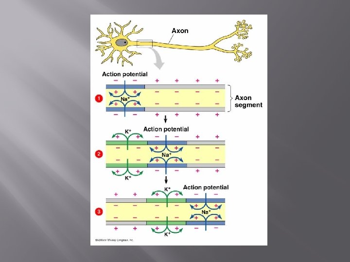
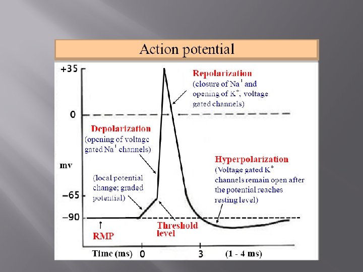
Electrical Conditions of a Resting Neuron’s Membrane � Plasma membrane in polarized � The inner surface is negatively charged � The outer surface is positively charged � Major positive ions in the cell are potassium � Major positive ions outside the cell are sodium
Action Potential Initiation and Generation � Stimulus initiates local depolarization �A stimulus changes the permeability of a section of the membrane � Sodium channels open and ions diffuse rapidly into the cell � The polarity at the site is now switched with the inside more positive and the outside more negative � Propagation of the action potential � Depolarization of the first membrane patch causes permeability changes in the adjacent membrane � The action potential propagates rapidly along the entire length of the axon
Action Potential Initiation and Generation � Repolarization � Immediately after opening the sodium channels close and are unable to reopen for a length of time This keeps the action potential moving down the axon � At this time potassium channels open allowing potassium to diffuse out of the cell � This cell now returns to having negative charge in the cell and a positive charge out � Initial ionic conditions restored � The ionic conditions of the resting state are restored later by the activity of the sodium-potassium pump
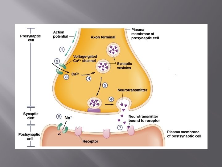
Transmission of the Signal at Synapses � � When the action potential reaches the axon terminal the depolarization causes calcium channels to open Influx of calcium causes the vesicles containing the neurotransmitters to fuse with the plasma membrane and release their contents into the synaptic cleft Neurotransmitters bind to receptors on the next neuron and the action potential is propagated again Neurotransmitters are removed from the synapse quickly either by reuptake into the axon terminal or enzymatic breakdown
Reflexes � � Rapid, predictable, and involuntary responses to stimuli Somatic reflexes � include all reflexes that stimulate the skeletal muscles � Autonomic reflexes � regulate the activity of smooth muscles, the heart, and glands
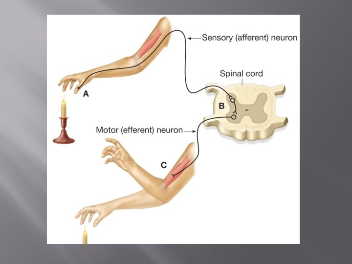
Reflex Arc � Have a minimum of five elements � Sensory receptor � Sensory neuron � Integration center (interneuron in the CNS) � Motor neuron � Effector organ � Whenever reflexes are exaggerated, distorted, or absent, nervous system disorders are indicated
Reflex Arc
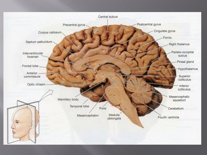
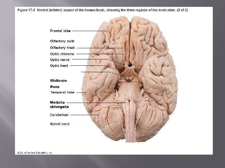
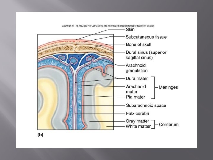
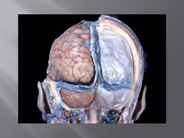
Central Nervous System � Functional Anatomy of the Brain � Cerebral Hemispheres (cerebrum) Gyri �tops of wrinkles Sulci �valleys between wrinkles Fissures �deep groves � Cerebral Cortex Area of brain that keeps speech, memory, logical and emotional response, consciousness, sensation, voluntary movement
Central Nervous System cont. � Diencephalon � Composed of the thalamus, hypothalamus, and epithalamus � Enclosed by the cerebral hemispheres � Thalamus Relay station for sensory impulses passing to the sensory cortex �Triage pleasant or unpleasant sensations � Hypothalamus Regulation of body temperature, water balance and metabolism, and emotions Limbic system �Thirst, appetite, sex, pain, pleasure centers �Regulates the pituitary gland (hormones)
Central Nervous System cont. � Epithalamus � Consists of the pineal body and choroid plexus Hormones Cerebral spinal fluid
Brain Stem � � Midbrain, pons, and medulla oblongata Autonomic behaviors necessary for survival � Breathing and blood pressure � Midbrain Reflex centers for vision and hearing � Pons Control breathing � Medulla oblongata Heart rate, blood pressure, swallowing, and vomiting
Cerebellum � Skeletal muscle activity, balance, equilibrium � Houses proprioceptors
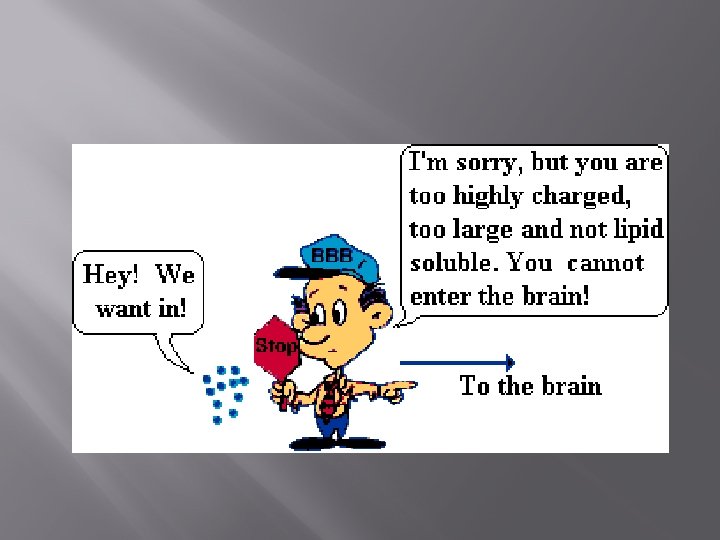
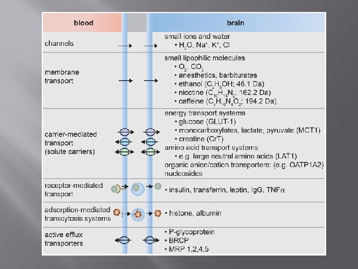
Blood Brain Barrier � Keeps most everything out of the brain � There is limited immune function � Brain needs a very constant environment � Water, oxygen, essential amino acids, glucose