Nervous System Compare the functions of the CNS


 impulses")


 - stress ◦ Parasympathetic")


 CNS is bathed in cerebrospinal fluid � Fills the space between")

 � Glucose")


- Slides: 34
Nervous System Compare the functions of the CNS & PNS
Objectives � Identify the principle parts of the nervous system � Describe the cells that make up the nervous system � Describe what starts and stops a nerve impulse (action potential) � The role of neurotransmitters � Compare the functions of the CNS & PNS � Identify the principle parts of the brain
Components of the Nervous System - Somatic
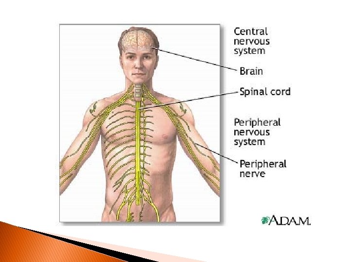
� CNS = spinal cord & brain � PNS = nerves carry (tissue) impulses to and from brain � Motor Output side of chart has 2 divisions: somatic and autonomic � Focus Somatic 1 st then Autonomic
Somatic division/Motor/PNS � Requires only one neuron system: CNS to cell � 12 pairs cranial nerves ◦ From brain’s underside/brain stem ◦ Brain to muscles, glands, head, neck, thorax, abdomen � 31 pairs spinal nerves ◦ Originate from spinal cord ◦ Dorsal root ganglia– sensory incoming AP from tissues to cord ◦ Ventral root ganglia– motor outgoing AP away from cord to body � Connects CNS to body parts
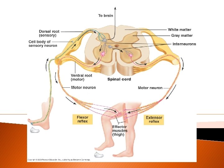
Reflexes � Spinal Reflexes – require no conscious thought – processes @ spinal cord only � E. g. flexor reflex – withdrawal of foot from something sharp � Knee-jerk reflex (check up) – tap below patella causes contraction of thigh and upward movement of foot and leg � Stretch (quadriceps) reflex – posture maintenance – stand move w/out having to think about it
Components of the Nervous System-autonomic
Autonomic Division � Sympathetic – stress / high activity � Parasympathetic – resting, homeostasis � 2 neuron system to transmit impulses to target cells � 1 st neuron - preganglionic in CNS � 2 nd neuron – postganglionic outside CNS & extending to the far reaches of the body (glands/organs) � Sympathetic & Parasympathetic oppose each other – work antagonistically for homeostasis
Sympathetic vs. Parasympathetic � Neurotransmitters ◦ Sympathetic – norepinephrine (adrenalin) - stress ◦ Parasympathetic – acetylcholine - relax
Somatic and Autonomic Divisions of the PNS
Components of the Nervous System
Brain & Spinal Cord � Central location & action � Integrating & processing of information � Info in CNS Complex Output
“Billions of action potentials travelling in millions of neurons all come together as a conscious thought”
Could this be what stress looks like? � Normal thoughts Dark thoughts
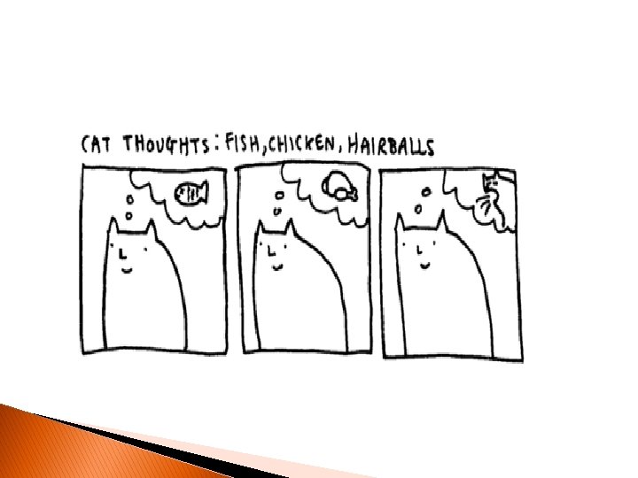
Comic thoughts…
Assignment 3. 23 � Reflexes and the reflex arc – terms 142 -143 � Learning Target #5 (Nervous System) p 135: Describe the structure of a reflex arc and the function of a reflex
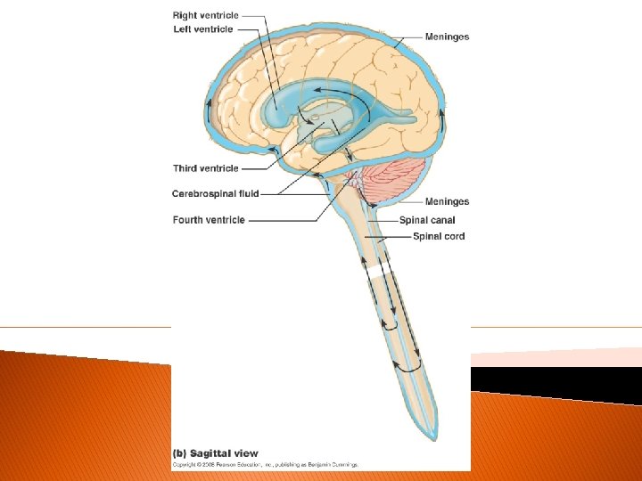
Protection of CNS � Bone, meninges & blood-brain barrier � Bone: skull & hollow vertebrae � Meninges: CNS enclosed by 3 membranous layers ◦ Out In ◦ Dura matter – arachnoid matter – pia matter
Figure 11. 14 ab
CNF (cerebrospinal fluid) CNS is bathed in cerebrospinal fluid � Fills the space between the arachnoid matter & pia matter � Functions � Isolates as a liquid shock absorber the CNS from infection (meningitis: bacterial or viral infection of meninges can spread to CNS)
More about CSF � CSF is like the interstitial fluid that bathes all cells but it does not exchange substances as freely with blood � Capillaries in this area are “tight” = not leaky & substances must pass through the actual capillary cells (vs. slipping between narrow slits of adjacent capillary cells) to get from blood to the brain
Blood-brain barrier � Lipid soluble substances pass easily (O & CO 2) � Glucose requires active transport � Larger molecules: proteins, viruses, bacteria kept out � What can pass through BBB? ◦ ◦ ◦ Alcohol Caffeine Nicotine Cocaine Anesthetics
Spinal cord � Information super highway for APs between the brain and the body � Recall – spinal reflexes don’t involve brain and therefore are considered “unconscious” � Size – about the diameter of your thumb � Location – runs from the base of your skull to the area of the 2 nd lumbar vertebra ~ 17 inches
Figure 11. 13 b
Figure 11. 10
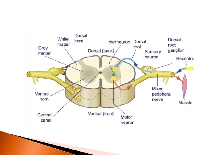
Inside the spine � Outer portions of the cord consist of bundles of axons = nerve tracts that are mylenated = white matter – ascending sensory nerves & descending motor nerves � Inner portions consist of cell bodies, dendrites, neuroglial cells that are unmylenated = gray matter – here sensory & motor neurons synapse & transmit to the brain…
Figure 11. 14