
Nervous System Chapter 31 Objectives Describe how nervous
Nervous System Chapter 31
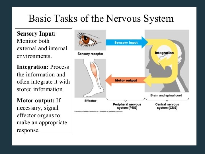
Objectives • • • Describe how nervous systems receive sensory input, interpret it (integration), and send out appropriate commands (motor output) Explain the role of sensory neurons, interneurons and motor neurons. Identify and explain the structure of a typical motor neuron. Explain how nerves send signals - action potential, resting potential, the role of the sodium potassium pump Explain how neurotransmitters are involved in carrying action potentials from a neuron to a receiving neuron or effector cell. Explain the role of effectors and how they relate to motor neurons. Describe how action potentials are affected by increased intensity of stimuli. Compare and contrast the anatomy and functions of the parasympathetic nervous system and the sympathetic nervous system Explain how multiple sclerosis relates to the nervous system. Explain the structure of vertebrate nervous systems. Explain the structure and function of the human brain
Types of Neurons 1. Sensory neurons – transmit impulses from receptor cells (receive stimuli) to the central nervous system (CNS) 2. Interneurons – transmit impules CNS 3. Motor neurons – transmit impulses from the CNS to the effector cells (respond to stimuli) Neurons (4 min): https: //www. youtube. com/watch? v=f. HRC 8 Sl. Lc. H 0
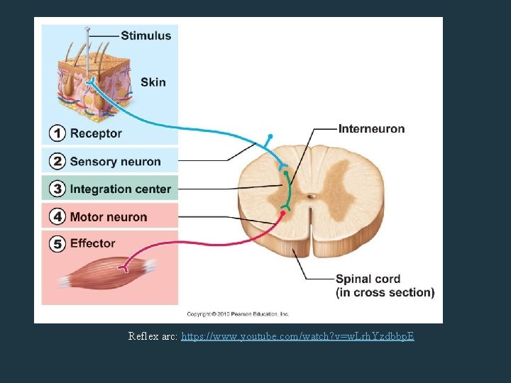
Reflex arc: https: //www. youtube. com/watch? v=w. Lrh. Yzdbbp. E
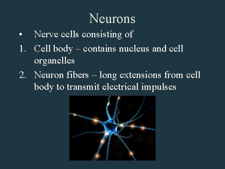
Neurons • Nerve cells consisting of 1. Cell body – contains nucleus and cell organelles 2. Neuron fibers – long extensions from cell body to transmit electrical impulses
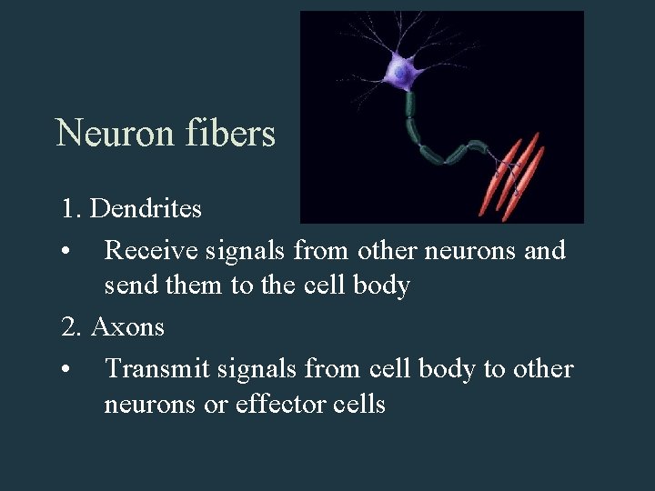
Neuron fibers 1. Dendrites • Receive signals from other neurons and send them to the cell body 2. Axons • Transmit signals from cell body to other neurons or effector cells
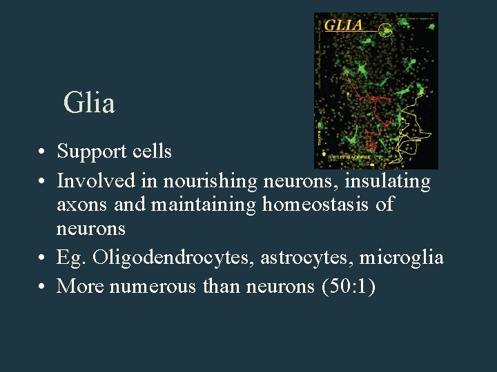
Glia • Support cells • Involved in nourishing neurons, insulating axons and maintaining homeostasis of neurons • Eg. Oligodendrocytes, astrocytes, microglia • More numerous than neurons (50: 1)
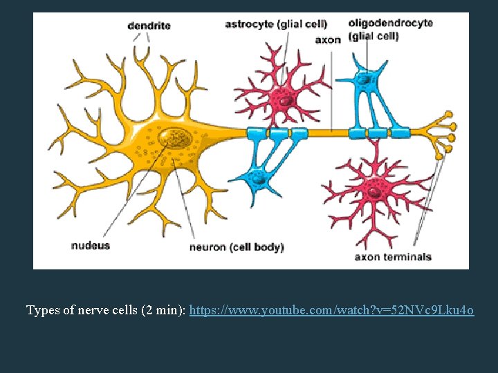
Types of nerve cells (2 min): https: //www. youtube. com/watch? v=52 NVc 9 Lku 4 o
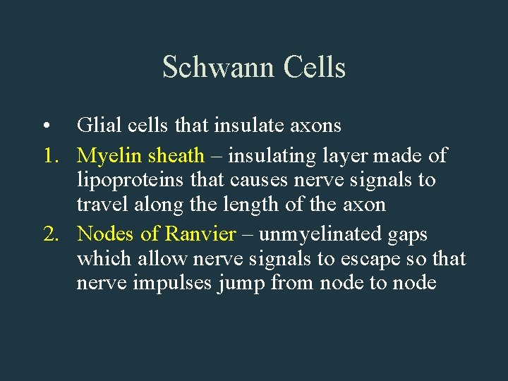
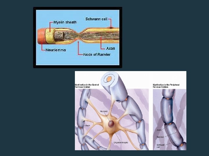
Schwann Cells • Glial cells that insulate axons 1. Myelin sheath – insulating layer made of lipoproteins that causes nerve signals to travel along the length of the axon 2. Nodes of Ranvier – unmyelinated gaps which allow nerve signals to escape so that nerve impulses jump from node to node
Nerve Signal Transmission • Membrane potential stores energy for impulse transmission • Due to the difference in electrical charge between the cytoplasm and outside of the cell
Resting Potential • At rest, the membrane stores energy by keeping the positive charge outside apart from the negative charge inside the neuron • The membrane is differentially permeable so that K+ diffuses out more readily than Na+ diffuses in • The Na+/K+ also helps maintain the ion distribution by pumping Na+ out while pumping K+ in
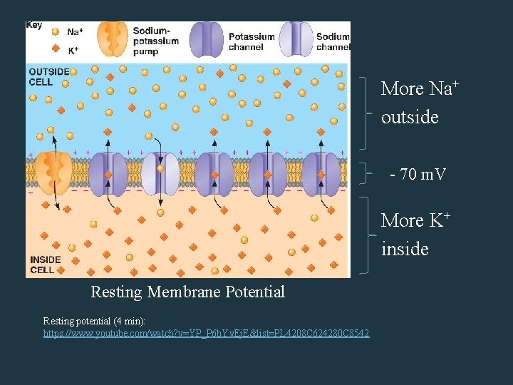
More Na+ outside - 70 m. V More K+ inside Resting Membrane Potential Resting potential (4 min): https: //www. youtube. com/watch? v=YP_P 6 b. Yv. Ej. E&list=PL 4208 C 624280 C 8542
Action Potential • Occurs when a nerve impulses reaches a Node of Ranvier • Requires a minimum change in membrane potential (threshold) before process occurs • Process of voltage-gated channels opening and closing due to changes in voltage • Occurs within 1 -2 milliseconds
• The axoplasm abruptly becomes positive in charge as Na+ channels open, allowing Na+ ions to flood into the cell (ie. Depolarization) • A split second later, Na+ channels close and K+ channels open, allowing K+ ions to flood out of the cell so that axoplasm becomes negative in charge (ie. Repolarization) • Because K+ channels close more slowly, K+ leaving the axon causes the membrane potential to become more negative than at rest (ie. Hyperpolarization)
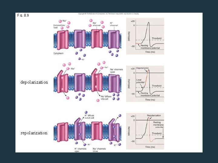
depolarization repolarization
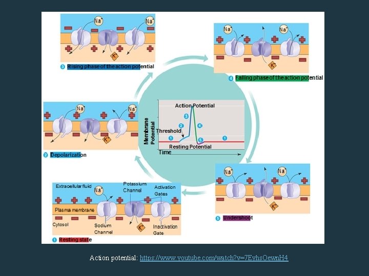
Action potential: https: //www. youtube. com/watch? v=7 Eyhs. Oewn. H 4
Synapse • Space between the axon knob of one neuron and the dendrite membrane of another neuron • Neurotransmitters need to be released across the synapse in order for the nerve impulse to be conducted to another neuron
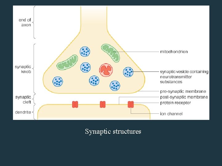
Synaptic structures
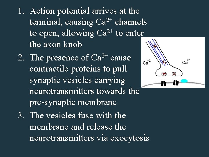
1. Action potential arrives at the terminal, causing Ca 2+ channels to open, allowing Ca 2+ to enter the axon knob 2. The presence of Ca 2+ cause contractile proteins to pull synaptic vesicles carrying neurotransmitters towards the pre-synaptic membrane 3. The vesicles fuse with the membrane and release the neurotransmitters via exocytosis
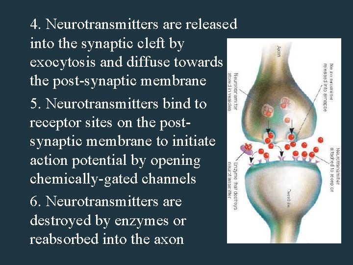
4. Neurotransmitters are released into the synaptic cleft by exocytosis and diffuse towards the post-synaptic membrane 5. Neurotransmitters bind to receptor sites on the postsynaptic membrane to initiate action potential by opening chemically-gated channels 6. Neurotransmitters are destroyed by enzymes or reabsorbed into the axon
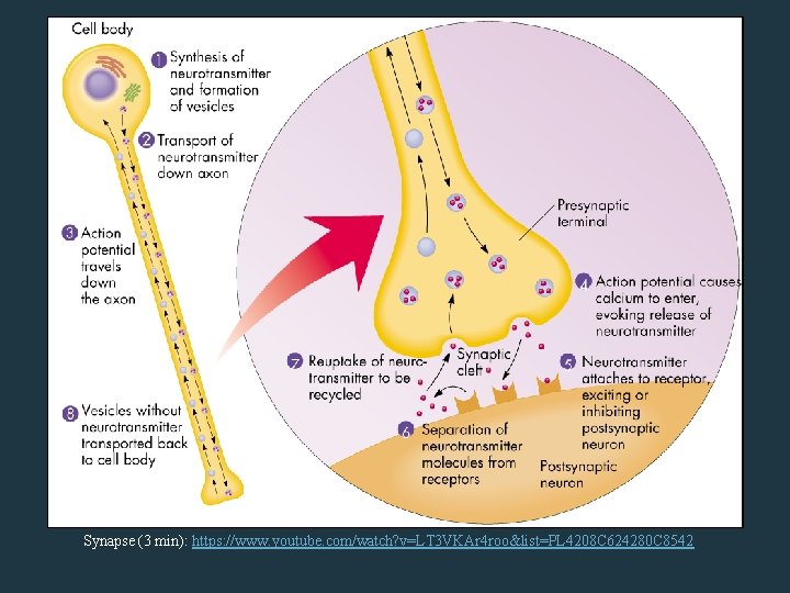
Synapse (3 min): https: //www. youtube. com/watch? v=LT 3 VKAr 4 roo&list=PL 4208 C 624280 C 8542
Neurotransmitters • Open ion channels when bound to receptor sites • May be excitatory (causes action potential) or inhibitory (inhibits action potential) • Eg. Acetylcholine is in the brain and between motor neurons and muscle cells • Eg. epinephrine, norepinephrine, serotonin and dopamine
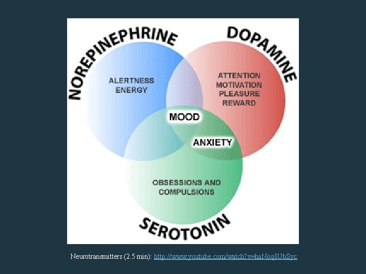
Neurotransmitters (2. 5 min): http: //www. youtube. com/watch? v=ha. Noq 8 Ub. Syc
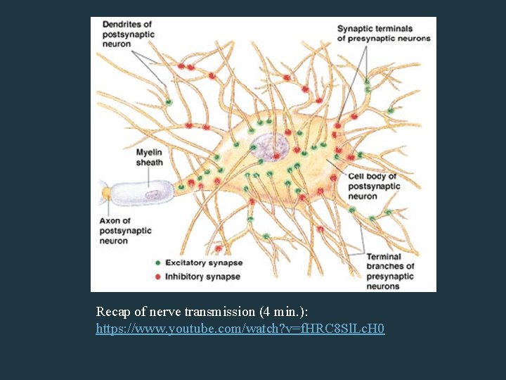
Recap of nerve transmission (4 min. ): https: //www. youtube. com/watch? v=f. HRC 8 Sl. Lc. H 0
The Nervous System • Divided into the Central Nervous System (CNS) and Peripheral Nervous System (PNS) • CNS consists of the brain, and the spinal cord • PNS consists of cranial and spinal nerves
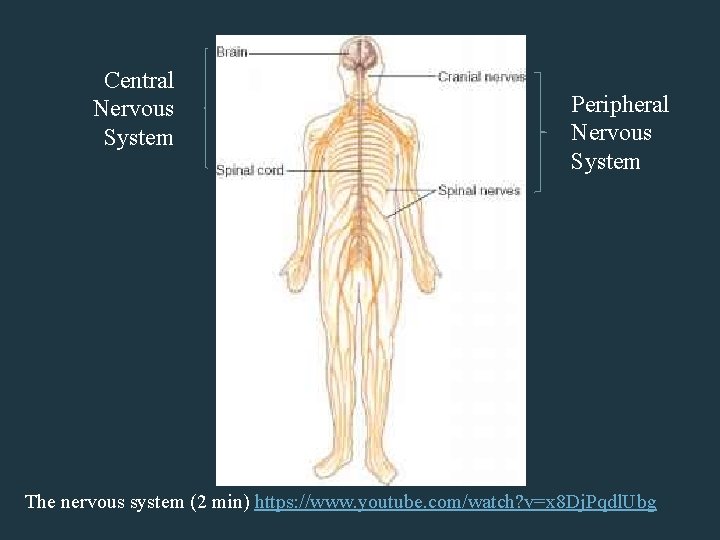
Central Nervous System Peripheral Nervous System The nervous system (2 min) https: //www. youtube. com/watch? v=x 8 Dj. Pqdl. Ubg
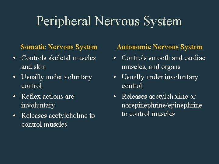
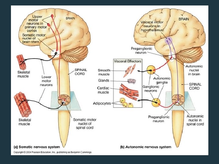
Peripheral Nervous System Somatic Nervous System • Controls skeletal muscles and skin • Usually under voluntary control • Reflex actions are involuntary • Releases acetylcholine to control muscles Autonomic Nervous System • Controls smooth and cardiac muscles, and organs • Usually under involuntary control • Releases acetylcholine or norepinephrine/epinephrine to control muscles
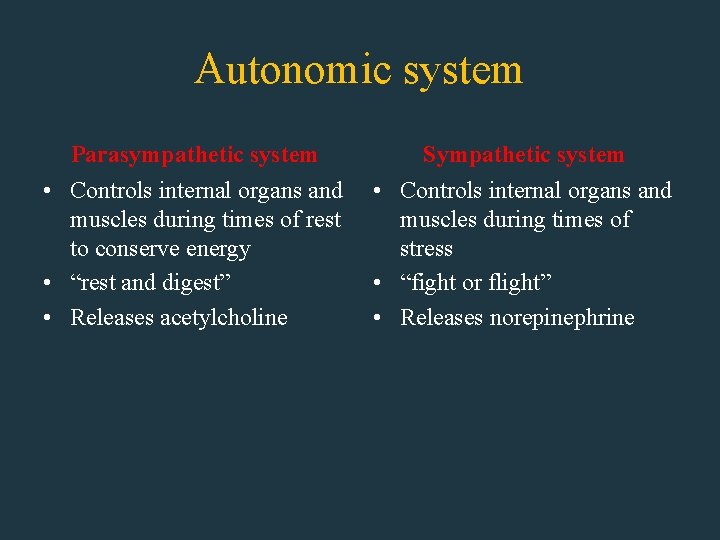
Autonomic system Parasympathetic system Sympathetic system • Controls internal organs and muscles during times of rest muscles during times of to conserve energy stress • “rest and digest” • “fight or flight” • Releases acetylcholine • Releases norepinephrine
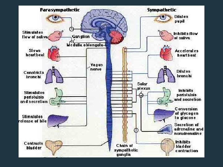
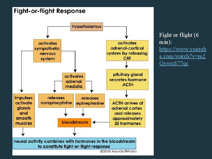
Fight or Flight Response Fight or flight (6 min): https: //www. youtub e. com/watch? v=m 2 Gywo. S 77 qc
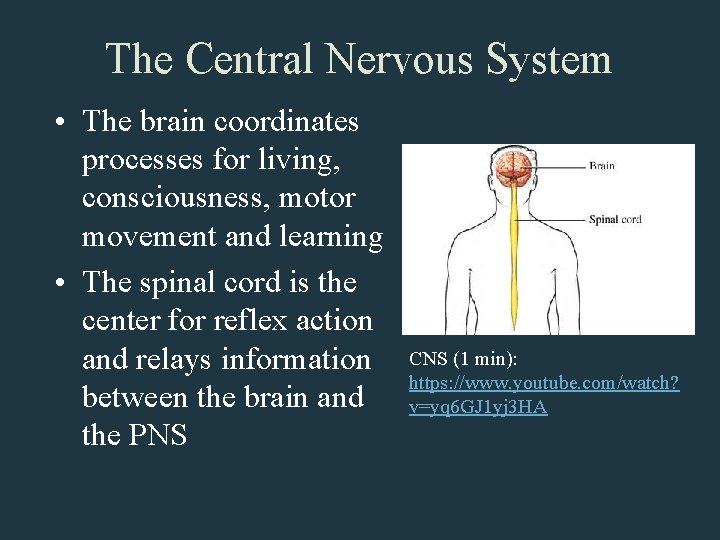
The Central Nervous System • The brain coordinates processes for living, consciousness, motor movement and learning • The spinal cord is the center for reflex action and relays information between the brain and the PNS CNS (1 min): https: //www. youtube. com/watch? v=yq 6 GJ 1 yj 3 HA
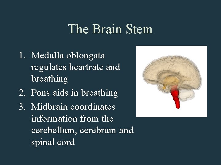
The Brain Stem 1. Medulla oblongata regulates heartrate and breathing 2. Pons aids in breathing 3. Midbrain coordinates information from the cerebellum, cerebrum and spinal cord
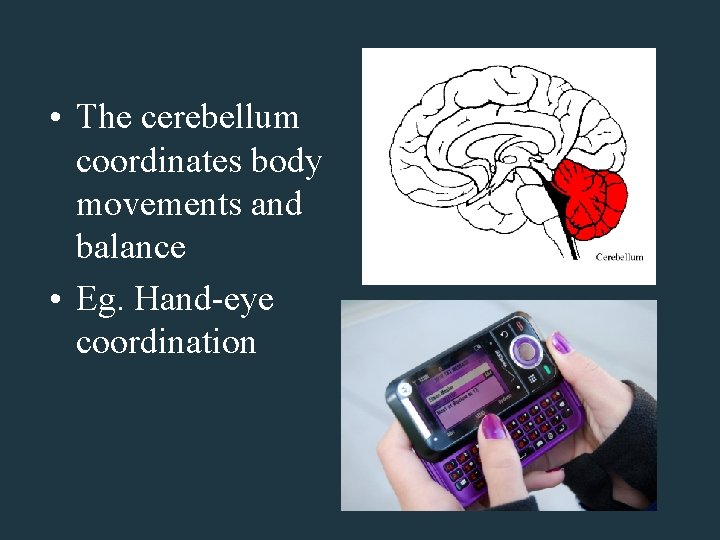
• The cerebellum coordinates body movements and balance • Eg. Hand-eye coordination
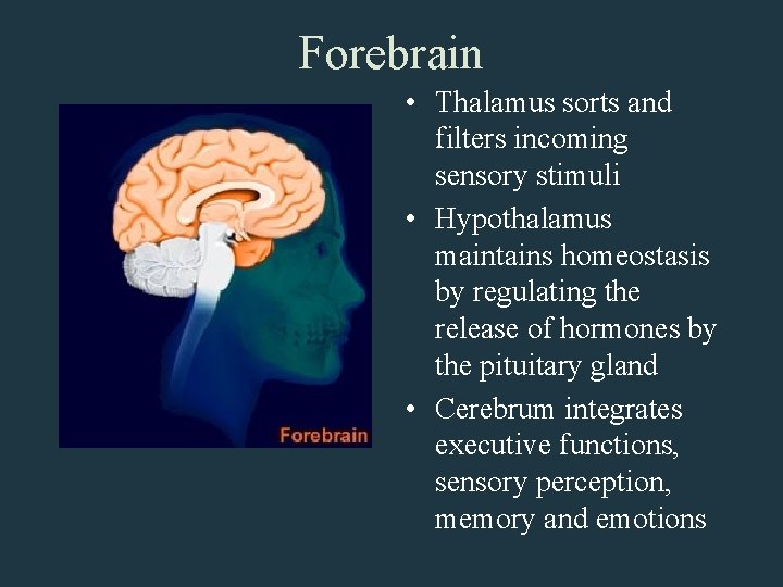
Forebrain • Thalamus sorts and filters incoming sensory stimuli • Hypothalamus maintains homeostasis by regulating the release of hormones by the pituitary gland • Cerebrum integrates executive functions, sensory perception, memory and emotions
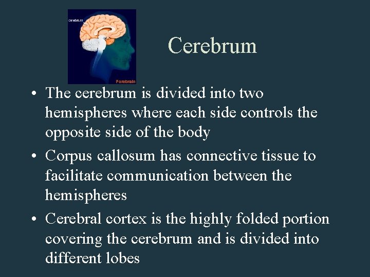
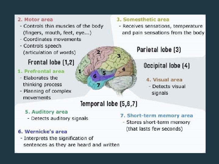
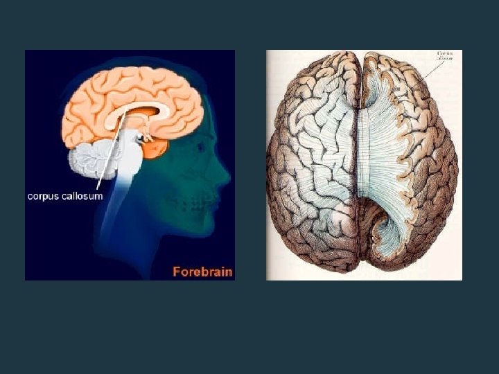
Cerebrum • The cerebrum is divided into two hemispheres where each side controls the opposite side of the body • Corpus callosum has connective tissue to facilitate communication between the hemispheres • Cerebral cortex is the highly folded portion covering the cerebrum and is divided into different lobes
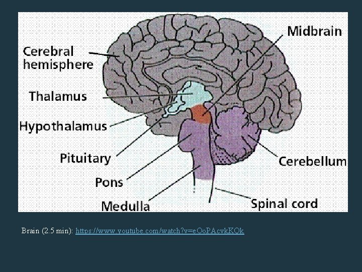
Brain (2. 5 min): https: //www. youtube. com/watch? v=e. Oo. PAcvk. KQk
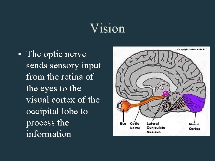
Vision • The optic nerve sends sensory input from the retina of the eyes to the visual cortex of the occipital lobe to process the information
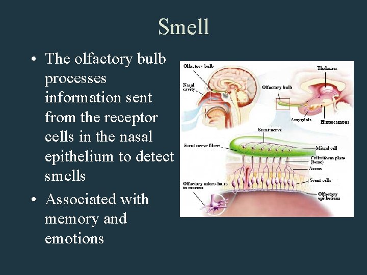
Smell • The olfactory bulb processes information sent from the receptor cells in the nasal epithelium to detect smells • Associated with memory and emotions
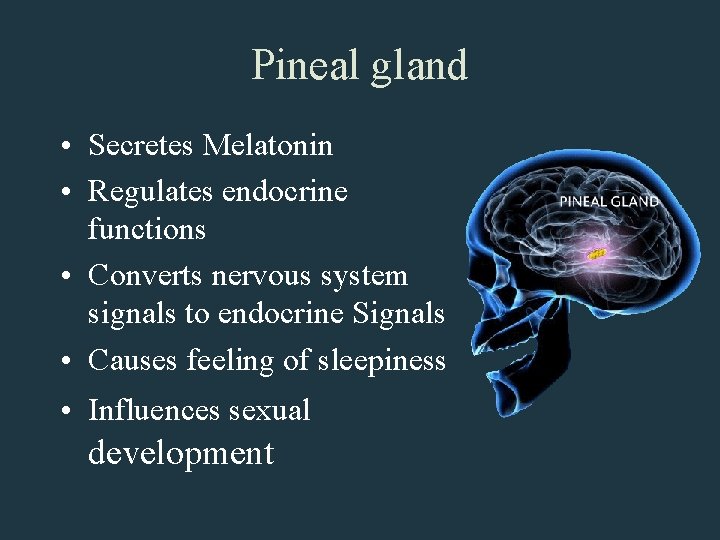
Pineal gland • Secretes Melatonin • Regulates endocrine functions • Converts nervous system signals to endocrine Signals • Causes feeling of sleepiness • Influences sexual development
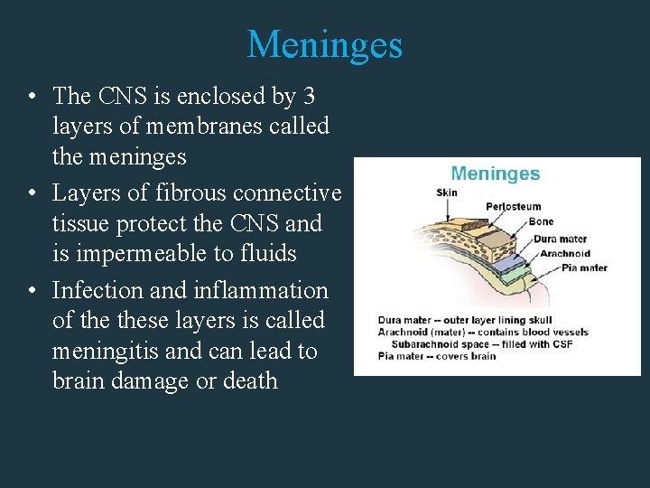
Meninges • The CNS is enclosed by 3 layers of membranes called the meninges • Layers of fibrous connective tissue protect the CNS and is impermeable to fluids • Infection and inflammation of these layers is called meningitis and can lead to brain damage or death
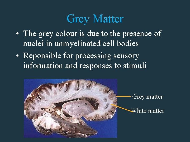
Grey Matter • The grey colour is due to the presence of nuclei in unmyelinated cell bodies • Reponsible for processing sensory information and responses to stimuli Grey matter White matter
White matter • The presence of myelin sheaths around axon fibers give white matter its white appearance • Responsible of communication within the grey matter and with the rest of the CNS • 95% of lesions that occur in Multiple Sclerosis occurs in the white matter
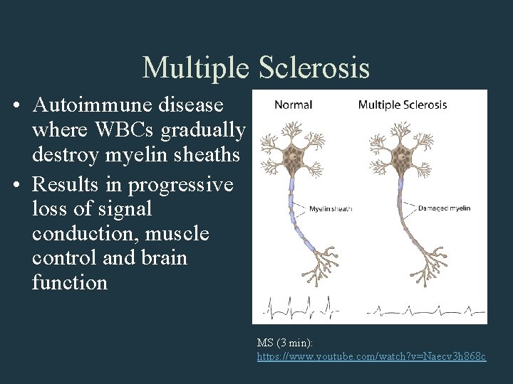
Multiple Sclerosis • Autoimmune disease where WBCs gradually destroy myelin sheaths • Results in progressive loss of signal conduction, muscle control and brain function MS (3 min): https: //www. youtube. com/watch? v=Naecv 3 h 868 c
- Slides: 48