Nervous System Brain Nervous System Brain Spinal cord

Nervous System Brain

Nervous System Brain, Spinal cord, and neurons

Nervous system organization • The nervous system has two major subdivisions: the central nervous system (CNS) and the peripheral nervous system (PNS). • The CNS consists of the brain and spinal cord. • The PNS informs the CNS of stimuli received from the external environment and carries appropriate responses to the glands and muscles. The PNS is composed mainly of the axons and dendrites of sensory and motor neurons. Axons and dendrites are extensions of nerve cells that transport signals throughout the body.

CNS-Spinal Cord • The spinal cord is approx. 18 inches long and is protected by the vertebrae. • The outside portion of the spinal cord is “white matter” b/c of the color. The inside portion of the spinal cord is “gray matter” due to its color. • The CNS is surrounded and protected by three layers that are collectively called meninges. The outer layer is a tough fibrous tissue called dura mater (translated “tough mother”), the middle layer is called the arachnoid mater which reabsorbs cerebrospinal fluid. The inner layer is called, pia mater. • Cerebrospinal fluid is found in a space between the arachnoid and pia mater. Cerebrospinal fluid give nutrients and other special needs for nerve cells.

Frontal lobe

Parietal lobe Frontal lobe

Parietal lobe Frontal lobe Occipital lobe

Parietal lobe Frontal lobe Occipital lobe Cerebellum

Parietal lobe Frontal lobe Occipital lobe Cerebellum Brain stem

Parietal lobe Frontal lobe Occipital lobe Cerebellum Temporal lobe Brain stem

Central Sulcus

Central Sulcus Longitudinal fissure

Central Sulcus Longitudinal fissure Transverse fissure

Central Sulcus Longitudinal fissure Transverse fissure Lateral Sulcus

Cerebrum • Controls all higher brain functions. Examples are: interpreting sensory impulses, muscle movements, memory, reasoning, judgments, intelligence. • Consists of two large hemispheres connected by the corpus callosum. The surface of the cerebrum contains grooves called gyri (singular gyrus) and a shallow groove is a sulcus, a deep groove is a fissure.

Cerebellum • Serves as a coordinating center for motor activity as well as a reflex center for skeletal muscle. • Ex. During walking the cerebellum determines the muscles used and the strength and sequence of the contractions.

Brainstem • Midbrain: contains reflex centers that move the eyes and head, and maintain posture. • Pons: Relays impulses sent to and from the medulla oblongata and cerebrum. Also helps regulate rate and depth of breathing. • Medulla Oblongata: Conducts impulses between brain and spinal cord. Controls heart beat, blood pressure, respiratory control and various other reflex controls such as sneezing, coughing, vomiting, and swallowing. • Diencephalon: Thalamus/hypothalamus: Takes sensory impulses from other parts of the nervous system and relays them to the cerebral cortex. Hypothalamus also produces hormones, as well as regulate hunger, body weight and temp. , and water balance.

Cerebrum
 Cerebrum Gryus (folds)")
Sulcus (grooves) Cerebrum Gryus (folds)
 Gryus (folds) Corpus callosum Cerebrum")
Sulcus (grooves) Gryus (folds) Corpus callosum Cerebrum
 Gryus (folds) Corpus callosum Cerebrum Pituitary gland")
Sulcus (grooves) Gryus (folds) Corpus callosum Cerebrum Pituitary gland
 Gryus (folds) Corpus callosum Cerebrum Pituitary gland Pons")
Sulcus (grooves) Gryus (folds) Corpus callosum Cerebrum Pituitary gland Pons
 Gyrus (upward folds) cerebrum Hypothalamus Thalamus Pituitary gland Epithalamus")
Corpus callosum Sulcus (downward grooves) Gyrus (upward folds) cerebrum Hypothalamus Thalamus Pituitary gland Epithalamus Diencephalon Pons Midbrain Medulla oblongata Cerebellum
 Gyrus (upward folds) cerebrum Hypothalamus Thalamus Pituitary gland Epithalamus")
Corpus callosum Sulcus (downward grooves) Gyrus (upward folds) cerebrum Hypothalamus Thalamus Pituitary gland Epithalamus Diencephalon Pons Midbrain Medulla oblongata Cerebellum
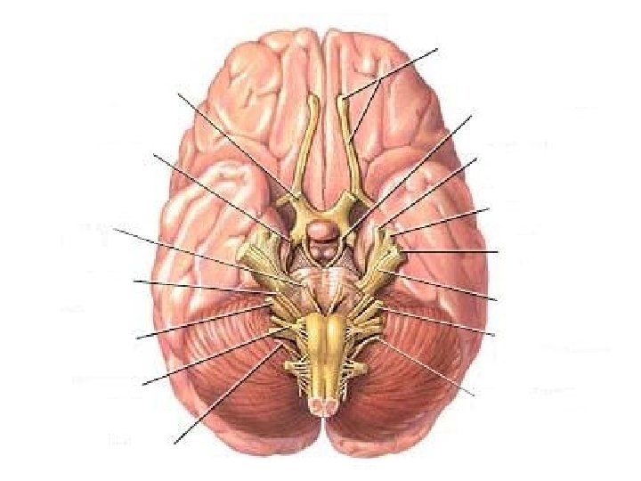
 Optic (II) Oculomotor (III) Trigeminal (V) Trochlear (IV) Abducens (VI) Facial (VII)")
Olfactory (I) Optic (II) Oculomotor (III) Trigeminal (V) Trochlear (IV) Abducens (VI) Facial (VII) Vestibulocochlear (VIII) Glossopharyngeal (IX) Vagus (X) Hypoglossal (XII) Spinal accessory (XI)

• • • Mnemonic Device I. Oh, III. Oh, IV. Touch VI. And VII. Feel VIII. Very IX. Good X. Velvet… XI. So XII. Heavenly!

Functions of cranial nerves • I. Olfactory: Smell • II. Optic: Vision • III. Oculomotor: Voluntary muscle movement in the eyes, pupil dialation • IV. Trochlear: Eye movements (smallest nerves) • V. Trigeminal: Sensory information (touch, pain) from the face and head; muscles for chewing • VI. Abducens: Eye movements • VII. Facial: Taste, sensory information from ear, controls muscles used in facial expression. • VIII. Vestibulocochlear: Hearing; Balance • IX. Glossopharyngeal: Taste; Swallowing • X. Vagus: digestion, heart rate • XI. Spinal accessory: Muscle movements of the head • XII. Hypoglossal: Controls muscles of tongue
,")
Functions of brain regions • All brain regions either perform functions of motor (movements), Sensory (sensations), or associations (intellectual/cognitive). • Frontal lobe: • Association-concentration, planning, problem solving, judgments, decision-making, understanding consequences to actions. • Motor-Movements of voluntary skeletal muscles.

Frontal lobe: • Association-concentration, planning, problem solving, judgments, decision-making, understanding consequences to actions. • Motor-Movements of voluntary skeletal muscles.

Parietal lobe • Association-understanding speech and using words to express thoughts and feelings. Awareness of the form of objects, including your own body parts. • Sensory-sensations of temperature, touch, pressure, and pain involving the skin.
 and store memories of")
Temporal lobe • Association-interpret sensory experiences (understand speech and reading) and store memories of visual scenes, music, and other complex sensory patterns. • Sensory-hearing
. •")
Occipital lobe • Association-Combine visual images with other sensory experiences (recognizing another person). • Sensory-Vision

Cerebellum • Sensory: Balance, coordination, depthperception,

Brain stem • Motor-Basic life functions such as heart beat, breathing, digestion, body temp.

Pop Quiz • 1. Name all lobes of the brain • 2. Name the fissure that separates the brain into left and right halves. • 3. Name the fissure that separates the brain to superior and inferior portions • 4. What are the downward grooves in the cerebrum called? • 5. What 3 parts make up the brain stem?

Nervous system • The central nervous system consists of the brain and spinal cord. • The peripheral nervous system consists of nerves (cranial and spinal) that connect the CNS to other body parts.

Neurons • Neurons are nerve cells that send and receive messages from the brain to parts of the body.

Dendrites: Receives info. Cell body: Made up of typical cell stuff… Nucleus, nucleolus, Mitochondria. Axon: Carries info away

Types of neurons • 1. Bipolar neurons: The cell body has only two processes. Even though they look similar, one is an axon and the other is a dendrite. They’re found in specialized parts of the eyes, nose, and ears. • Unipolar neurons: One process extends from the cell body. That process splits into two branches. One serves as an axon and the other connects to the brain or spinal cord. These are sometimes referred to as ganglia. • Multipolar neurons: Many processes branch off of the cell body. One process is an axon, the rest are dendrites. Most neurons in the brain and spinal cord are this type.
 that it is")
Synapse • Between the neuron and the cell (or other neuron) that it is communicating with is a space called a synapse. • The carriers of this information are biological messenger molecules called neurotransmitters.

• 1. A message travels along the nerve and when it approaches the nerve ending a neurotransmitter is released. • 2. The neurotransmitter is received by the next cell • 3. some of the neurotransmitter gets reabsorbed • 4. When enough neurotransmitter is received by the next nerve cell the message moves forward

Neurons • Neurons can be classified into different groups based on the job they do. Neurons either carry information into the CNS, within the CNS, or out of the CNS. The three groups are sensory neurons, interneurons, or motor neurons.

Sensory neurons • Carry nerve impulses from peripheral body parts into the brain or spinal cord. • Most are unipolar, some are bipolar • Specialize in detecting changes in the outside world (ex. Eyes, ears, touch receptors in skin) or within the body (ex. Temp. , blood pressure)

Interneurons • • Located within the brain or spinal cord Multipolar Make connections with other neurons Transmit impulses from one part of the brain to another. They direct incoming impulses from sensory neurons to appropriate regions of the brain

Motor neurons • Multipolar • Carry impulses out of the brain or spinal cord to muscles or glands. (when they reach muscles, the muscles contract. When they reach glands, they release secretions) • Some are under voluntary control, others are under involuntary control.
- Slides: 47