Nervous System Autonomic Nervous System involuntary Affects smooth

 Affects smooth muscles, cardiac muscle, glands Sympathetic Division - Activated")
 • Carries impulses from sensory receptors in skin and sensory")


: unipolar carry impulses from periphery to")


 Nerve impulse arrives at synaptic end bulb 2.")


 Meninges (3)")


 of gray matter Source of integrative functions")


- Slides: 56
Nervous System
Autonomic Nervous System (involuntary) Affects smooth muscles, cardiac muscle, glands Sympathetic Division - Activated when body is stressed (flight or fight response) speeds heart constricts blood vessels signals for secretions from sweat and adrenal glands slows digestion Parasympathetic Division - Activated under normal conditions slows heart relaxes blood vessels increases digestion
Somatic Nervous System (voluntary) • Carries impulses from sensory receptors in skin and sensory organs of the head • Carries impulses to motor neurons in skeletal muscles
Nerve Tissue 2 cell types: Neuroglia: provide structural framework insulate axons perform phagocytosis circulate cerebrospinal fluid Neurons: conduct impulses
Types of Glial Cells Name Astrocytes Location CNS Function anchors neurons to capillaries forms blood-brain barrier controls ion flow around neurons aids in impulse conduction Ependymal Cells Line cavities in CNS Help form and circulate cerebrospinal fluid Microglia CNS Phagocytize invading microorganisms and dead nerve tissue Oligodendrocytes CNS Form myelin sheath Insulate CNS axons Multiple Sclerosis affects these and impairs impulses Schwann cells Large nerves of PNS Insulate large PNS axons Possibly assist in regeneration
Neuron Anatomy Neurons have a wide variety of shapes and sizes Generalized Structure: Dendrites: short, branched - receive impulses - transmit impulses towards cell body Cell Body: middle section - holds organelles typical of other cells Nissl bodies: similar to rough ER Neurofibrils: similar to microtublules No spindle fibers = NO MITOSIS Axon: nerve fiber, very long (up to 1 m) - wrapped in myelin sheath - transmit impulses away from cell body
Made from tight layers of Schwann cells little cytoplasm cell membrane of high lipid concentration high insulation outer layer of myelin sheath contains cytoplasm and nuclei of Schwann cells = neurilemma Gaps between Schwann cells = nodes of Ranvier
Fibers that are enclosed by tightly wrapped Schwann cells to form the myelin sheath Fibers that may be enclosed by Schwann cells but lack myelin sheath
Neuron types Structural Differences: Multipolar Neurons: many dendrites one axon carry impulses from CNS to skeletal muscles Bipolar Neurons: one dendrite one axon found in special sensory areas (eyes, ears, nose) Unipolar Neurons: single nerve fiber extends from cell body, then branches into one axon and one dendrite carry impulses from skin receptors to spinal cord
Neuron types Functional Differences: Sensory Neurons (afferent neurons): unipolar carry impulses from periphery to CNS Association Neurons (interneurons): multipolar located within CNS relay impulses from one region of CNS to another conduct impulses from sensory neurons to motor neurons Motor Neurons (efferent neurons): multipolar carry impulses from CNS to responders (muscles, glands)
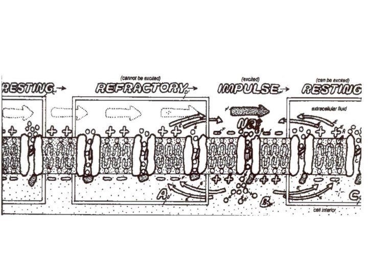
Nerve Impulses Resting Membrane Potential - separation of charge between extracellular and intracellular environments - due to unequal distribution of ions across the cell membranes - created by ion channels integral proteins of membrane guard openings
Resting Membrane Potential High concentration of sodium ions + outside cell High concentration of potassium ions inside cell + High concentration of chlorine ions inside cell - Net positive charge outside cell Sodium channel Potassium channel Net negative charge inside cell Polarized State
Action Potential Stimulus must reach a threshold Sodium ion channels open Sodium ions flow into the cell Briefly reverse electrical charges in cell and outside cell membrane Net negative Net positive charge outside cell Sodium channel Potassium channel Net positive Net negative charge inside cell Depolarized State
Action Potential Sodium ion channels close Potassium ion channels open Potassium ions flow out of the cell = net movement of + ions out of cell Restores resting potential Net positive charge outside cell Sodium channel Potassium channel Net negative charge inside cell Repolarization
Action Potential Sodium-Potassium Pump switches locations of sodium and potassium ion concentrations Impulse is further transmitted down the neuron as depolarized areas stimulate adjoining areas to depolarize (positive feedback) Impulse only travels in one direction because preceding area is busy repolarizing. Sodium gates remain closed despite stimuli. Refractory Period
Saltatory conduction: Speed of impulse in unmyelinated nerve fiber = 10 m/sec Myelin blocks continuous flow of ions Impulse jumps across myelin sheath from one node of Ranvier to next. cut distance = decreased time Speed = 130 m/sec
All – or –None Response Action potential does not occur unless stimulus is strong enough (threshold) Action potential always occurs at its maximum strength
Summation Series of subthreshold stimuli are applied quickly and have a cumulative effect that can lead to an action potential
Cell to Cell Transmission Synapse = junction between cells Synaptic End Bulb: rounded end of presynaptic neuron’s axon contains synaptic vesicles (hold neurotransmitters) Synaptic cleft = gap between synaptic end bulb and postsynaptic neuron
Cell to Cell Transmission 1. ) Nerve impulse arrives at synaptic end bulb 2. ) Signals calcium channels to open Calcium flows into presynaptic cell 3. ) Ions cause vesicles to fuse with plasma membrane and release neurotransmitters by exocytosis 4. ) Neurotransmitters diffuse across synaptic cleft 5. ) Contact membrane of postsynaptic neuron 6. ) Results in excitation or inhibition of postsynaptic neuron 7. ) Result is limited to a fraction of a second 8. ) Enzymes inactivate or transport neurotransmitters away
Excitatory Transmission Neurotransmitters increase membrane permeability to sodium ions Facilitation: each molecule of neurotransmitter received by postsynaptic neuron allows more sodium ions into cell = partial polarization
Inhibitory Transmission Neurotransmitters increase membrane permeability to potassium ions Hyperpolarization: More and more potassium ions flow out of the cell while chlorine ions flow in. Creates an even larger positive charge outside the cell and larger negative charge inside the cell.
Common Neurotransmitters Acetylcholine – released in spinal cord and at neuromuscular junctions Norepinephrine Dopamine Serotonin Endorphins Enkephalins Affect sleep, mood, motor functions, and pleasure recognition Natural pain killers In spinal cord and brain
Cell to Cell Transmission Postsynaptic dendrites may receive thousands of signals from thousands of presynaptic neurons at the same time Cells may receive conflicting signals at the same time Overall effect is determined by the sum of the total incoming signals
CNS – Spinal Cord Extends from base of brain, through foramen magnum, down vertebral canal to the 1 st or 2 nd lumbar vertebra
CNS – Spinal Cord Spinal cord is protected by 3 layers: Vertebral Column Cerebrospinal Fluid (CSF) Meninges (3)
Meninges Epidural space filled with fat and areolar C. T. found between dura mater and vertebral column Dura Mater tough outer layer Arachnoid thick, cobweb like layer of collagen fibers Subarachnoid Space filled with CSF Pia Mater attached to spinal cord’s outer surface; thin, delicate
Spinal Cord Anatomy 31 segments - 1 pair of spinal nerves branches from each - spinal nerves relay info between spinal cord and peripheral body
Spinal Cord Anatomy Cervical and Lumbosacral Enlargements - thickened areas of spinal cord -serve upper and lower appendages Conus medullaris: tapered end of spinal cord Cauda equina: collection of spinal nerves that continue beyond conus medullaris Filium Terminale: extension of pia mater continuing beyond the spinal cord, other meninges, to the back of the coccyx Anterior Median Fissure: Posterior Median Fissure: : Grooves that partially divide spinal cord into right and left portions
Spinal Cord Cross Section Gray Matter: - central section - unmyelinated Posterior Horn Sensory neuron cell bodies lie in clusters (ganglia) outside spinal cord Lateral Horn Anterior Horn Contain cell bodies of motor neurons Contain terminal endings of sensory neurons Found only in thoracic and first two lumbar segments Gray Commissure -surrounds central canal - filled with CSF Front
Spinal Cord Cross Section White Matter: - Outer section - myelinated Posterior Column Lateral Columns = bundles of myelinated fibers that represent major nerve pathways extending up and down the spinal cord (Nerve Tracts) Anterior Column
Spinal Cord Functions Conduction Pathways: Ascending Tracts – carry sensory info to brain Descending Tracts – carry motor info away from brain
Spinal Cord Functions Reflex Centers: simplest pathways an impulse can take involve few neurons impulse does not travel to higher levels of brain = NO THINKING Reflex Arc: Sensory Neuron Receptors have the ability to generate action potentials based on changes in their environments Association Neuron In CNS Quickly process info and send it to the appropriate motor neurons Motor Neuron Association neurons also link to other parts of the nervous system so that you can further process info after the crisis has passed. Effector
Common Reflex Types Somatic Reflexes: - Effectors are skeletal muscles - Often cause large muscle movements Withdrawal Reflex protective minimizes injuries due to rapid response Patellar Reflex involves only sensory and motor neurons used to diagnose nervous disorders Visceral Reflexes: - effectors are smooth and cardiac muscles - Responses include heart and breathing rate changes, vomiting, sneezing, and coughing
CNS – Brain 3 major regions: Forebrain: largest section; contains cerebrum and diencephalon Midbrain: smallest section Hindbrain: contains pons, medulla oblongata, cerebellum Midbrain + pons + medulla oblongata = brain stem
CNS – Brain is protected by 3 layers: Cranium Cerebrospinal Fluid (CSF) Meninges (3)
Clear, colorless Circulates within and around spinal cord and brain Cushions, nourishes, removes metabolic wastes Most is found in ventricles Selectively filters blood plasma Choroid Plexus + neuroglia (capillaries within lateral ventricles) (astrocytes) = blood brain barrier Continuous production of CSF creates pressure Circulation results Reabsorption occurs in arachnoid by arachnoid villi Empty into superior sagittal sinus (vein)
Ventricles - Cavities within brain - Continuous with central canal of spinal cord and subarachnoid space (2) Lateral Ventricles one in each cerebral hemisphere connect to third ventricle by foramen of Monro Third Ventricle middle of deincephalon connect to fourth ventricle by cerebral aqueduct Fourth Ventricle between cerebellum and medulla oblongata
Cerebrum Largest structure of the brain “higher brain” responsible for various complex thinking, learning and memorization functions receives and interprets sensations initiates responses
Cerebrum – Structural Characteristics Convolutions: wrinkles occur as embryo’s brain rapidly develops Gyri: ridges Sulci: shallow grooves Fissures: deep grooves Longitudinal Fissure: divides right and left cerebral hemispheres across brain’s midline Transverse Fissure: divides lower margin of the cerebrum from the cerebellum
Cerebrum - 4 Functional Divisions Frontal Lobe: Primary Motor Area controls specific muscles of groups of muscles Premotor Area controls coordinated, precise movements of skeletal muscles that are usually learned Broca’s Area controls speech muscles
Cerebrum - 4 Functional Divisions Parietal Lobe: General Sensory Area Mainly receives sensations from skin; pinpoints locations Somesthetic Association Area receives impulses from thalamus and general sensory area interpretes nature of sensations stores memories of past sensory experiences Primary Gustatory Area interprets taste
Cerebrum - 4 Functional Divisions Occipital Lobe: Primary Visual Area Visual Association Area Interpret and analyze images
Cerebrum - 4 Functional Divisions Temporal Lobe: Primary Auditory Area receives impulses from ears and interprets the nature of sound Auditory Association Area translates sounds of speech into thought Primary Olfactory Area interprets smell Gnostic Area (usually only the left temporal lobe) integrates all sensory incoming signals into conscious thought or understanding activates other parts of cerebrum to cause proper responses
CNS - Brain Central Sulcus separates frontal and parietal lobes Lateral Sulcus separates frontal and temporal lobes
Cross section revels external layer (2 mm) of gray matter Source of integrative functions Billions of cell bodies and synapses
CNS - Brain Lies underneath gray matter Extends in 3 directions: - hemisphere to hemisphere = corpus callosum - one region in a hemisphere to another - one hemisphere to other brain parts Interrupted by basal ganglia/nuclei - Masses of gray matter embedded in white matter
CNS - Brain Diencephalon: Found below corpus callosum Composed of gray matter Thalamus Hypothalamus Pineal Gland Pituitary Gland
CNS – Brain Diencephalon Principle relay station for: - Sensory impulses traveling to the cerebral cortex - Involuntary motor impulses traveling outward Recognizes primitive (survival) sensations quickly Pineal Gland is attached to its posterior
CNS – Brain Diencephalon Controls involuntary body activities that effect homeostasis Controls ANS Regulates visceral activities Body’s thermostat Regulates food and water intake Maintains sleeping patterns Stimulates or inhibits pituitary gland Associated with emotion (along with the rest of the limbic system (thalamus, cerebral cortex, basal ganglia, other nuclei)
Midbrain 2 portions: ANTERIOR Cerebral Peduncles - bundles of myelinated fibers - connects motor pathways of cerebrum and cerebellum POSTERIOR Corpora Quadrigemina - contains reflex centers for rapid eye, head and trunk movements
Pons - Regulates breathing rhythm - Relays sensory impulses from peripheral nerves to the cerebral cortex
Medulla Oblongata Cardiac Center - regulates heart rate Vasomotor Center - regulates blood pressure Respiratory Center - controls the depth of breathing - involved in consciousness Pyramids - locations where descending motor fibers cross to opposite sides
Cerebellum Vermis - connects 2 hemispheres Arbor Vitae - tree-like pattern of white matter Thin shell of gray matter Convolutions = folds Folia = ridges Sulci = grooves Function: motor refinement - coordination - precision - timing - posture - balance
PNS Ganglia - Clusters of neuron cell bodies in PNS Nerves: parallel bundles of nerve fibers (axons) enclosed in 3 sheaths of C. T. Epineurium: tough, fiberous outer covering of nerve Fascicles: groups of nerve fibers wrapped together in smaller bundles wrapped in perineurium Endoneurium: innermost wrapping, surrounds each fiber separately 3 nerve types, dependent on type of fibers it contains: - Sensory (afferent) - Motor (efferent) - Mixed