NERVOUS SYSTEM AND ANS OVERVIEW OF THE NERVOUS


 � � There are six types of supportive cells Four occur in")

 Spider-like phagocytes Dispose of debris Ependymal cells (CNS) Line cavities of the")
 Produce myelin sheath around nerve fibers in the central nervous system")


")


")


White rami communicans Spinal cord T 1 -L 2(3)")


- Slides: 49
NERVOUS SYSTEM AND ANS
OVERVIEW OF THE NERVOUS SYSTEM � The nervous system has two major anatomical subdivisions; � The central nervous system (CNS) � The peripheral nervous system (PNS)
SUBDIVISION OF THE NERVOUS SYSTEM
CELLS OF THE NERVOUS SYSTEM � � � There are two cells of the nervous system. These are; üNeuron üNeuroglia The functional unit of the nervous system is the nerve cell, or neuron Neuroglia or glial are supportive cells in the nervous system that aid the function of neurons
Neurons = nerve cells Cells specialized to transmit messages Major regions of neurons Cell body – nucleus and metabolic center of the cell Processes – fibers that extend from the cell body (dendrites and axons)
Anatomy of neuron � Cell body Nucleus � Large nucleolus � Extensions outside the cell body Dendrites – conduct impulses toward the cell body Axons – conduct impulses away from the cell body �
STRUCTURE OF A NEURON
CLASSIFICATION OF NEURONS � Neurons may be classified according to structure or function. Function • Sensory (afferent) neuron • Interneuron (association) • Motor neuron Structure • Unipolar neurons • Bipolar neurons • Multipolar neurons • Anaxonic neurons
FUNCTIONAL CLASSIFICATION
STRUCTURAL CLASSIFICATION � � � Multipolar neurons – many extensions from the cell body Bipolar neurons – one axon and one dendrite Unipolar neurons – have a short single process leaving the cell body
NEUROGLIA (Glia) � � There are six types of supportive cells Four occur in the CNS Oligodendrocytes ü Ependymal cells ü Microglia ü Astrocytes ü � Two occur in the PNS Schwann cells (Neurilemmocytes) ü Satellite cells ü
Astrocytes Abundant, starshaped cells Brace neurons Form barrier between capillaries and neurons Control the chemical environment of the brain (CNS)
Microglia (CNS) Spider-like phagocytes Dispose of debris Ependymal cells (CNS) Line cavities of the brain and spinal cord Circulate cerebrospinal fluid
Oli godendrocytes(CNS) Produce myelin sheath around nerve fibers in the central nervous system
Satellite cells Protect neuron cell bodies Schwann cells Form myelin sheath in the peripheral nervous system
SPINAL CORD � � � elongated cylindrical structure In adults, it averages about 1. 8 cm thick and 45 cm long continuation of the medulla oblongata at the level of the foramen magnum The spinal cord serves three principal functions: Conduction, Locomotion and Reflexes 31 pairs of spinal nerve, part supplied by each pair of spinal nerves is called a segment. The spinal cord is divided into cervical, thoracic, lumbar, and sacral regions.
STRUCTURE OF THE SPINAL CORD (Surface Anatomy)
GRAY AND WHITE MATTER
� � � White matter contains an abundance of myelinated axons, which give it a bright, pearly white appearance. It is composed of bundles of axons called tracts or fascicule It carry signals from one part of the CNS to another. The spinal cord has two tracts; Ascending and descending tract Ascending tracts carry sensory information up the cord and descending tracts conduct motor impulses down
CRANIAL NERVES � � � Cranial nerves are nerves that emerge directly from the brain and the brainstem. There are 12 pairs of cranial nerves, numbered I to XII They relay information between the brain and parts of the body. Considers to be parts of both CNS and PNS They are traditionally classified as sensory, motor or mixed based on their functions.
CRANIAL NERVES
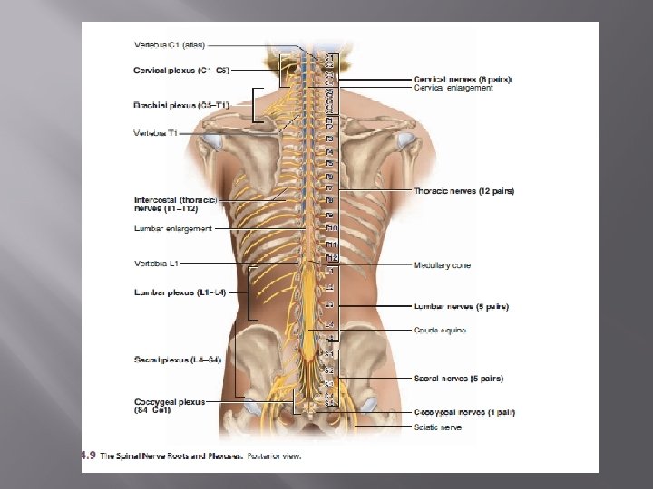
SPINAL NERVES � There are 31 pairs of spinal nerves: 8 cervical (C 1 –C 8), 12 thoracic (T 1–T 12), 5 lumbar (L 1–L 5), 5 sacral (S 1–S 5), and 1 coccygeal (Co). � The first cervical nerve emerges between the skull and atlas � The others emerge through intervertebral foramina, including the anterior and posterior foramina of the sacrum and the sacral hiatus.
Structure of a Nerve � � � Endoneurium surrounds each fiber Groups of fibers are bound into fascicles by perineurium F ascicles are bound together by epineurium
AUTONOMIC NERVOUS SYSTEM (ANS)
Defination � is the part of the nervous system that acts as a control system, functioning largely below the level of consciousness, and controls visceral functions. � Concerned with control of the internal environment through innervation of secretory glands and with cardiac and smooth muscle.
Difference between Somatic and Autonomic nervous System
DIVISIONS 3 subdivisions : Sympathetic Nervous System Parasympathetic Nervous System Enteric Nervous System
Sympathetic Division Fright, Fight or Flight reaction � Larger of the two parts � Called as thoracolumbar division � � Function - prepare the body for an emergency
Fright, Fight or Flight reaction
� Cell bodies in the lateral horns of the spinal cord gray matter between the first thoracic(T 1) and the second lumbar (L 2) segments (sometimes L 3)
Sympathetic trunks 2 ganglionated nerve cords � Extends from the cranial base to the coccyx � The ganglia are joined to spinal nerve by short connecting nerves called as white ramus & grey ramus communicantes. �
Neck posterior to carotid sheath and anterior to the transverse processes of the cervical vertebrae. Thorax anterior to heads of the ribs Abdomen anterolateral to bodies of L vertebrae Pelvis anterior to the sacrum Anterior to coccyx 2 trunks meet as terminal ganglion.
Sympathetic ganglia Cervical 3 Thoracic 11 -12 Lumbar 4 -5 Sacral 4 -5
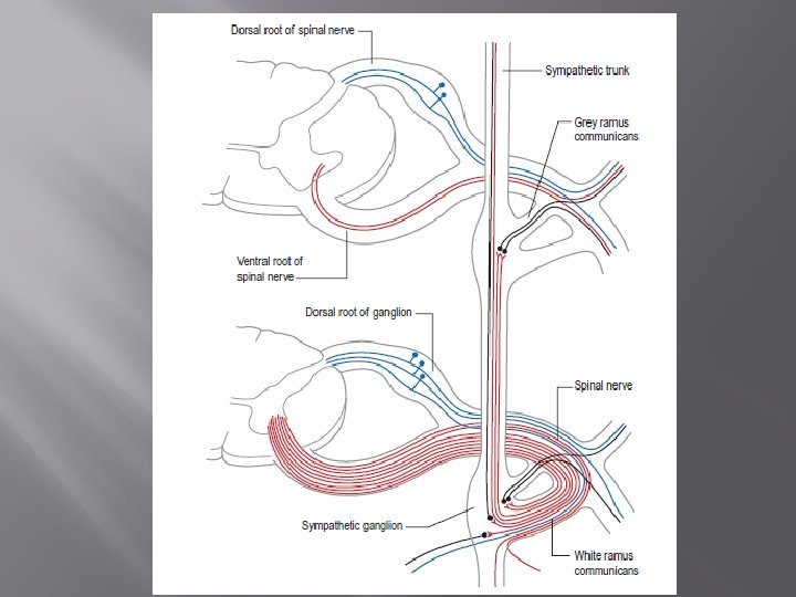
Preganglionic neurons � Myelinated, short � Cell bodies intermediolateral gray column � Pregnaglionic neurons ventral root white ramus communicans synapse with postganglionic axon in paravertebral ganglion � Neurotransmitter acetylcholine
Ramus communicantes Latin connecting branch 1)White rami communicans Spinal cord T 1 -L 2(3) Carry preganglionic nerve fibers from cord to paravertebral ganglion 2)Grey rami communicans Carry postganglionic fibers from paravertebral ganglion to effector organ Also carry preganglionic fibers which enter paravertebral ganglion but do not synapse
white rami Grey rami 1. Lateral in position 1. Medial in position 2. Convey preganglionic motor And viscero sensory fibres 2. Convey postganglionic fibres only 3. Connected to lateral ganglia from T 1 – L 2 spinal nerves 3. Connected to all 31 pairs of spinal nerve 4. Inresegmental in distribution 4. Segmental in distribution and supply Sudomotor, Pilomotor and vasoconstrictor fibres of the body wall.
Postganglionic neurons � Unmyelinated, long � Cell bodies paravertebral ganglia extend to visceral organs � Distribution widespread � Neurotransmitter norepinephrine
Splanchnic nerves -Fibers not synapsed at paravertebral ganglia -T 5 to L 2 enter abd. cavity as Splanchnic nerve -Synapse with prevertebral ganglia Types 1)Cardiopulmonary nerve-postsynaptic & sym. 2)Thoracic splanchnic nerve 3)Lumbar splanchnic nerve 4) Sacral splanchnic nerve 5)Pelvic splanchnic nerve-Parasym. fibers
The parasympathetic system The preganglionic motor neurons are partly located in the brainstem in connection with -3 rd cranial nerve(oculomotor) -7 th cranial nerve(facial) -9 th cranial nerve(glossopharyngeal) -10 th cranial nerve(vagus) Partly in the lateral horn cells of 2 nd to 4 th sacral segments of the spinal cord. It is also known as cranio-sacral outflow Postganglionic para-sympathetic neurons consists of collateral and terminal ganglia.
Rest, Recovery , Relaxation
Parasympathetic � Rest, Recovery , Relaxation � Craniosacral �Cranial nerves III, VII, IX and X �In anterior horn of gray matter from S 2 -S 4 �Leave Sacral nerve & forms pelvic splanchnic nerve cranial part 4 peripheral ganglia— Ciliary, Pterygopalatine, Submandibular and Otic
Preganglionic neurons � Myelinated, long � Synapse with postganglionic axons in ganglia close to organs � Neurotransmitter acetylcholine. Postganglionic axons � Unmyelinated, short � Neurotransmitter acetylcholine
Ganglia � � Parasympathetic ganglia are the autonomic ganglia of the parasympathetic nervous system. Most are small terminal ganglia or intramural ganglia, so named because they lie near or within (respectively) the organs they innervate.
Enteric Nervous System nerve plexuses in the wall of the digestive tract, gallbladder and bile ducts, and the pancreas � derived from the neural crest �
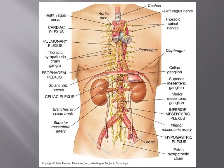
Autonomic Plexuses Thorax, abdomen, and pelvis � Branches from the plexuses innervate the viscera Thorax Cardiac, Pulmonary, and Esophageal Abdomen Celiac, Superior &Inferior mesenteric and Aortic Pelvis Superior & Inferior Hypogastric �