NERVE INJURIES OF UPPER LIMB By Dr Mujahid

NERVE INJURIES OF UPPER LIMB By: Dr. Mujahid Khan
 Ø These are caused by the excessive displacement of")
Brachial Plexus Injuries (upper lesions) Ø These are caused by the excessive displacement of the head to the opposite side Ø Depression of the shoulder on the same side Ø This causes excessive traction of C 5 and C 6 roots of the plexus
 Ø Infraspinatus (lateral rotator of")
Muscles to be Paralyzed Ø Supraspinatus (Abductor of shoulder) Ø Infraspinatus (lateral rotator of shoulder) Ø Biceps brachii (flexor of elbow) Ø Coracobrachialis (flexor of shoulder) Ø Deltoid (Abductor of shoulder) Ø Teres minor (lateral rotator of shoulder)

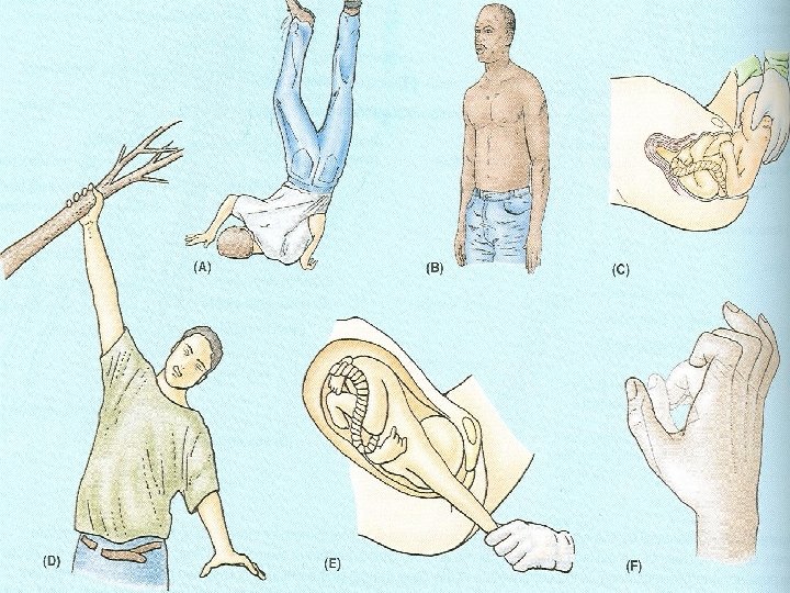
Erb-Duchenne Palsy Ø The limb hangs limply by the side likened to a waiter or porter hinting for a tip Ø There will be a loss of sensation down the lateral side of arm
 Ø Are usually a traction injuries caused by excessive")
Brachial Plexus Injuries (Lower lesions) Ø Are usually a traction injuries caused by excessive abduction of the arm Ø The first thoracic nerve is usually torn Ø The hand has a clawed appearance caused by hyperextension of metacarpophalangeal joints & flexion of interphalangeal joints
 Ø Loss of sensation will occur along the medial")
Brachial Plexus Injuries (Lower lesions) Ø Loss of sensation will occur along the medial side of the arm Ø Lower lesions can also be produced by a presence of a cervical rib or malignant metastases from the lungs in the lower deep cervical lymph nodes

Axillary Sheath Ø A brachial plexus nerve block can be obtained by injecting a local anesthetic Ø The position of the sheath can be verified by feeling the pulsations of the 3 rd part of the axillary artery

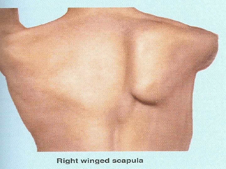
Injuries of Long Thoracic Nerve Ø Can be injured by blows to or pressure on the posterior triangle of the neck Ø Serratus anterior is paralyzed Ø The patient feels difficulty in raising the arm Ø The vertebral border & inferior angle of scapula protrude posteriorly Ø Known as winged scapula

Injuries of Axillary Nerve Ø Can be injured by the pressure of a badly adjusted crutch pressing upward into the armpit Ø It is vulnerable during the downward displacement of the humeral head in shoulder dislocations or fractures of the surgical neck of the humerus Ø Paralysis of deltoid and teres minor muscles results

Axillary Nerve Ø Loss of skin sensation over the lower half of the deltoid muscle Ø Paralyzed deltoid wastes rapidly Ø Underlying greater tuberosity can be palpated Ø Abduction of the shoulder is impaired Ø Paralysis of teres minor is not recognized clinically

Injuries of Radial Nerve Can be injured by: Ø Pressure of badly fitting crutches Ø Drunkard falling asleep with one arm over the back of a chair Ø Fractures or dislocation of the proximal end of the humerus

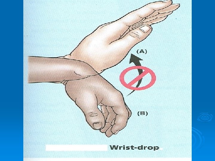
Findings in Radial N. Injury Ø Triceps, anconeus and long extensors of the wrist are paralyzed Ø Unable to extend the elbow joint, wrist joint and fingers Ø Wrist drop or flexion of wrist occurs Ø Unable to flex the fingers firmly for gripping Ø Brachioradialis & supinator are paralyzed

Sensory Findings Ø Little loss of skin sensation over posterior surface of lower part of the arm Ø Sensory loss on the lateral part of dorsum of the hand Ø Sensory loss on the dorsal surface of the roots of the lateral 3 & ½ fingers

In the Spiral Groove Ø Radial nerve can be injured in the spiral groove at the time of fracture of shaft of the humerus Ø Wrist drop occurs Ø Sensory loss on the dorsal surface of the roots of the lateral 3 & ½ fingers

Deep Branch of Radial Nerve Ø Can be damaged in the fracture of the proximal end of radius or during dislocation of the radial head Ø No wrist drop as extensor carpi radialis longus is undamaged Ø No sensory loss as this is a motor nerve

Injuries of Musculocutaneous Nerve Ø Rarely injured due to its protected position beneath the biceps brachii muscle Ø If injured high up in the arm, the biceps & coracobrachialis are paralyzed & brachialis is weakened Ø Sensory loss along the lateral side of the forearm occurs

Injuries of Median Nerve Can be injured: Ø Occasionally in the elbow region in supracondylar fractures of the humerus Ø Commonly injured by stab wounds or broken glass just proximal to the flexor retinaculum Ø Here it lies between the tendons of flexor carpi radialis and flexor digitorum superficialis
 Ø Pronator muscles of forearm, long flexor muscles of the")
Injury at Elbow (motor) Ø Pronator muscles of forearm, long flexor muscles of the wrist & fingers will be paralyzed Ø Forearm is kept in supine position Ø Wrist flexion is weak & accompanied by adduction Ø No flexion at interphalangeal joints of index & middle fingers
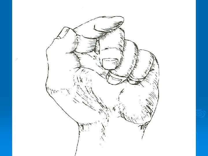
 Ø When the patient tries to make a fist, the")
Injury at Elbow (motor) Ø When the patient tries to make a fist, the index & middle fingers tend to remain straight Ø Only ring & little fingers flex Ø Flexion in these fingers is weakened by the loss of the flexor digitorum superficialis
 Ø Flexion of terminal phalanx of thumb is lost because")
Injury at Elbow (motor) Ø Flexion of terminal phalanx of thumb is lost because of paralysis of flexor policis longus Ø The thumb is laterally rotated and adducted Ø Muscles of thenar eminence are paralyzed Ø The hand looks flattened and ape like
 Ø Skin sensation is lost on the palmar aspect of")
Injury at Elbow (sensory) Ø Skin sensation is lost on the palmar aspect of the lateral 3 & ½ fingers Ø Sensory loss occurs on the skin of the distal part of the dorsal surfaces of the lateral 3 & ½ fingers Ø Total area of anesthesia is less
 Ø The skin areas involved in sensory loss are")
Injury at Elbow (vasomotor changes) Ø The skin areas involved in sensory loss are warmer and drier than normal Ø Arteriolar dilatation and absence of sweating resulting from loss of sympathetic control
 In long standing cases: Ø Skin is dry and")
Injury at Elbow (Trophic changes) In long standing cases: Ø Skin is dry and scaly Ø Nails crack easily Ø Atrophy of the pulp of the fingers

Injury at Wrist Ø Almost all the clinical findings are same as injury of the median nerve at elbow Ø In addition a delicate pincer like movement is not possible

Carpal Tunnel Syndrome Ø The carpal tunnel is formed by the concave anterior surface of carpal bones and closed by flexor retinaculum Ø Clinically, the syndrome consists of a burning pain or pins & needles along the distribution of the median nerve Ø Lateral 3 & ½ fingers are involved

Carpal Tunnel Syndrome Ø The exact cause is difficult to determine Ø Condition is relieved by decompressing the tunnel by making a longitudinal incision through the flexor retinaculum
 Ø Flexor carpi ulnaris & medial")
Injury to the Ulnar Nerve (motor at elbow) Ø Flexor carpi ulnaris & medial half of flexor digitorum profundus are paralyzed Ø In a tightly clenched fist the tightening of the tendon of profundus is absent Ø Profundus tendon to the ring & little fingers will be functionless Ø Terminal phalanges of these fingers fail to flex properly
 Ø Flexion of wrist joint will")
Injury to the Ulnar Nerve (motor at elbow) Ø Flexion of wrist joint will result in abduction due to paralysis of flexor carpi ulnaris Ø Small muscles of hand will be paralyzed except the muscles of thenar eminence and first 2 lumbricals Ø Adductor pollicis longus is paralyzed so the adduction of thumb is not possible
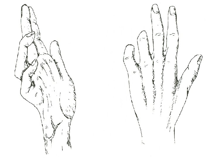
 Ø Metacarpophalangeal joints become hyperextended due")
Injury to the Ulnar Nerve (motor at elbow) Ø Metacarpophalangeal joints become hyperextended due to the paralysis of lumbrical and interosseous muscles Ø Interphalangeal joints are flexed due to the same reason as mentioned above Ø Dorsum of hand will show hollowing due to the wasting of dorsal interosseous muscles
 Ø Loss of skin sensation of")
Injury to the Ulnar Nerve (sensory at elbow) Ø Loss of skin sensation of anterior & posterior surfaces of the medial 3 rd of the hand medial 1 & ½ fingers Ø The skin areas involved in sensory loss are warmer and drier than normal Ø Arteriolar dilatation and absence of sweating resulting from loss of sympathetic control
 Ø Small muscles of the hand")
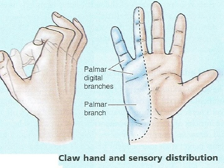
Injury to the Ulnar Nerve (motor at wrist) Ø Small muscles of the hand will be paralyzed Ø Claw hand is more obvious as flexor digitorum profundus is not paralyzed Ø Marked flexion of the terminal phalanges occur
 Ø The sensory loss is usually")
Injury to the Ulnar Nerve (sensory at wrist) Ø The sensory loss is usually confined to the palmar surface of medial 3 rd of the hand the medial 1 & ½ finger Ø Trophic changes are same as that injuries of ulnar nerve at elbow Ø Unlike median nerve injuries, lesions of ulnar nerve leave a relatively efficient hand Ø Pincer like action is good
- Slides: 38