Nerve Action Potential Dr B Kalpana At the

Nerve Action Potential Dr. B. Kalpana

At the end of class student should be able to: • Apppreciate the role of ion channels in the generation of nerve impulse • Understand the stages of Action Potential • Explain the ionic basis of Action Potential of nerve fiber. • List the blockers of Ion channels

How neurons communicate?
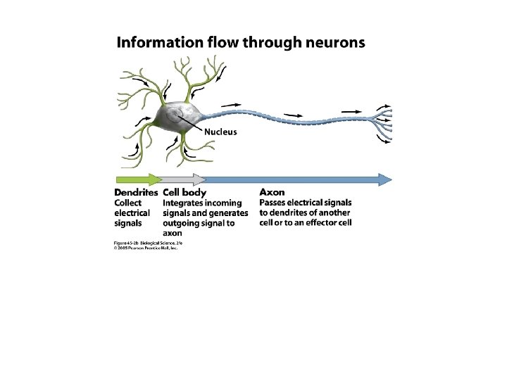

The Nerve Impulse or Action Potential • • Is the electrical current moving from the dendrites to cell body to axon. It results from the movement of ions (charged particles) into and out a neuron through the plasma membrane.

How is a nerve impulse generated?

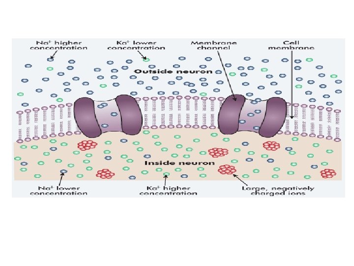
Excitability: The ability of the cell to generate the Action Potential Excitable cells: Cells that generate Action Potential during excitation. Ø Nerve and Muscle cells
 environmental condition of the cell.")
STIMULUS: A sudden change of the (internal or external) environmental condition of the cell. TYPES OF STIMULUS: ØThreshold: The stimulus with the intensity equal to threshold ØSubthreshold: The stimulus with the intensity weaker than the threshold ØSuprathreshold: The stimulus with the intensity greater than the threshold.

9

Definition • Sudden change in the membrane potential from normal RMP to a positive potential and coming back to Resting stage. Phases of AP • Resting stage • Depolarization stage • Repolarization stage

Recording of action potential in a nerve fiber

RESTING STAGE Neuron is polarized at rest RMP = -70 mv

DEPOLARIZATION STAGE Permeable to Na +ions • Mp becomes positive • opening of voltage gated Na + Channels •

Depolarization

DEPOLARISATION STAGE: • Reduction of Membrane Potential from Negative to Positive

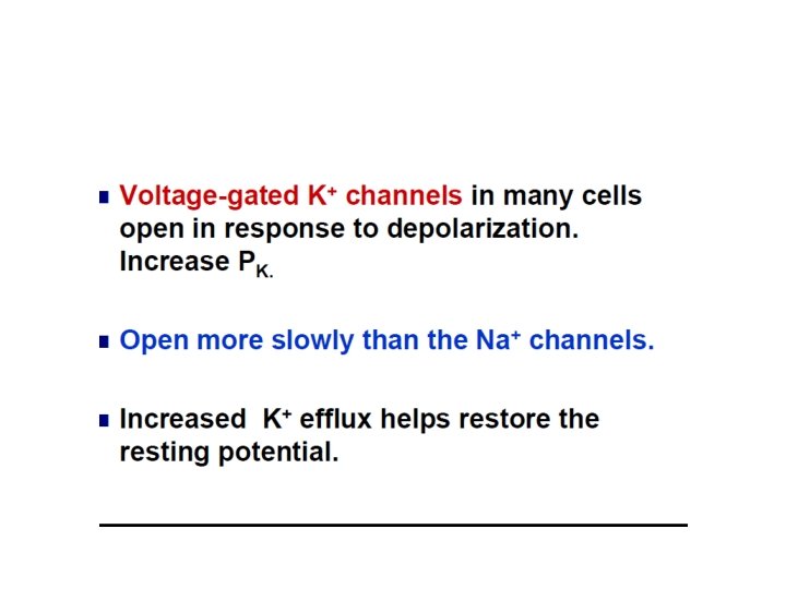
Repolarisation stage • Permeable to K+ • Membrane Potential becomes negative • Opening of voltage gated K+ channel

Repolarization

REPOLARISATION STAGE: v. K+ efflux due to opening of Voltage Gated K+ channels

Phase of Repolarization • Rapid falling phase • After-depolarization Phase of After-hyperpolarization • More negative than RMP • MP returns to resting level
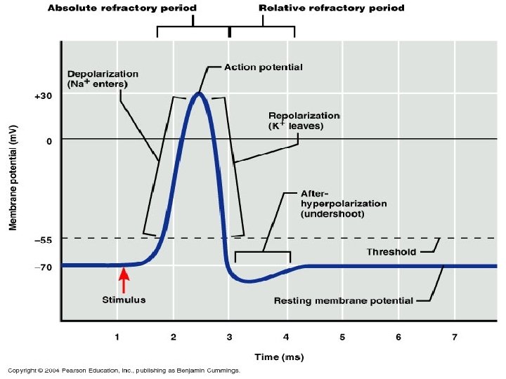
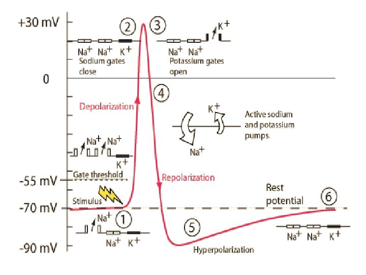

Ionic basis of action potential Stimulus artifact • Irregular deflection of baseline due to current leakage from stimulating electrode to recording electrode Latent period • Time taken for the impulse to travel from stimulating electrode to recording electrode.

Depolarization • Reduction of membrane potential from negative to zero Firing level • Initial 15 mv of depolarization • Opening of voltage gated Na +channels

Rapid depolarization • When partial depolarization reaches the activation threshold, voltage-gated sodium ion channels open. • Sodium ions rush in. • The membrane potential changes from -70 m. V to +35 m. V. • Autoactivation of sodium channels + - - Na+ Na+ +

Overshoot • From the firing level , the curve reaches the zero potential rapidly and then overshoots the zero line up to +35 m. V. Spike potential • The phase of rapid rise in potential in depolarisation and a rapid fall in Repolarisation phase

Repolarisation • K+ efflux due to opening of voltage gated K+ channels.

Repolarization • • Sodium ion channels close and become refractory Rapid autodeactivation Depolarization triggers opening of voltage-gated potassium ion channels. K+ ions rush out of the cell, repolarizing and then hyperpolarizing the membrane. Na+ K+ K+ + -

After depolarization • Slow k+ efflux After hyper polarization • Na+-k+ pump is activated to achieve the ionic composition

Voltage-gated Na+ channels Activation gate-m gate Inactivation gate-h gate

Stimulation Positive feedback loop Reach “threshold”? If YES, then. . . 33

Voltage-gated K+ channels • Voltage-gated K+ channels have only one gate. • This gate is also activated by depolarization. • However, this gate is much slower to respond to the depolarization.

Andrew Fielding Huxley “for their discoveries concerning the ionic mechanisms involved in excitation and inhibition in the peripheral and central portions of the nerve cell membrane” 35 The Nobel Prize in Physiology or Medicine (1963) Alan Lloyd Hodgkin

Effects of extracellular ionic changes: 1. ECF sodium concentration decreased- amplitude of AP becomes smaller. 2. ECF- potassium concentration decreased MP-more negative and its concentration increases-excitable 3. ECF- Calcium is decreased-RMP-firing level-increase in exciitability of the cell.

Ion Channel blocker Local anesthetics attach to Na+ channels, preventing Na+ inflow General anesthetics (ether, chloroform) Open K+ channels: clamp potential Scorpion Venom: Keeps Na+ channels open and K+ channels closed Tetrodotoxin (TTX, from puffer fish) blocks Na+ channels Cyanide blocks ATP-dependent Na+-K+ pump TEA- blocks K+ channels closed

Summary
 G K Pal")
References • Comprehensive Textbook of Medical physiology (Vol 2 first edition) G K Pal • Text book of medical physiology (Vol 2 6 th edition) A K Jain • Essentials of medical physiology (6 th edition) K Sembulingam and Prema Sembulingam AEJ 39

- Slides: 40