Nephrotic and Nephritic syndrome Dr Kriti mohan Assistant


 �Spot protein/creatinine")


 § Low serum complement")


) �Symptoms resolves")


- Slides: 43
Nephrotic and Nephritic syndrome Dr Kriti mohan Assistant Professor, Pediatrics
Learning objectives �Definition of Nephrotic syndrome �Etiopathogenesis of nephrotic syndrome �Clinical manifestation �Evaluation �Management �outcome �Post streptococcal GMN
Introduction �Important chronic disease in children � 80% children show remission with steroid �Most patients have multiple relapses
Definition �Heavy proteinurea>3. 5 gm/24 hr or >40 mg/m 2/hr in children �Hypoalbunemia <2. 5 gm% �Oedema �Hyperlipidemia (serum cholestrol>200 mg/dl)
Nephrotic range proteinurea �Early morning protein is 3+/4+ (dipstick or boiling test) �Spot protein/creatinine ratio >2 mg/mg or �Urine albumin excretion >40 mg/m 2 per hr (on a timed-sample).
Etiology �Idiopathic: 90% � minimal change 85%, mesangial proliferation , FSG , membranoproliferative, congenital (Finnish type) �Secondary: 10% �SLE, anaphylactoid purpura, sickle cell disease, Hodgkin lymphoma, diabetes mellitus, amyloidosis, malaria (P. malariae), intrauterine infections (syphilis, toxoplasmosis, cytomegalovirus) and other infections like HIV, parvovirus B 19, hepatitis B and C virus, drugs like d-penicillamine, gold and toxins or allergies (bee sting, poison ivy, food allergy).
Pathophysiology �Increase in permeability of glomerular BM �T- cell dysfunction �Mechanism of edema: �Urine protein loss leads to hypoalbuminemia decreased oncotic pressure transudation of fluid �Reduction in intravascular volume and decrease renal perfusion pressure
Pathophysiology �Mechanism of lipid elevation: �Hypoalbuminemia stimulates generalized hepatic protein synthesis including lipoprotein �Lipid catabolism is diminished due to decrease in lipoprotein lipase
Clinical Features clinical Minimal change disease Mesangial proliferation Focal segmental glomerulosclero sis Incidence 85% 10% 5% Age at presentation 2 -6 years 2 -10 years Hypertension 10% 10 -45% 35 -45% Microscopic Hematuria 10 -20% 45 -90% 60 -80% Response to prednisolone 95% 50 -60% 20 -30% Likelihood of maintaining renal function 95% 50 -60% 20 -30%
Cont… clinical Minimal change disease Mesangial proliferation Focal segmental glomerulosclero sis Light Microscopy Normal Increase in mesangial cells Focal or segmental glomerular hyalinization Immunofluoresce- Normal nt microscopy Negative or variable Ig. M and C 3 deposition Focal or segmental deposition of Igm and C 3 Electron microscopy Increase in mesangial cells and matrix, small scattered electron dense deposits in mesangium Fine granular deposits in subendothelial regions Fusion of foot processes of podocytes
Initial evaluation �Detailed evaluation �The height, weight and blood pressure should be recorded �Regular weight record �Physical examination is done to detect infections and underlying systemic disorder � Infections should be treated before starting therapy with corticosteroids.
Investigations �Urinalysis �Complete blood count �Blood levels of Proteins, lipids, urea and creatinine and electrolytes �ASLO and C 3: gross hematuria �Appropriate test –Hb. SAg, HIV and tuberculosis �Renal biopsy
Indications for kidney biopsy �At Onset � Age of onset <1 year or >10 years �Gross hematuria, persistent microscopic hematuria or low serum C 3. �Sustained hypertension. �Renal failure not attributable to hypovolemia. �Suspected secondary causes of nephrotic syndrome. �After Initial Treatment �Proteinuria persisting despite 4 -weeks of daily corticosteroid therapy. �Before treatment with cyclosporin A or tacrolimus.
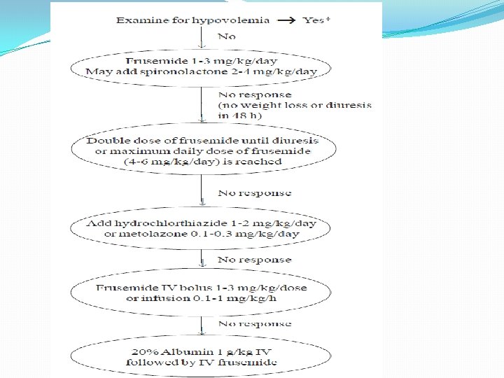
Management of Nephrotic syndrome �Relief of edema �Hypertension �Identify and treat infection �Specific treatment regimen �Complication
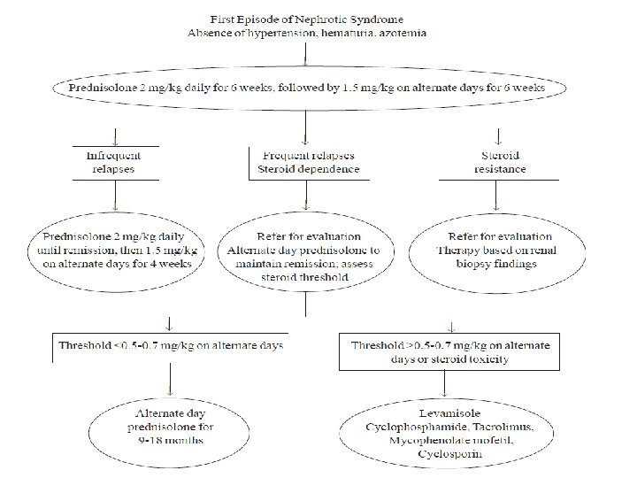
Definition related to nephrotic syndrome �Remission: Urine albumin nil or trace (or proteinuria <4 mg/m 2/h) for 3 consecutive early morning specimens. �Relapse: Urine albumin 3+ or 4+ (or proteinuria >40 mg/m 2/h) for 3 consecutive early morning specimens, having been in remission previously. �Infrequent relapses: <2 relapses in 6 months of initial response or <4 relapses within any 12 months period. �Frequent relapses: Two or more relapses in initial six months or more than three relapses in any twelve months.
Definition related to nephrotic syndrome �Steroid dependence Two consecutive relapses when on alternate day steroids or within 14 days of its discontinuation. �Steroid resistance Absence of remission despite therapy with daily prednisolone at a dose of 2 mg/kg per day for 4 weeks
Treatment of initial episode �Oral prednisolone � 2 mg/kg/day 6 weeks � 1. 5 mg/kg/EOD 6 weeks
Treatment of infrequent relapse �Prednisolone 2 mg/kg/day till remission and then �Prednisolone 1. 5 mg/kg/day for 4 weeks
Treatment of frequent repalse or steroid dependent �Low dose steroids with�Levamisole �Cyclophosphamide �Calcineurin inhibitor : Cyclosporin, Tacrolimus �Mycophenolate mofetil (MMF)
Toxicity of drugs Side effects of prednisolone �Hirsutism �Obesity �Hypertension �Behavioral problems �Cataracts �Striae �Growth failure
Side effects of Levamisole �The chief side effect of levamisole is leukopenia �Flu-like symptoms, �Liver toxicity �Convulsions and skin rash are rare � The leukocyte count should be monitored every 12 -16 weeks.
Side effects of Cyclophosphamide �Leucopenia �Hemorrhagic cystitis �Alopecia �Skin rash �Nausea
Side effects of Cyclosporin �Hypertension �Cosmetic symptoms �Gum hypertrophy �Hirsutism �Nephrotoxicity �hypercholesterolemia and elevated transaminases may occur �Estimation of blood levels of creatinine is required every 2 -3 months and a lipid profile annually.
Side effects of MMF �Gastrointestinal discomfort, diarrhea and leukopenia. �Leukocyte counts should be monitored every 1 -2 months �Treatment is withheld if count falls below 4000/mm 3.
Choice of agent �Few studies comparing one study with another �Levamisole has a modest steroid sparing effect and is a satisfactory initial choice �Treatment with cyclophosphamide is preferred in patients showing: I. significant steroid toxicity II. severe relapses with episodes of hypovolemia or thrombosis, and III. poor compliance or difficult follow up
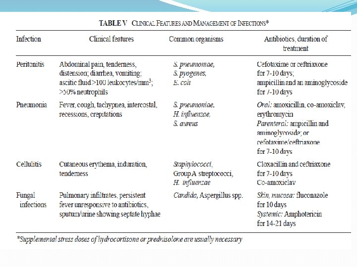
Complications �Infection �Thrombosis �Hypertension �Hypovolumic shock �Corticosteroid side effects �Malnutrition
Outcome �Steroid responsive - >90% �Relapses- >70% �Mortality – 2 -5%
Patient and parents education �Urine examination at home �Maintain diary showing result of urine protein �Ensure normal activity and school attendance �Appropriate immunization
Acute glomerulophritis �Glomerulonephritis refers to a group of glomerular diseases characterise by inflammatory changes in the glomeruli and manifesting as acute nephritic syndrome which is characterized by�Abrupt onset of hematuria �Oligouria �Edema �Hypertension �Subnephrotic range proteinuria �Azotemia
Causes of Acute GMN �Post infectious: Bacterial-Streptococcal, staphylococcal, pnemococcal, meningococcal. Bacterial endocarditis, infected ventriculoatrial shunt and prosthesis can cause acute GMN. Viral- Hepatitis B and C, mumps, HIV, varicella, infectious mononucleosis. Parasitic- malaria and toxoplasmosis �Systemic vasculitis: HSP, SLE, PAN, Wegner’s granulomatosis
Pathogenesis Immune complex mediated disease i. Immune complex Glomerulonephritis (70%) § Low serum complement C 3 - poststreptococcal, rapidly progressive, mesangioproliferative glomerulonephritis, SLE, bacterial endocarditis, cryoglobulinemia § Normal serum complement C 3 - Ig. A nephropathy, H-S purpura ii. Pauci-immune glomerulonephritis (30%) Anti-neutrophil cytoplasmic antibody positive wegener’s granulomatosis, polyarteritis nodosa iii. Anti GBM disease(<1%) Anti-glomerular basement membrane antibody positive Good pasture syndrome.
Post streptococcal Glomerulonephritis �Following group A beta-hemolytic streptococci �School age children �Boys are more frequently affected
Etiology �Follows a pharyngeal or cutaneous infection by the nephritogenic strains of β hemolytic Group A streptococcus 1 to 4 week preceding streptococcal throat/skin infection �Strain M type 1, 4 and 12 causing pharyngitis and 49, 55, 57 and 60 causing pyoderma �Typical example of immune complex disease
Pathogenesis �Immune complex deposition �Glomeruli enlarged �Ischemia �Capillary wall narrowing �Deposits of Ig. G and C 3
Clinical feature �Rare below 3 years of age �Acute onset �Latent period: Following pharyngitis- 1 to 2 weeks and following cutaneous infection- 2 to 4 weeks �Puffiness around eye and pedal edema �Cola colored urine �Oliguria �Hypertension �Atypical presentation : Convulsion, Pul edema, ARF, CHF �Course of the disease- acute phase: 4 -10 days, azotemia and hypertension: persist for 2 weeks, gross hematuria: 1 -2 weeks
Laboratory investigations � Urine : 1+/2+ protein, dysmorphic RBC’s, and red cell, leukocyte or granular cast, nephrotic range poteinuria in < 5% cases � Hemogram: Anemia, mild leucocytosis, ESR↑ � Biochemistry: C 3 (normal- 77 -195 mg/ d. L) becomes normal in 6 to 8 weeks. � Evidence of streptococcal infection: Throat swab culture, elevated ASO ( for pharyngeal infection+ve in 80%), elevated antideoxyribonucleases-B antihyaluronidase antibodies ( for cutaneous infection), streptozyme test � Others- X- ray chest, ECG � renal biopsy- to exclude other diseases in patients withØ ARF Ø normal C 3 level Ø without evidence of preceding streptococcal infection Ø persistant gross hematuria and hypertension (>3 weeks) Ø prolonged diminished renal functions (> 2 weeks) Ø persistent low serum C 3 (>8 weeks)
Management Presence of ARF and Hypertension requires hospitalisation �Bed rest �Diet �Weight �Fluid restriction �Antibiotics �Diuretics �Alkalinization of urine �Hypertension �LVF �ARF
Outcome and prognosis �Overall excellent prognosis( >95% complete recovery, <1% develop RPGN)) �Symptoms resolves within 1 month �Gross hematuria and proteinuria disappear within 2 weeks �Microscopic hematuria may last for years �Recurrence rare
Difference between acute nephritis and nephrotic syndrome Acute nephritis Nephrotic syndrome � Characterized by hematuria, edema, hypertension, oligouria � Characterized by heavy proteinuria, hpo albuminemia, edema, hyperlipidemia � 90% post infective, immune complex � 90% idiopathic � Usually only 1 attack � Immune complex deposition � Relapses common � Retraction of epithelial foot process � Urine: Alb 1+/2+, hematuria, RBC cast � Blood urea/creat raised � Urine: Selective proteinuria, No RBC � Blood urea/ creat normal
Thank you