NEOPLASMS OF THE PERITONEUM By Dr Mustafa Usama

NEOPLASMS OF THE PERITONEUM By Dr Mustafa Usama General and laparoscopic surgeon

� This is a common terminal event in many cases of carcinoma of the stomach, colon, ovary or other abdominal organs and also of the breast and bronchus. � The peritoneum, both parietal and visceral, is studded with secondary growths and the peritoneal cavity becomes filled with clear, strawcoloured or blood-stained ascitic fluid.
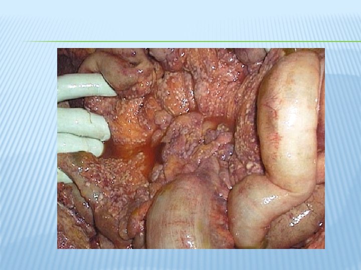

THE MAIN FORMS OF PERITONEAL METASTASES ARE: � • discrete nodules – by far the most common variety. � • plaques varying in size and colour; � • diffuse adhesions – this form occurs at a late stage of the disease and gives rise, sometimes, to a ‘frozen pelvis’.

TREATMENT �Ascites caused by carcinomatosis of the peritoneum may respond to systemic or intraperitoneal chemotherapy or to endocrine therapy in the case of hormone receptor-positive tumours.

PSEUDOMYXOMA PERITONEI �This rare condition occurs more frequently in women. �The abdomen is filled with a yellow jelly, large quantities of which are often encysted. The �condition is associated with mucinous cystic tumours of the appendix.

� It is often painless and there is frequently no impairment of general health. � Pseudomyxoma peritonei does not give rise to extraperitoneal metastases. � Although an abdomen distended with what seems to be fluid that cannot be made to shift. � the diagnosis is more often suggested by ultrasound and CT scanning or made at operation.

� The appendix, if present, should be excised � Unfortunately, recurrence is inevitable, but patients may gain symptomatic benefit from repeated ‘debulking’ surgery. Occasionally, the condition responds to radioactive isotopes or intraperitoneal chemotherapy.

MESENTERIC CYSTS Cysts may occur in the mesentery of either the small intestine (60 per cent) or the colon (40 per cent) and can be classified as: � chylolymphatic; � enterogenous; � urogenital remnant (actually retroperitoneal but project intoperitoneum); � dermoid.

CHYLOLYMPHATIC CYST � This is the most common variety, probably arising in congenitally misplaced lymphatic tissue. � The thin wall of the cyst, which is composed of connective tissue lined by flat endothelium � enucleation is possible without the need for resection of gut.

ENTEROGENOUS CYSTS � These are believed to be derived either from a diverticulum of the mesenteric border of the intestine that has become sequestrated from the intestinal canal during embryonic life or from a duplication of the intestine. � An enterogenous cyst has a thicker wall than a chylolymphatic cyst and it is lined by mucous membrane, sometimes ciliated. The content is mucinous

� The muscle in the wall of an enteric duplication cyst and the bowel with which it is in contact have a common blood supply; consequently, removal of the cyst always entails resection of the related portion of intestine.

RETROPERITONEAL CHRONIC INFLAMMATION/ FIBROSIS
- Slides: 13