NEOPLASIA II CHARACTERISTICS OF BENIGN MALIGNANT TUMORS CHARACTERISTICS


NEOPLASIA II CHARACTERISTICS OF BENIGN & MALIGNANT TUMORS

CHARACTERISTICS OF BENIGN & MALIGNANT � Differentiation and Anaplasia � Rates of Growth � Cancer stem cells & cancer cell lineages � Local invasion � Metastasis

DIFFERENTIATION AND ANAPLASIA Differentiation – extent to which neoplastic parenchymal cells resemble the corresponding normal parenchymal cells both morphologically & functionally � Benign tumors – cells differentiated , closely resemble normal cells �

DIFFERENTIATION AND ANAPLASIA � Malignant poorly diff tumors – well diff, moderately diff,

DIFFERENTIATION AND ANAPLASIA Anaplasia – “to form backwards” � Anaplasia – lack of differentiation ; considered a hallmark of malignancy Associated morphologic features: � Pleomorphism � Abnormal nuclear morphology (N: C ratio 1: 1) �

ANAPLASIA � Mitoses : ↑mitosis , atypical mitosis

OTHER FEATURES OF MALIGNANCY/ANAPLASIA � Loss of polarity Tumor giant cells � Necrosis �

Dysplasia : disordered growth � loss in � uniformity of individual cells � their architectural orientation

Functional capabilities: � Benign tumors & well diff Cas – retain � Poorly diff & Anaplastic Cas – don’t retain � New Unanticipated functions may emerge Fetal proteins � Hormone production by Nonendocrine Ca �

RATES OF GROWTH � 3 major factors: � Doubling time of tumor cells � Fraction of tumor cells in replicative pool � Rate of cell death Schematic representation of tumor growth

RATES OF GROWTH � � � Growth fraction : the proportion of cells in replicative pool Rate of growth of tumors is determined by an excess of cell production over cell loss Fast growing tumors have a high cell turnover Growth fraction of tumor cells has profound effect on their susceptibility to chemotherapy Tumor with low growth fraction : 1 st shift tumor cells from GO into cell cycle – debulking the tumor by radiotherapy or surgery & then plan chemo

RATES OF GROWTH � � Growth rate of tumors correlate with their level of differentiation Rate of growth may not be constant � � � Hormonal influencies Adequacy of blood supply Emergence of aggressive subclones

CANCER STEM CELLS & CA CELL LINEAGES Resident population of tissue stem cells that are longlived and capable of self-renewl � Cancers also must contain cells with “stemlike”properties � Practical implication : if Cancer stem cells are “essential” for tumor progression they must be eliminated by therapy; �

CANCER STEM CELLS & CA CELL LINEAGES � T- ICs : Cells that allow a human tumor to grow and maintain itself indefinitely when transplanted into an immunodeficient mouse. In future it will be imp to identify T-Ics in each tumor to direct therapy against tumor stemcells

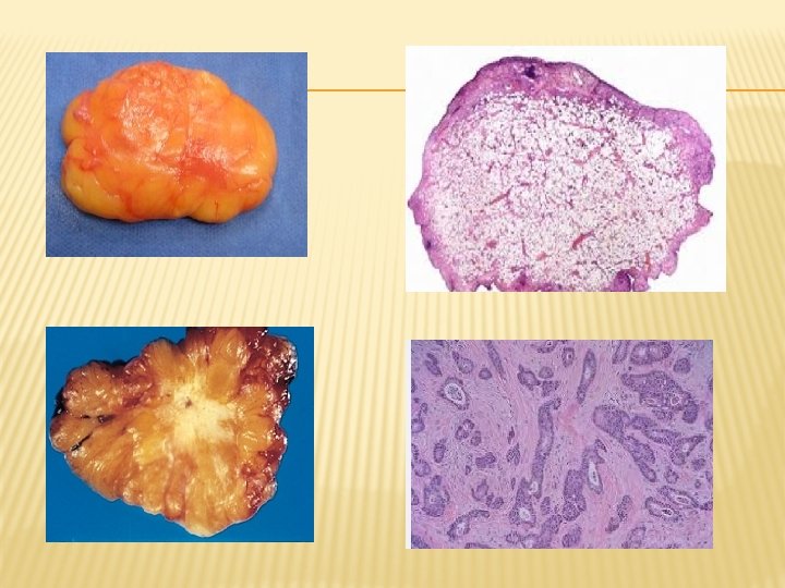
LOCAL INVASION Benign tumors - cohesive expansile masses a rim of fibrous capsule � Malignant tumors – � a crab-like pattern of growth � infiltration, invasion & destruction of tissues � No capsule � � Invasiveness makes surgical resection difficult

Fibroadenoma – encapsulated small tumor Invasive ductual carcinoma – infiltrating tumor

LOCAL INVASION � In-situ epithelial cancers have cytologic features of malignancy without invasion of basement membrane

METASTASIS Tumor implants discontinuous with the primary tumors � Unequivocal sign of malignancy � Exceptions – locally invasive rarely metastasize � Malignant neoplasms of glial cells in CNS � Basal cell carcinoma skin �

METASTASIS � Pathways of spread : � Direct seeding of body cavities and surfaces � Lymphatic spread � Hematogenous spread

Seeding of body cavities and surfaces : May occur whenever a malignant tumor penetrates into a natural “open field” � Peritoneal cavity most oftenly involved � Particularly characteristic of Ca ovaries � Pseudomyxoma peritonei � Colon carcinoma invading pericolonic adipose tissue

Lymphatic spread : � Most common pathway for initial spread of tumor � Lymphatic vessels located at tumor margin � Pattern of L node involvement follows the natural routes of lymphatic drainage

Metastatic Ductal Carcinoma Breast

LYMPHATIC SPREAD Regional L nodes - effective barriers to further dissemination, at least for sometime � “Sentinel lymph node” biopsy � “Skip metastasis” – local lymph nodes are bypassed � Nodal ↑is due to � growth of tumor � reactive changes �

Hematogenous Spread : � Typical of sarcoma, also seen with Ca � Arteries less readily penetrated than veins � Arterial spread may occur � � � tumor cells pass pul-monary capillary bed or pulm A/V shunts pul mets give rise to additional tumor emboli

Hematogenous Spread : � With venous invasion blood-borne cells follow venus flow draining the site of neoplasm , and usually settle in the 1 st capillary bed they encounter

METASTATIC CASCADE 2 Phases of cascade: � Invasion of ECM � Vascular dissemination & Homing of tumor cells

INVASION OF ECM : � Loosening of I/cellular junction � � � Degradation of ECM � � � E – cadherins Β – catenin Proteases MMPs ECM sequestered growth F Ameboid migration Attachment of tumor cells to novel ECM components Migration of tumor cells � � � Tumor cell aggregates PLATELETS CD 44

� � Liver and lung are the most frequently involved Tumors in close proximity to vertebral column (Ca thyroid & prostate) embolise through paravertebral plexus Skeletal muscle and spleen are rarely the site of metastasis despite enormous blood flow Certain tumors have propensity for invasion of veins : Renal cell carcinoma & Hepatocellular Ca

� Molecular genetics of metastasis � Mechanisms of metastasis development in primary tumour


- Slides: 33