Neonatal surgical jaundice Done by Mahmoud toubeh Jaundice

Neonatal surgical jaundice Done by: Mahmoud toubeh

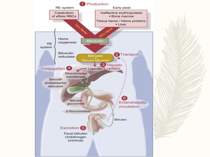
– Jaundice presents during the first week of life in 60% of term infants and 80% of preterm infants – In a majority cases this hyperbilirubinemia is due to elevated unconjugated bilirubin and resolves spontaneously with no need for surgical consultation or intervention ( normal transitional phenomenon ) NOTE: Unconjugated bilirubin is neurotoxic (kernicterus) – Jaundice is the most common condition that require medical attention in newborns
 High initial Hematocrit and decreased RBCs life span ( 90")
Neonatal Risk Factor 1) High initial Hematocrit and decreased RBCs life span ( 90 days ) 2)Frequent occurrence of haematomas ( e. g : cephalhaematoma ) and bruising 3)The newborn liver metabolism in utero occurs via the placenta and maternal liver, At birth there is a sudden increased bilirubin load 4)Decreased Ligandin concentration 5)Decreased activity of the glucuronidation pathway 6)Increased enterohepatic circulation 7)Relative dehydration in the first few days concentrates the bilirubin 8)Relative calorie deprivation in the first few days decreases glucuronidation 9)Binding sites competitors ( e. g: diazepam, sulfonamides, oxacillin, ceftriaxone, etc… ), especially if the pt. is jaundiced they may displace bilirubin from albumin binding sites and precipitate Kernicterus

Any preterm jaundiced baby should be on serious observation. Why ? – Immature BBB – Both term and preterm babies have hypoalbuminemia but is more in preterm. So the free bilirubin is more and more cross of BBB – RDS due to respiratory & metabolic acidosis ( risk factor for Kernicterus ) – Hypothermia is risk factor for Kernicterus due to increased permeability of BBB – They are at high risk for Sepsis

Types of neonatal jaundice Physiologic Pathologic

– Physiologic jaundice is evident by the second or third day of life and usually resolves within approximately 5 to 7 day – jaundice that persists beyond 2 weeks is considered pathologic. – Jaundice in the first day of life always pathological not physiological Pathologic jaundice may be due to : 1 -Biliary obstruction 2 -Increased hemoglobin load 3 -Liver dysfunction

the following possibilities: v Obstructive disorders including : – Biliary atresia : The most important surgical cause – Choledochal cyst – Inspissated bile syndrome : increased viscosity of bile leading to obstruction of the canaliculi ( seen in infants receiving parenteral nutrition also in cystic fibrosis and hemolysis )(colangiography) v Hematologic disorders, including : ABO incompatibility, Rh incompatibility, and spherocytosis v Metabolic disorders, including : α 1 -antitrypsin deficiency, galactosemia, and pyruvate kinase deficiency v Congenital infection, including syphilis and TORCH infection

Biliary Atresia

Pathogenesis -Biliary atresia is a rare disease associated with significant morbidity and mortality. -This disease is characterized by a fibroproliferative obliteration of the biliary tree, which progresses toward hepatic fibrosis, cirrhosis, and end-stage liver Failure -The obliterative process involves: common bile duct, cystic duct, one or both hepatic ducts & gallbladder, in a variety of combinations. -Biliary atresia occurs in between 1 in 20. 000

Etiology – The exact etiology of biliary atresia is likely Multifactorial. – the cause of biliary atresia as an arrest of development during the solid stage of bile duct formation

-previously proposed theories of cause focused on defects in hepatogenesis, prenatal vasculogenesis, immune dysregulation, infectious agents, and exposure to toxins. -More recently, genetic mutations in the cfc 1 gene identified in patients with biliary atresia-splenic malformation syndrome. -perinatal exposure to reovirus or rotavirus.

Congenital Anomalies Associated with Biliary Atresia – 25% of biliary atresia cases are associated with other coincidental malformations 1) Intestinal Malrotation 2) Preduodenal portal vein : is a rare anomaly in which the portal vein passes anterior to the duodenum rather than posteriorly. Generally asymptomatic, PDPV may rarely cause duodenal obstruction or may coexist with other anomalies 3) Polysplenia : multiple small accessory spleens, rather than a single, full-sized, normal spleen 4) Interrupted inferior vena cava with Azygous continuation In this rare condition the IVC terminates below the hepatic vein and lower limb venous drainage is completed via the azygos and hemiazygos veins into the superior vena cava. The hepatic veins drain directly into the right atrium.

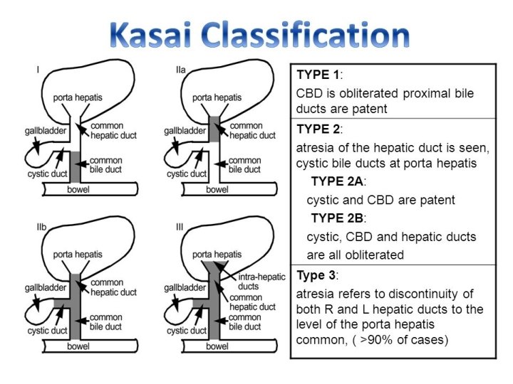
The Japanese Association of Pediatric Surgeons proposed that cases should be classified according to the location of atresia type I atresia at the level of the common bile duct type IIa atresia is at the level of the common hepatic duct type IIb atresia in the cystic , common bile and hepatic ducts type III the most frequent type, atresia occurs at the porta hepatis.

type I atresia at the level of the common bile duct

type IIa atresia is at the level of the common hepatic duct

type IIb atresia in the cystic , common bile and hepatic ducts

type III the most frequent type, atresia occurs at the porta hepatis.

The histopathology Inflammatory changes within the parenchyma of the liver, as well as fibrous deposition at the portal plates

SIGNS AND SYMPTOMS 1 -progressive jaundice at birth or shortly thereafter 2 -pale gray stools 3 -dark urine, 4 -firm hepatomegaly. v signs of advanced liver disease such as palpable hepatomegaly and splenomegaly, ascites, failure to thrive and malnutrition become present. If not treated, progress to develop stigmata of liver failure, portal HTN & esophageal varices Biliary atresia is fatal within the first 2 years of life with one study reporting a survival rate of less than 10% at 3 years

Case scenario Hx ; 4 weeks old female who presents to the office with parental reports of increasing jaundice over the last week. Her parents report that 2 weeks ago, she began to have yellowing of her eyes with subsequent yellowing of her skin when she was diagnosed with physiologic jaundice. After persistent jaundice for 5 days, her parents changed her from breast-feeding to a commercial formula. Since the jaundice appears to be worsening, her parents decided to bring her in for re-evaluation. Her stools have been acholic pale gray in color for the past 10 days along with darker urine.

Exam: VS Normal. Weight and height are at the 60 th percentile. She is awake, alert, in no acute distress and is easily comforted by her mother during the exam. Her skin is jaundiced, most notably in the cephalic and truncal areas, with scleral icterus. Her liver is slightly enlarged without nodularity. No splenomegaly is noted. The remainder of her exam is normal.
 DDx?")
– 1) DDx?

DDx –Biliary atresia – Choledochal cyst – Inspissated bile syndrome – Neonatal hepatitis
invstigations ?")
– 2)invstigations ?

1 -BLOOD TESTS – Laboratory examinations reveal a total bilirubin of 15 mg/d. L, direct bilirubin of 12. 3 mg/d. L, ALT 45 U/L, AST 52 U/L, and an alkaline phosphatase of 2007 U/L.

1 -BLOOD TESTS liver function tests : In biliary atresia both the direct and indirect bilirubin levels are elevated Alkaline phosphatase levels are often elevated in infants and children due to contribution from bone remodeling. For this reason Gammaglutamyl transpeptidase (GGTP) level more specific indicator of hepatobiliary disease

tests of hepatic synthetic function : such as the clotting cascade and serum albumin are Normal In order to rule out conditions that can mimic biliary atresia, screening for perinatal infections due to members of the TORCH family (Toxoplasmosis, Other viruses, Rubella, Cytomegalovirus, and Herpes Simplex Virus screening for the presence of alpha-1 antitrypsin deficiency should occur

2 - ULTRASOUND – The absence of a gallbladder

2 - ULTRASOUND to assess the presence of other causes of biliary tract obstruction including choledochal cyst. The absence of a gallbladder is highly suggestive of the diagnosis of biliary atresia the presence of a gallbladder does not exclude the diagnosis of biliary atresia It is important to note that the – intrahepatic bile ducts are never dilated in patients with biliary atresia

3 - nuclear medicine scan – The patient undergoes a DISIDA scan after 5 days of phenobarbital therapy. The scan showed normal uptake by the liver but no excretion of the isotope (i. e. , no bile flow) into the bowel even after 24 hours.
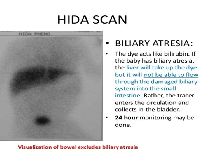

3 - nuclear medicine scan using technetium- 99 m iminodiacetic acid, performed after pretreatment of the patient with phenobarbital, If radionuclide appears in the intestine, there is patency of the biliary tree and the diagnosis of biliary atresia is excluded Delayed assessment of isotope excretion at 24 hours is warranted

4 - percutaneous liver biopsy To distinguish between biliary atresia and other sources of jaundice such as neonatal hepatitis The presence of varying degrees of inflammation with ductular proliferation is considered compatible with the diagnosis of biliary atresia because these findings are not seen in other nonobstructive cholestatic Syndromes

At surgery, a cholangiogram may be performed if possible, using the gallbladder as a of access. This may be performed using a laparoscope. The cholangiogram demonstrates the anatomy of the biliary tree. The cholangiogram may demonstrate hypoplasia of the extrahepatic biliary system When these tests point to or cannot exclude the diagnosis of biliary atresia, surgical exploration is warranted

DIAGNOSIS – No single test can reliably be used to differentiate biliary atresia from other causes of jaundice in the infant a combination of data obtained from a complete clinical assessment, blood tests, imaging studies, and pathologic evaluation – however, is always confirmed at surgical exploration.


Biliary atresia treatment Tareq Abo-Lebdeh

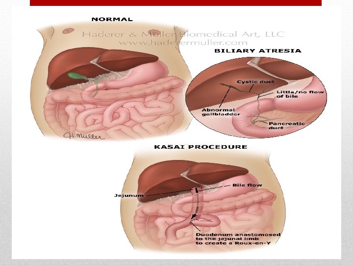
If the diagnosis of biliary atresia is confirmed intraoperative , then surgical treatment is undertaken at the • same setting. : lntrahepatic Liver transplantation. • Extrahepatic : (Removal of extrahepatic stenosis & its replacement by small intestine.

• Patent segments of proximal bile duct are found in 10% of type I lesions. A direct Roux-en-Y hepaticojejunostomy will achieve bile flow in 75%, but progressive fibrosis results in disappointing long-term results.

• A simple biliary–enteric anatomosis is not possible in the majority of cases in which the proximal hepatic ducts are either very small (type II) or atretic (type III). These are treated by the Kasai procedure in 90% of cases

radical excision of all bile duct tissue up to the liver capsule is performed. A Roux-en-Y loop of jejunum is anastomosed to the exposed area of liver capsule above the bifurcation of the portal vein, creating a portoenterostomy. • The chances of achieving effective bile drainage after portoenterostomy are maximal when the operation is performed before the age of 8 weeks, and approximately 90% of children whose bilirubin falls to within the normal range can be expected to survive for 10 years or more.

* liver biopsy is performed at the time of surgery to determine the degree of hepatic fibrosis that is present. • The diameter of bile ducts at the portal plate is predictive of likelihood of long-term success of biliary drainage through the portoenterostomy. • After this procedure, infants are usually in the hospital for seven to 10 days to heal. Long-term antibiotic therapy is given to reduce the risk of infection, and additional medications may be used to promote bile flow and maximize the success of the operation • Liver transplantation should be considered in children in whom a portoenterostomy is unsuccessful. Results are improving, with 70– 80% alive 2– 5 years following transplant

A common postoperative complication is : • Bacterial cholangitis • Repeated attacks lead to hepatic fibrosis • portal hypertension

Choledochal cyst Congenital cystic dilatation of intra and / or extrahepatic biliary system • Several hypotheses exist as to the etiology of choledochal cysts including pancreaticobiliary maljunction. • Three patterns of presentation : ü The first is a cystic mass in the abdomen identified prenatally. ü The second is jaundice presenting in infancy. ü The third is ascending cholangitis, obstructive jaundice, or pancreatitis presenting later in childhood or in adulthood.

�Regardless of the time of presentation, surgical intervention is indicated to: - ü Reduce potential damage to the liver. ü Prevent jaundice. ü Prevent pancreatitis and cancer.

• The incidence of choledochal cyst ranges from 1 in 100, 000 to 150, 000 live births in Western populations with the incidence in the United States reportedly as high as 1 in 13, 500. • Choledochal cysts are significantly more common in Asia. • There is a well-documented female dominance (3 to 4: 1) that contributes to the belief that choledochal cyst is sex-linked.
 of choledochal cysts are diagnosed in the first decade of")
� The majority (60%) of choledochal cysts are diagnosed in the first decade of life. � Roughly 20% remain undiagnosed until later in childhood or adulthood. � the remaining 20% to 25% of cases are diagnosed prenatally. � The etiology of choledochal cyst remains unknown.

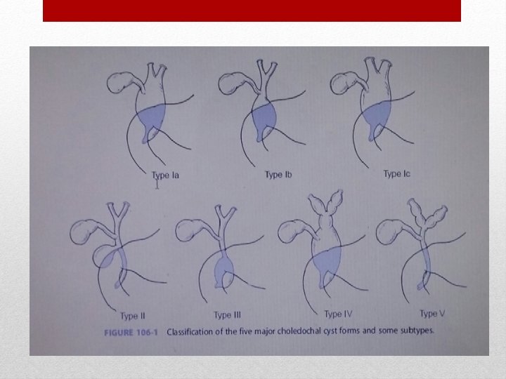
Anatomic Classification After review of cholangiograms, Todani and colleagues broadened the classification into five types with some subtypes.

Type 1: - It is the most common, accounting for 90%-95% of cases. Constitutes the cystic/saccular or fusiform dilation of the common bile duct. Type I is divided into three subtypes (types Ia, Ib, and Ic).

• Type Ia consists of cystic dilation of the entire common bile duct. • Type Ib is cystic dilation of a segment of the common bile duct. • Type Ic is fusiform dilation of the common bile duct.

Type 2: - • It is a diverticulum of the common bile duct with no dilation of the common bile, extrahepatic , or intrahepatic ducts.

Type 3: - • also referred to as a choledochocele , usually has a normal common bile duct and main pancreatic duct with cystic dilation of the distal common bile duct that is either intraduodenal or intrapancreatic in location. • The choledochocele is usually stenotic at their openings due to chronic inflammation

Type 4: - • is composed of multiple cysts located intrahepatically, extrahepatically, or in both locations.

Type 5 : - comprises single or multiple intrahepatic cysts without extrahepatic duct dilation. Type V cysts in conjunction with hepatic fibrosis are commonly referred to as Caroli disease.


Choledochal cyst Baha’ Alshamali
 • a fairly uncommon")
Background • Cystic dilatation of the common bile duct (CBD) • a fairly uncommon anomaly of the biliary tract (1: 50. 000)

Background • Abnormality in the pancreaticobiliary junction • The earliest reported choledochal cyst was detected in a fetus at 15 weeks' gestation, which may correspond to the timing of the formation of pancreatic enzymes.

Clinical presntation • Two distinct clinical groups of patients are recognized with regard to age at presentation. • Infantile form of choledochal cyst consisting of babies younger than 1 year : – with or without obvious hepatomegaly. – with obstructive jaundice. – acholic stools.

• Adult form of choledochal cyst generally have one or more components of the classic triad: – Pain – jaundice – palpable mass. The entire triad is present in fewer than 30% of patients.

• Undiagnosed choledochal cysts can lead to choledocholithiasis, cirrhosis with portal hypertension, cyst rupture, or biliary carcinomas.

Workup • Laboratory studies: – direct bilirubin – alkaline phosphatase – serum aspartate aminotransferase (AST) – serum alanine aminotransferase (ALT) – gamma-glutamyl transferase (GGT) – coagulation profiles. – A complete blood count (CBC) should also be obtained to exclude any associated or underlying anemia prior to surgery.

• Imaging Studies: cornerstone of diagnosis of choledochal cysts.
 Ultrasonography : is the best initial study. can demonstrate changes in the bile")
1) Ultrasonography : is the best initial study. can demonstrate changes in the bile ducts as well as in the liver
 Endoscopic retrograde cholangiopancreatography : the criterion standard diagnostic study.")
2) Endoscopic retrograde cholangiopancreatography : the criterion standard diagnostic study.
 Magnetic resonance cholangiopancreatography : has largely supplanted ERCP as the diagnostic test of")
3) Magnetic resonance cholangiopancreatography : has largely supplanted ERCP as the diagnostic test of choice for choledochal cysts : – offers high resolution detailed images of relevant anatomy. – Noninvasive – does not suffer from complications such as postprocedure pancreatitis – with sensitivities from 90 -100% and specificities from 73 -100%,
 of a type IV")
MRCP shows dilated hepatic ducts and common bile duct (CBD) of a type IV cyst.
 Computed tomography : may also be useful to delineate the cyst and its")
4) Computed tomography : may also be useful to delineate the cyst and its relationship to surrounding structures.

Treatment • Radical excision of cyst is the treatment of choice, with reconstruction of the biliary tract using a Rouxen-Y loop of jejunum.

• Complete resection of the cyst is important because of association with the development of cholangiocarcinoma. Resection and Roux-en-Y reconstruction are associated with a reduced indcidence of stricture formation and recurrent cholangitis.

Long-Term Monitoring • Long-term follow-up is necessary to detect any late complications, especially the development of malignancy.

• Thank you
- Slides: 77