Neonatal skull mandible By Dr Usama Mohammed Aldaghir

Neonatal skull & mandible By Dr. Usama Mohammed Aldaghir Oral & maxillofacial surgeon C. A. B. M. S

Neonatal Skull The newborn skull, compared with the adult skull, has a disproportionately large cranium relative to the face. In childhood, the growth of the mandible, the maxillary sinuses, and the alveolar processes of the maxillae results in a great increase in length of the face.

Neonatal Skull The bones of the skull are smooth and unilaminar, there being no diploë present. Most of the skull bones are ossified at birth, but the process is incomplete, and the bones are mobile on each other, being connected by fibrous tissue or cartilage.

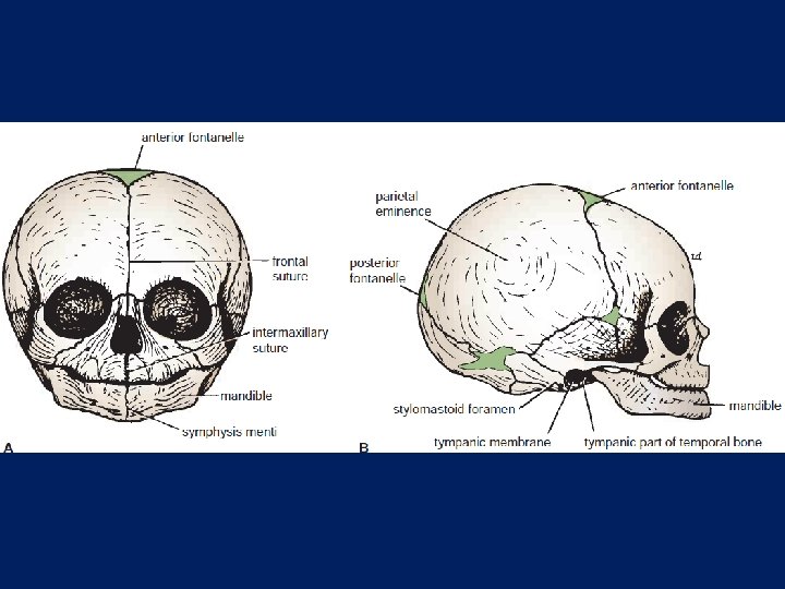
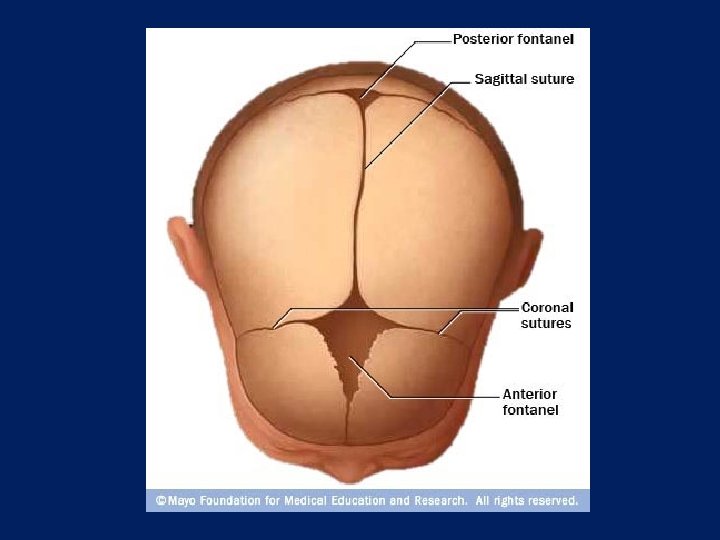
Neonatal Skull The bones of the vault are ossified in membrane; the bones of the base are ossified in cartilage. The bones of the vault are not closely knit at sutures, as in the adult, but are separated by unossified membranous intervals called fontanelles. Clinically, the anterior and posterior fontanelles are most important and are easily examined in the midline of the vault.

Neonatal Skull The anterior fontanelle is diamond shaped and lies between the two halves of the frontal bone in front and the two parietal bones behind. The fibrous membrane forming the floor of the anterior fontanelle is replaced by bone and is closed by 18 months of age.

Neonatal Skull The posterior fontanelle is triangular and lies between the two parietal bones in front and the occipital bone behind. By the end of the 1 st year, the fontanelle is usually closed and can no longer be palpated.

Neonatal Skull The tympanic part of the temporal bone is merely a C-shaped ring at birth, compared with a C-shaped curved plate in the adult. This means that the external auditory meatus is almost entirely cartilaginous in the newborn, and the tympanic membrane is nearer the surface.
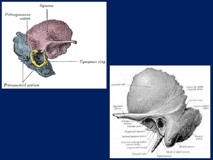

Neonatal Skull Although the tympanic membrane is nearly as large as in the adult, it faces more inferiorly. During childhood, the tympanic plate grows laterally, forming the bony part of the meatus, and the tympanic membrane comes to face more directly laterally.

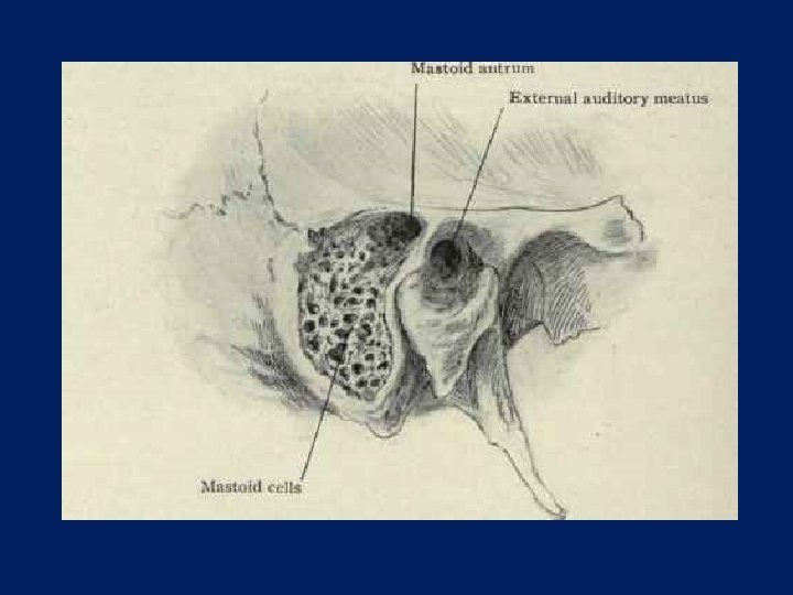
Neonatal Skull The mastoid process is not present at birth and develops later in response to the pull of the sternocleidomastoid muscle when the child moves his or head. At birth, the mastoid antrum lies about 3 mm deep to the floor of the suprameatal triangle. As growth of the skull continues, the lateral bony wall thickens so that at puberty the antrum may lie as much as 15 mm from the surface.

Neonatal Skull The mandible has right and left halves at birth, united in the midline with fibrous tissue. The two halves fuse at the symphysis menti by the end of the 1 st year. The angle of the mandible at birth is obtuse, the head being placed level with the upper margin of the body and the coronoid process lying at a superior level to the head. It is only after eruption of the permanent teeth that the angle of the mandible assumes the adult shape and the head and neck grow so that the head comes to lie higher than the coronoid process.

Neonatal Skull In old age, the size of the mandible is reduced when the teeth are lost. As the alveolar part of the bone becomes smaller, the ramus becomes oblique in position so that the head is bent posteriorly


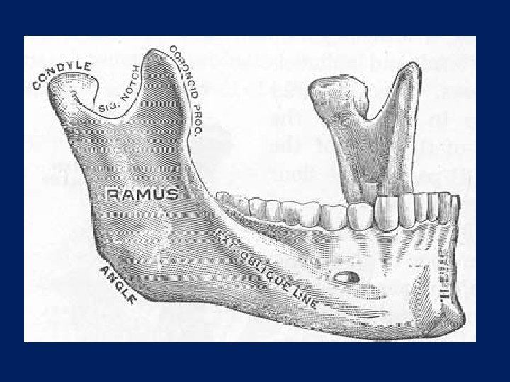
The Mandible The mandible or lower jaw is the largest and strongest bone of the face, and it articulates with the skull at the temporomandibular joint. The mandible consists of a horseshoe-shaped body and a pair of rami. The body of the mandible meets the ramus on each side at the angle of the mandible.
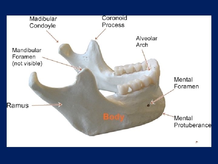

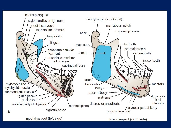
The Mandible The body of the mandible, on its external surface in the midline, has a faint ridge indicating the line of fusion of the two halves during development at the symphysis menti. The mental foramen can be seen below the second premolar tooth; it transmits the terminal branches of the inferior alveolar nerve and vessels.
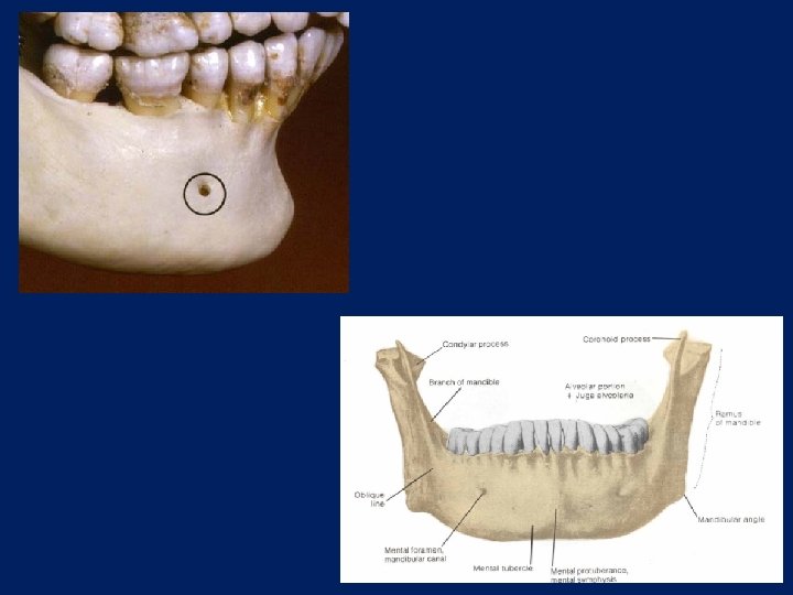

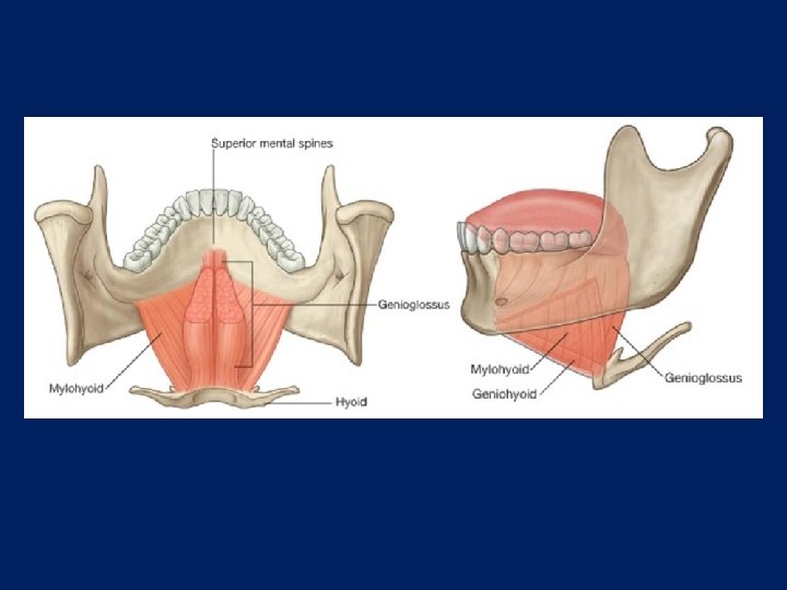
The Mandible On the medial surface of the body of the mandible in the median plane are seen the mental spines; these give origin to the genioglossus muscles above and the geniohyoid muscles below. The mylohyoid line can be seen as an oblique ridge that runs backward and laterally from the area of the mental spines to an area below and behind the third molar tooth.

The Mandible The submandibular fossa, for the superficial part of the submandibular salivary gland, lies below the posterior part of the mylohyoid line. The sublingual fossa, for the sublingual gland, lies above the anterior part of the mylohyoid line.
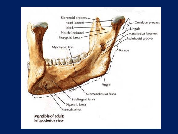

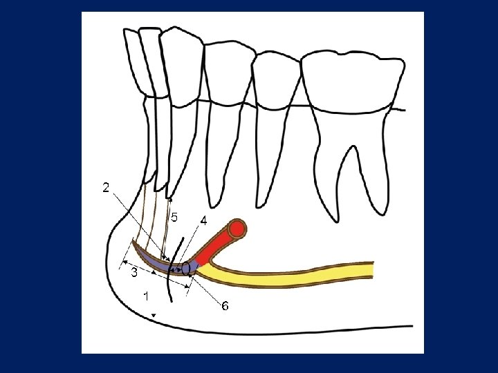
The Mandible The upper border of the body of the mandible is called the alveolar part; in the adult, it contains 16 sockets for the roots of the teeth. The lower border of the body of the mandible is called the base. The digastric fossa is a small, roughened depression on the base, on either side of the symphysis menti. It is in these fossae that the anterior bellies of the digastric muscles are attached.

The Mandible The ramus of the mandible is vertically placed and has an anterior coronoid process and a posterior condyloid process, or head; the two processes are separated by the mandibular notch. On the lateral surface of the ramus are markings for the attachment of the masseter muscle. On the medial surface is the mandibular foramen for the inferior alveolar nerve and vessels.


The Mandible In front of the foramen is a projection of bone, called the lingula, for the attachment of the sphenomandibular ligament.

The Mandible The foramen leads into the mandibular canal, which opens on the lateral surface of the body of the mandible at the mental foramen. The incisive canal is a continuation forward of the mandibular canal beyond the mental foramen and below the incisor teeth.

The Mandible The coronoid process receives on its medial surface the attachment of the temporalis muscle. Below the condyloid process, or head, is a short neck.

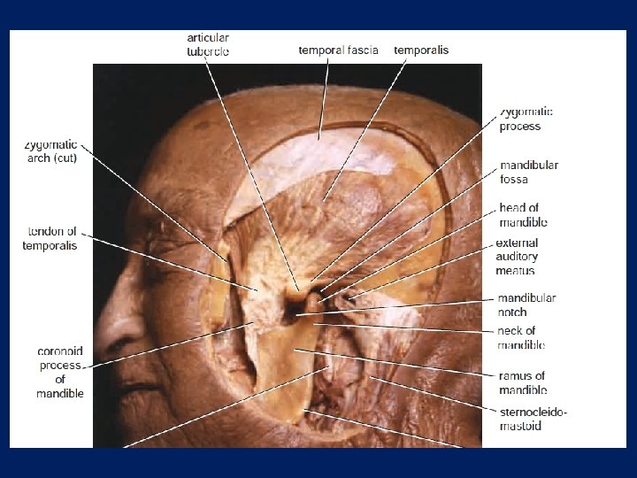
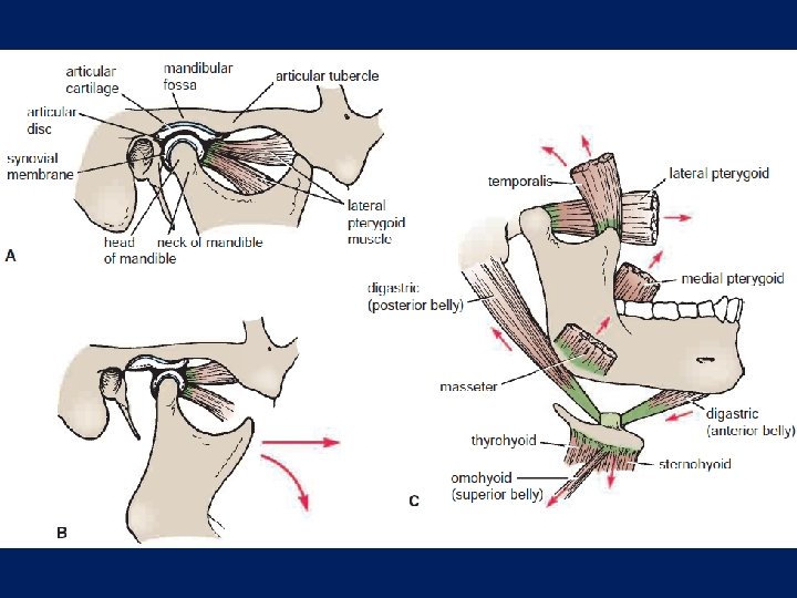
Temporomandibular Joint Articulation Occurs between the articular tubercle and the anterior portion of the mandibular fossa of the temporal bone above and the head (condyloid process) of the mandible below. The articular surfaces are covered with fibrocartilage.

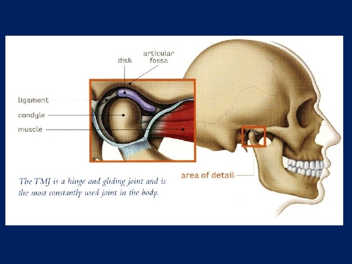
Temporomandibular Joint Type of Joint The temporomandibular joint is synovial. The articular disc divides the joint into upper and lower cavities.

Temporomandibular Joint Capsule The capsule surrounds the joint and is attached above to the articular tubercle and the margins of the mandibular fossa and below to the neck of the mandible.
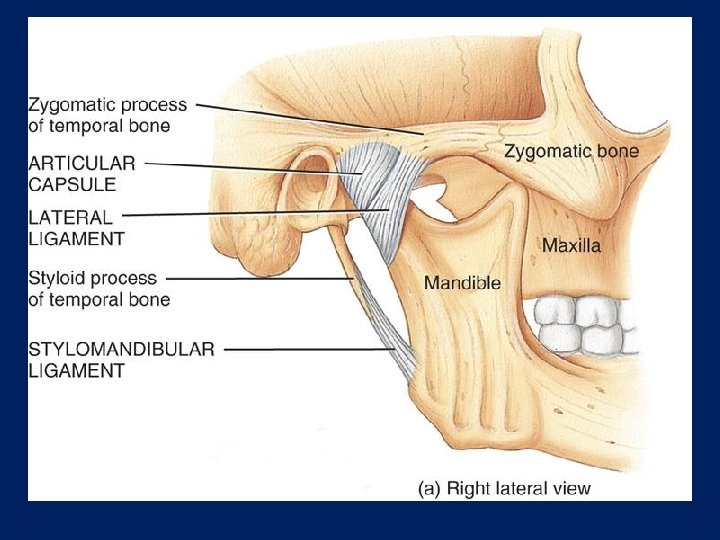

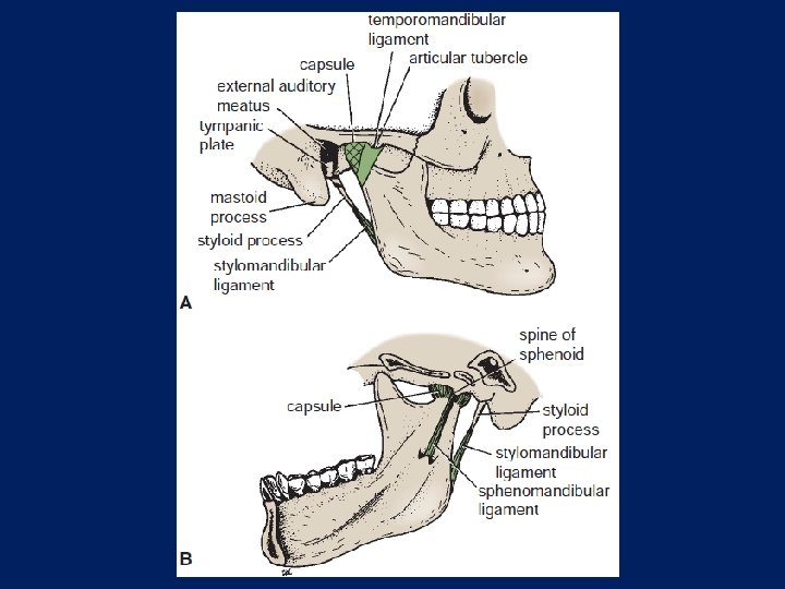
Temporomandibular Joint Ligaments The lateral temporomandibular ligament strengthens the lateral aspect of the capsule, and its fibers run downward and backward from the tubercle on the root of the zygoma to the lateral surface of the neck of the mandible. This ligament limits the movement of the mandible in a posterior direction and thus protects the external auditory meatus.


Temporomandibular Joint The sphenomandibular ligament lies on the medial side of the joint. It is a thin band that is attached above to the spine of the sphenoid bone and below to the lingula of the mandibular foramen. It represents the remains of the first pharyngeal arch in this region.


Temporomandibular Joint The stylomandibular ligament lies behind and medial to the joint and some distance from it. It is merely a band of thickened deep cervical fascia that extends from the apex of the styloid process to the angle of the mandible.


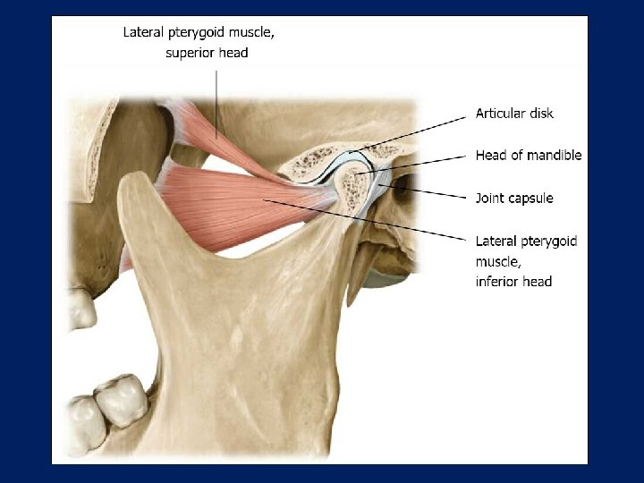
Temporomandibular Joint The articular disc divides the joint into upper and lower cavities. It is an oval plate of fibrocartilage that is attached circumferentially to the capsule. lateral pterygoid muscle and by fibrous bands to the head of the mandible. These bands ensure that the disc moves forward and backward with the head of the mandible during protraction and retraction of the mandible.

Temporomandibular Joint The upper surface of the disc is concavoconvex from before backward to fit the shape of the articular tubercle and the mandibular fossa; the lower surface is concave to fit the head of the mandible.

Temporomandibular Joint Synovial Membrane This lines the capsule in the upper and lower cavities of the joint.

Temporomandibular Joint Nerve Supply Auriculotemporal and masseteric branches of the mandibular nerve.

Temporomandibular Joint Movements The mandible can be depressed or elevated, protruded or retracted. Rotation can also occur, as in chewing. In the position of rest, the teeth of the upper and lower jaws are slightly apart. On closure of the jaws, the teeth come into contact.

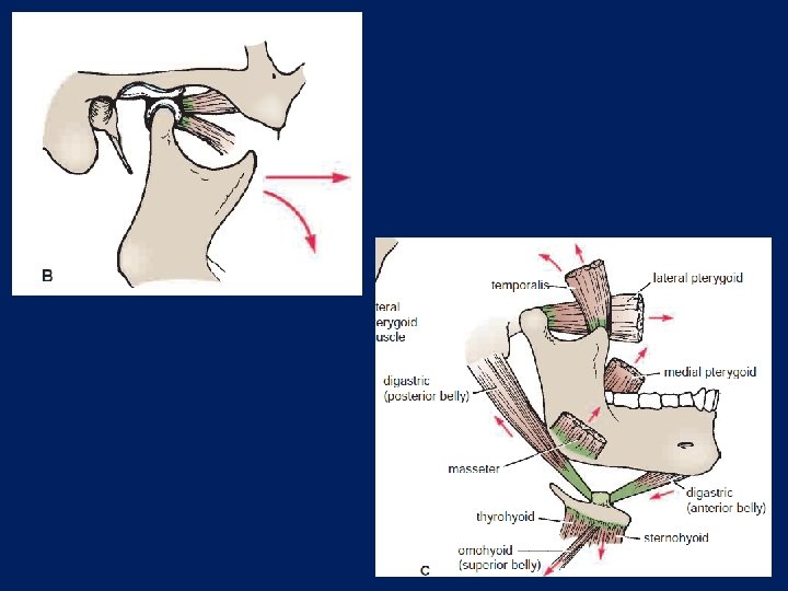
Temporomandibular Joint Depression of the Mandible As the mouth is opened, the head of the mandible rotates on the undersurface of the articular disc around a horizontal axis. Depression of the mandible is brought about by contraction of the digastrics, the geniohyoids, and the mylohyoids; the lateral pterygoids play an important role by pulling the mandible forward.

Temporomandibular Joint To prevent the angle of the jaw impinging unnecessarily on the parotid gland the sternocleidomastoid muscle, the mandible is pulled forward. This is accomplished by the contraction of the lateral pterygoid muscle, which pulls forward the neck of the mandible and the articular disc so that the latter moves onto the articular tubercle. The forward movement of the disc is limited by the tension of the fibroelastic tissue, which tethers the disc to the temporal bone posteriorly.
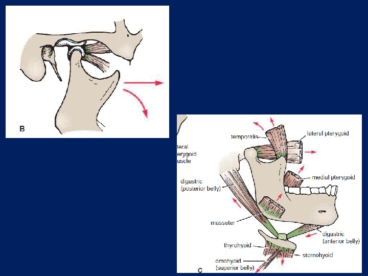

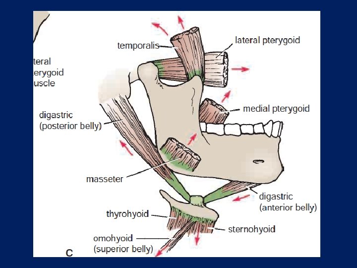
Temporomandibular Joint Elevation of the Mandible Elevation of the mandible is brought about by contraction of the temporalis, the masseter, and the medial pterygoids. The head of the mandible is pulled backward by the posterior fibers of the temporalis. The articular disc is pulled backward by the fibroelastic tissue, which tethers the disc to the temporal bone posteriorly.

Temporomandibular Joint Protrusion of the Mandible The articular disc is pulled forward onto the anterior tubercle, carrying the head of the mandible with it. All movement thus takes place in the upper cavity of the joint. In protrusion, the lower teeth are drawn forward over the upper teeth, which is brought about by contraction of the lateral pterygoid muscles of both sides, assisted by both medial pterygoids.

Temporomandibular Joint Retraction of the Mandible The articular disc and the head of the mandible are pulled backward into the mandibular fossa. Retraction is brought about by contraction of the posterior fibers of the temporalis.

Temporomandibular Joint Lateral Chewing Movements These are accomplished by alternately protruding and retracting the mandible on each side. For this to take place, a certain amount of rotation occurs, and the muscles responsible on both sides work alternately and not in unison.

Important Relations of the Temporomandibular Joint Anteriorly: The mandibular notch and the masseteric nerve and artery.

Important Relations of the TMJ Posteriorly: The tympanic plate of the external auditory meatus and the glenoid process of the parotid gland

Important Relations of the TMJ Laterally: The parotid gland, fascia, and skin

Important Relations of the TMJ Medially: The maxillary artery and vein and the auriculotemporal nerve

- Slides: 68