
Neonatal Shock Sneha Sood MD Objectives To discuss
Neonatal Shock Sneha Sood, MD
Objectives To discuss the causes of neonatal shock To discuss management of shock in the delivery room and nursery To understand the placement of a stabilization umbilical vein catheter in emergency management of shock
Definition Circulatory failure with inadequate organ and tissue perfusion resulting in impaired delivery of oxygen and substrates as well as impaired excretion of metabolic waste products. Can result in cellular dysfunction and death May be accompanied by hypotension

Most Common Causes of Shock Hypovolemia Cardiogenic Sepsis
Intrapartum Hemorrhage Placenta previa/abruption Umbilical cord injury Twin-twin transfusion Maternal-fetal hemorrhage Fetal hemorrhage

Postpartum Brain hemorrhage Lungs Adrenal glands Scalp


Subgaleal Hemorrhage
Cardiogenic Shock Congenital heart disease Arrhythmia Severe hypoglycemia Asphyxia Bacterial or viral infection Severe metabolic/electrolyte abnormalities Hypoxemia and metabolic acidosis
Septic Shock Bacterial or viral Extremely ill Usually require significant respiratory and blood pressure support High risk of development of Persistent Pulmonary Hypertension of the Newborn (PPHN)
Other Causes of Shock Vasoactive shock without sepsis due to endothelial injury, mediator release Obstruction due to cardiac tamponade, pneumothorax Inadequate oxygen releasing capacity such as severe anemia or methemoglobinemia.
Signs of Shock Observation of overall status of neonate: Does baby appear sick or well based on respiratory status, color, activity, tone. Are there any obvious abnormalities. Problem focused physical examination Extent of exam may be limited by degree of distress
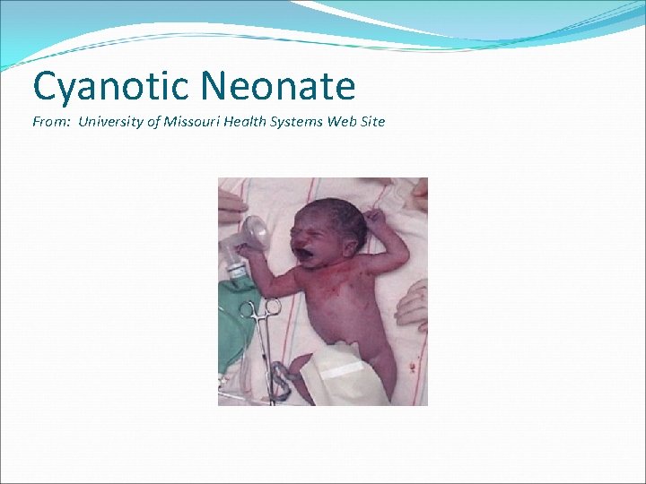
Cyanotic Neonate From: University of Missouri Health Systems Web Site
![Pale Baby From: University of Missouri Health Systems]and New York Presbyterian Morgan Stanley Children’s](http://slidetodoc.com/presentation_image/4743fa2d932b8fa00d5bb531562c92ac/image-14.jpg "Pale Baby From: University of Missouri Health Systems]and New York Presbyterian Morgan Stanley Children’s")
Pale Baby From: University of Missouri Health Systems]and New York Presbyterian Morgan Stanley Children’s Hospital
Signs of Shock: Cardiorespiratory Examine heart and lungs first; important to identify and manage cardiorespiratory problems as one of the first priorities per NRP and basic ABCs.
Signs of Shock: Respiratory Assessment on physical exam: Breathing comfortably or signs of respiratory distress Chest symmetry Auscultation for equality of breath sounds, aeration, quality of breath sounds Respiratory manifestations of shock could include: Respiratory distress/failure Apnea Gasping respirations
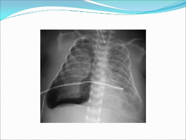
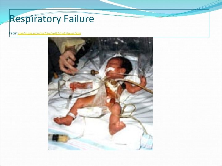
Respiratory Failure From: tumj. tums. ac. ir/archive/vol 65/no 2/issue. html
Signs of Shock: Cardiac exam to include heart rate, heart sounds, pulses, perfusion. Blood pressure if able to measure.
Signs of Shock: Cardiac Heart Rate Cardiac output determined: stroke volume x HR; neonates with little capacity to increase stroke volume so more likely to compensate via HR. Normal: 120 -160 bpm; may range 80 -200 Bradycardia: < 100 Cardiac: Congenital Heart block Metabolic derangements: Metabolic acidosis, Hypoxemia, hypotension may depress myocardium Well baby may have low resting HR Tachycardia: > 180 (sustained) Cardiac arrhythmia Volume depletion Heart failure, decreased cardiac output Activity can increase HR in well baby
Signs of Shock: Cardiac Rhythm: normal sinus vs arrhythmia Presence of heart murmur Innocent vs. pathologic due to CHD Not all CHD associated with heart murmur
Signs of shock Perfusion Prolonged capillary refill > 3 seconds Cool Mottling

Capillary Refill From: EMS Responder. com

Mottled skin in Neonate
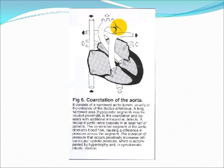
Signs of Shock: Cardiac Pulses Weak pulses Differential between upper and lower extremity pulses could suggest coarctation or hypoplasia of aorta
Hypotension Mean blood pressure (MBP) used for reference in Neonates Definition of hypotension: < 5 th percentile for gestational age Good estimate of lower limit of MBP is gestational age, especially in premature infant ≥ 30 mm. Hg by 72 hours, even in the premature infant Controversy as to whether to treat if MBP low but perfusion and pulses good. Blood pressure may not be abnormal in the early stage of shock

Blood Pressure Parameters Picture from Kliegman, Nelson Textbook of Pediatrics, 18 th ed

Neurological Status Activity Tone Cry Symmetry of movements

Management
Management Important to recognize neonatal shock based on brief physical exam. Volume expansion will be the primary therapy. Normal saline is the volume expander of choice. Other volume expanders included Ringer’s lactate and O- PRBCs if fetal anemia is expected. Limited physical exam will focus on general observation, auscultation, and perfusion/pulses. Accept limitations based on physical environment in DR. BP cannot be immediately measured. Address basics of NRP first: Stimulation, drying, warmth, airway stabilization. A cold, wet baby can mimic one in shock. Assess need for chest compressions and emergency drugs. Babies may also manifest signs of shock in the postresuscitation phase.
Management Place stabilization UVC in the delivery room if baby too unstable to transfer to the nursery. Obviously a baby requiring chest compressions or other intensive resuscitation cannot be taken to the nursery. Stabilization UVC can provide a site to give emergency drugs and volume expanders.
Management For shock give NS, 10 ml/kg over 5 -10 min; repeat x 2 every 5 -10 minutes depending on clinical response. In small babies at risk for IVH consider giving 10 ml/kg NS over 20 -30 min. Consider early use of dopamine in consultation with the Pediatrician and Neonatologist in small babies at risk for IVH, especially when < 1000 grams. Obviously if small baby is in shock in the DR and deteriorating then volume must be given more rapidly.
Management O- blood can be pushed in DR in emergency situations for acute blood loss; give in 10 ml/kg aliquots; avoid giving blood electively in the nursery unless at a level II or tertiary care center; blood may not be CMV negative. Base need for transfusion on discussion with Neonatologist.
Management in the Nursery CR monitor and pulse oximeter immediately on arrival in the nursery. Pulse oximeter can be placed in the DR. Maintain oxygen saturations*: 85 -93% if < 32 weeks or < 1500 grams 90 -98% in all other babies Consider higher saturations over 95% in late preterm or term/posterm infants if PPHN suspected. *These are only recommendations and based on personal practice and practice guidelines at Kapi’olani Medical Center
Management in the Nursery Check VS to include BP, examine baby. PIV, UVC and UAC if needed can be if skilled personnel are available. Stabilization UVC not usually left in place for long periods time; however, if unable to place other lines do not remove the stabilization UVC. Line placement very important before running fluids; obtain abdomen/CXR. Do not run fluids through UVC if placement in liver.
Management If blood pressure or perfusion still poor after 3 boluses of NS consider starting dopamine. Epinephrine should be reserved for babies refractory to treatment with dopamine and in consultation with the physician or transport team.
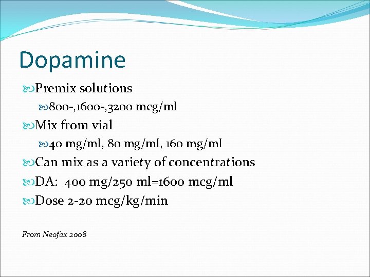
Dopamine Premix solutions 800 -, 1600 -, 3200 mcg/ml Mix from vial 40 mg/ml, 80 mg/ml, 160 mg/ml Can mix as a variety of concentrations DA: 400 mg/250 ml=1600 mcg/ml Dose 2 -20 mcg/kg/min From Neofax 2008
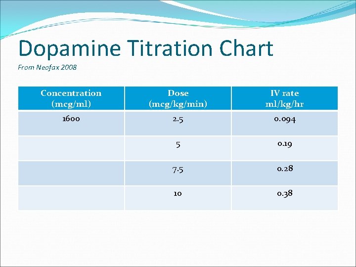
Dopamine Titration Chart From Neofax 2008 Concentration (mcg/ml) Dose (mcg/kg/min) IV rate ml/kg/hr 1600 2. 5 0. 094 5 0. 19 7. 5 0. 28 10 0. 38
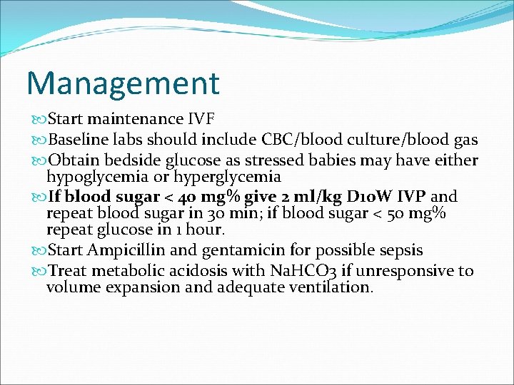
Management Start maintenance IVF Baseline labs should include CBC/blood culture/blood gas Obtain bedside glucose as stressed babies may have either hypoglycemia or hyperglycemia If blood sugar < 40 mg% give 2 ml/kg D 10 W IVP and repeat blood sugar in 30 min; if blood sugar < 50 mg% repeat glucose in 1 hour. Start Ampicillin and gentamicin for possible sepsis Treat metabolic acidosis with Na. HCO 3 if unresponsive to volume expansion and adequate ventilation.
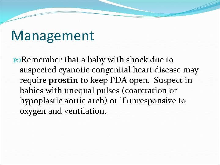
Management Remember that a baby with shock due to suspected cyanotic congenital heart disease may require prostin to keep PDA open. Suspect in babies with unequal pulses (coarctation or hypoplastic aortic arch) or if unresponsive to oxygen and ventilation.

Placement of Stabilization UVC Picture from American Journal of Roentgenology, Schlesinger et al, 2003
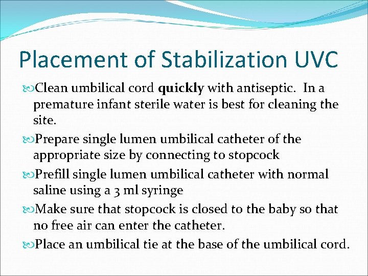
Placement of Stabilization UVC Clean umbilical cord quickly with antiseptic. In a premature infant sterile water is best for cleaning the site. Prepare single lumen umbilical catheter of the appropriate size by connecting to stopcock Prefill single lumen umbilical catheter with normal saline using a 3 ml syringe Make sure that stopcock is closed to the baby so that no free air can enter the catheter. Place an umbilical tie at the base of the umbilical cord.
Placement of Stabilization UVC Cut the umbilical cord leaving about 1 -2 cm from the skin line after tightening umbilical tie. Insert the catheter into the single umbilical vein until you see blood return when you open the stopcock and aspirate with the syringe. This is usually about 24 cm (less in a preterm baby). In a stabilization UVC only insert the catheter far enough to get blood return. You do not want the catheter to be in the liver. After giving epinephrine or volume expander give 0. 51 ml NS to clear the drug from the catheter.
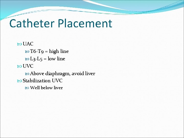
Catheter Placement UAC T 6 -T 9 = high line L 3 -L 5 = low line UVC Above diaphragm, avoid liver Stabilization UVC Well below liver
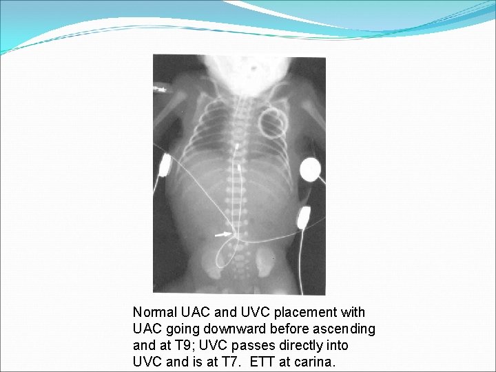
Normal UAC and UVC placement with UAC going downward before ascending and at T 9; UVC passes directly into UVC and is at T 7. ETT at carina.
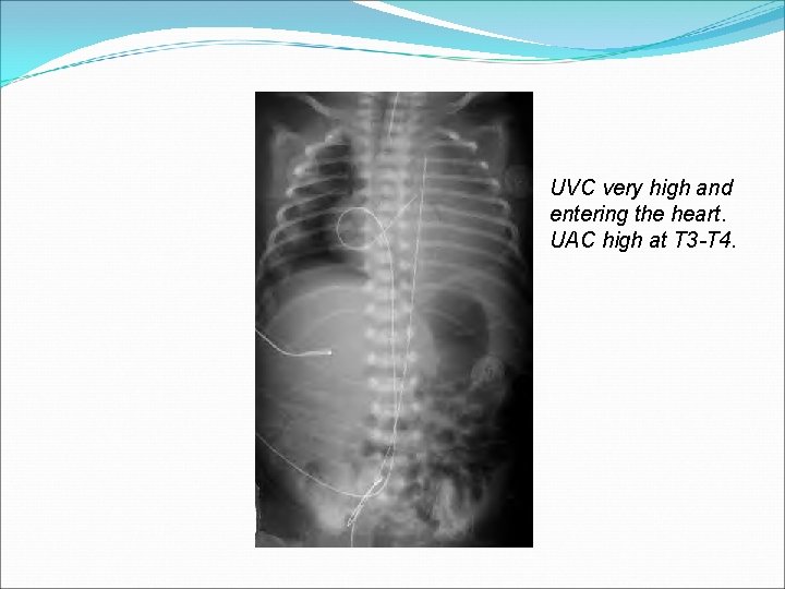
UVC very high and entering the heart. UAC high at T 3 -T 4.
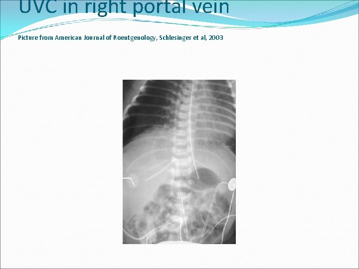
UVC in right portal vein Picture from American Journal of Roentgenology, Schlesinger et al, 2003
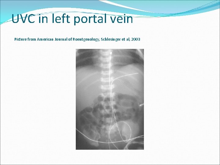
UVC in left portal vein Picture from American Journal of Roentgenology, Schlesinger et al, 2003
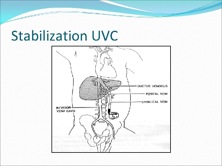
Stabilization UVC
- Slides: 50