Neonatal examination Learning Objectives Assess growth measurements Assess

Neonatal examination

Learning Objectives Assess growth measurements Assess vital signs examine the different body systems of newborn Recognize normal findings in the newborn examination Recognize common newborn problems

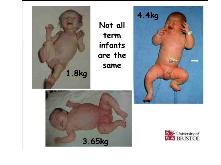
Classification of newborn Classification by Gestational Age Preterm <37 wks Full term 37 -42 Postterm >42 Wks Classification By Birth Weight Low Birth Weight < 2500 g Very Low birth weight < 1500 g Extreme low birth weight < 1000 g

Classification By Weight Percentiles AGA SGA LGA 10 th-90 thpercentile for GA < 10 thpercentile for GA >90 th percentile for GA

Small for Gestational Age • Symmetric – HC, length, weight all <10 percentile – 33% of SGA infants – Cause: Infection, chromosomal abnormalities, inborn errors of metabolism, smoking, drugs • Asymmetric – Weight <10 percentile, HC and length normal – 55% of SGA infants – Cause: Uteroplacental insufficiency, Chronic hypertension or disease, Preeclampsia, Hemoglobinopathies, altitude, Placental infarcts or chronic abruption • Combined – Symmetric or asymmetric – 12% of SGA infants – Cause: Smoking, drugs, Placental infarcts or chronic abruption, velamentous insertion, circumvallate placenta, multiple gestation

Large for Gestational Age • Etiologies – Infants of diabetic mothers – Beckwith-Wiedemann Syndrome • characterized by macroglossia, visceromegaly, macrosomia, umbilical hernia or omphalocele, and neonatal hypoglycemia – Hydrops fetalis – Large mother
 Color 0")
APGAR Score Heart Rate Respiratory effort Muscle tone Reflex irritability (nose suction) Color 0 1 2 Absent <100 bpm >100 bpm Absent, irregular Slow, crying Good Limp Some flexion of extremities Active motion No response Grimace Cough or sneeze Blue, pale Acrocyanosis Completely pink

Apgar Score Assess the physical condition of newborns after delivery at 1, 5 m and every 5 m. until its value is > 7 A value > 7 indicate the baby’s condition is good to excellent A value less than 4 necessitate continued resuscitation Apgar score is a good predictor of survival but using it to predict long-term outcome is inappropriate

Examination of newborn complete physical exam. should be done within 24 h. after birth Include the following: 1. Vital signs 2. Physical exam 3. Neurological exam 4. Estimation of gestational age

Temperature Heart rate Respiratory rate Blood pressure Capillary refill time

1. Temperature should be taken axillary The normal temperature for infant is 36. 5 - 37 -50 C. Axillary temp. is 0. 5 -1 0 c lower than rectal

Heart rate It should be obtained by auscultation and counted for a full minute Normal heart rate is 120 -160 beat /m. If the infant is tachycardic (heart rate >170 BPM), make sure the infant is not crying or moving vigorously

3. Respiratory rate Normal respiratory rate is 40 – 60/minute Respiratory rate should be obtained by observation for one full minute Newborns have periodic rather than regular breathing

4. Blood pressure It is not measured routinely Normal blood pressure varies with gestational and postnatal ages

5. Capillary refill time Normally < 3 seconds over the trunk May be as long as 4 seconds on extremities Delayed capillary refill time indicates poor perfusion


Physical examination 1 stexamination in delivery room or as soon as possible after delivery 2 ndand more detailed examination within 24 h of life Discharge examination with 24 h of discharge from hospital

1 - Measurements There are three components for growth measurements in neonates Weight Length Head circumference All should be plotted on standardized growth curves for the infant’s gestational age

1 - Weight • Weight of F. T infants at birth is 2. 6– 3. 8 kg. • Babies less than 2. 5 kg are considered low birth weight. • Babies loose 5 – 10% of their birth weight in the first few days after birth and regain their birth weight by 7 – 10 days. • Weight gain varies between 15 -20 gm/day.

2. Length Crown to heel length should be obtained on admission and weekly Acceptable newborn length ranges from 48 -52 cm at birth

2. Length

3. Head Circumference Head circumference should be measured on admission and weekly Using the measuring paper tape around the most prominent part of the occipital bone and the frontal bone Acceptable head circumference at birth in term newborn is 33 -38 cm

3. Head Circumference

GENERAL EXAMINATION

1 -Colour – Pallor: associated with low hemoglobin or shock – Cyanosis: associated with hypoxemia – Plethora: associated with polycythemia – Jaundice: elevated bilirubin

Cyanosis

Acrocyanosis

Jaundice

2 -skin • Purpura, echymosis • Mottling • Vernix caseosa • Edema • Mongolian spots • Collodion infant

Vernix Caseosa A lubricant found on the skin or skin fold Disappears as the fetus ages Almost absent in post- term

Purpura red or purple discolored spots on the skin that do not blanch on applying pressure

Mottling skin that has patchy and irregular colors. The skin may have red and purple marks, streaks, or spots. I

Edema

Mongolian spots Dark blue bruise-like macular spots usually over sacrum In 90% of blacks and Asians Disappear by 4 yrs

Collodion Baby infant who is born encased in a tight shiny membrane that resembles plastic wrap

3 - rashes • Milia • Erythema toxicum • Bullous impetigo • Diaper rash • nevi

Milia White papules < 1 mm in diameter scattered across the forehead, nose, cheeks Sebaceous retention cysts disappear within wks

Erythema toxicum White vesicles with a red base Contain esinophils 48 h after birth Transient Benign

Bullous impetigo: Pemphigus neonatorum

Candida diaper dermatitis

Port Wine stain ”Nevus flammeus“ Flat, deep red, do not blanch with pressure May be associated with retinal and intracranial hematomas

4 - Head and Neck • Skull – – – Macrocephaly and microcephaly Caput succedaneum cephalhematoma, subgaleal hemorrhage Fontanelle

Hydrocephalus

Microcephaly

Caput Succedaneum Edema of scalp skin, crosses suture lines

Cephalhematoma • Subperiosteal • Not cross suture lines

Cephalhematoma Complications: • Underlying linear skull fracture • Jaundice • Calcification • Infection • Intracranial Hge

Subgaleal hemorrhage Under the aponeurosis of the scalp Cross suture lines

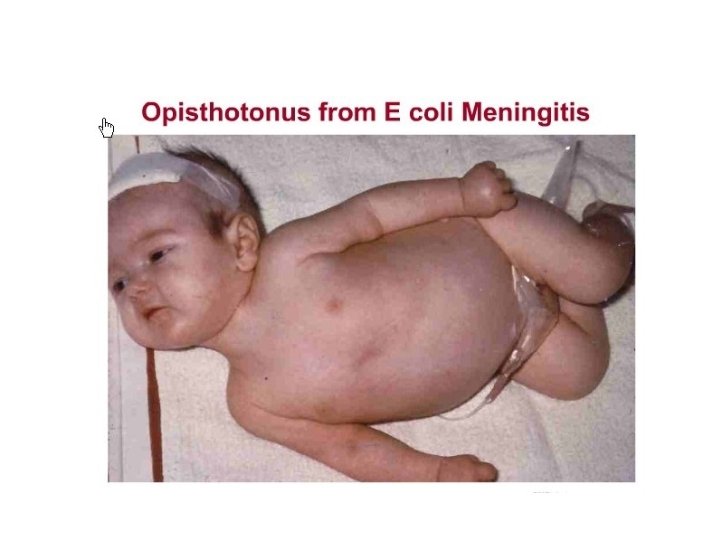
Anterior and posterior fontanelle • Large anterior fontanelle is seen in hypothyroidism, osteogenesisimperfecta, hydrocephalus • Small ant. fontanelle in microcephaly and craniostenosis • Bulging ant. fontanelle in menigitis and hydrocephalus Intracranial hemorrhage • Depressed ant. fontanelle in dehydration • Large post. fontanelle : suspicious of hypothyroidism

Eyes Pupils: equality, reactivity to light. Squint Cornea Conjunctiva Iris

Subconjunctival hemmorrhage Benign condition Resolve by 2 -4 wks

Congenital cataract: rubella

Glaucoma

Dysconjugate Eye Movements

Ear Examination Assess for asymmetry or irregular shape – Note presence of auricular or pre-auricular pits, fleshy appendages, lipomas, or skin tags. – Low set ears • Below lateral canthus of eye • Associated with genitourinary anomalies, because these areas develop at similar times. – Malformed ears • Can be associated with Downs or Turners Syndromes

Ear Tag

Nose Patency of each nostril: exclude choanal atresia Flaring of nostrils

Dislocated Nasal Septum

Mouth Cleft lip and palate Tongue tie Natal teeth Tongue size

Cleft Lip

Unilateral Cleft Lip and cleft palate

Bilateral Cleft Lip and cleft palate

Epstein Pearls & cheeks • small white cysts which contain keratin • frequently found on either side of the median raphe of the palate. • Resolves in 1 -2 months

Mouth • Ranulas – small bluish-white swellings of variable size on the floor of the mouth representing benign mucous gland retention cysts

Normal Tongue Ankyloglossia

Ankyloglossia

Natal Tooth Precarious teeth

Macroglossia

Oral thrush

Neck Cysts: Thyroglossal cyst Cystic hygroma Masses: Sternomastoid tumor Thyroid Webbing

Sternomastoid tumor Hematoma in the middle third of the sternomastoid muscle Torticolis, Limitation of lateral rotation of the neck

Webbed Neck

Muskloskletal Fractures Dislocations Polydactyly Syndactyly Deformities

Erb’s Palsy paralysis of the arm caused by injury to the upper group of the arm's main nerves, specifically the severing of the upper trunk C 5–C 6 nerves.

Polydactyly

Syndactyly • Simple – involves soft tissue attachment only • Complex – involves fusion of bone or nail • Partial - web extends from base partially • Complete - web from base to tip of finger • Radiographs needed to determine degree of fusion. • Should refer to orthopedics.
")
Talipes Equinovarius (Clubfoot)

Spine and hips • Inspect back for meningeocele • Examine for dislocation hip: expected if there is assymetry of skin folds of the thigh and shortening of the affected leg

Meningiomyelocele

Meningiomyelocele & meningeocele

DDH
 – Breast")
Chest/Lung Examination • Inspection – Supernumerary breast or nipple is common (10%) – Breast enlargement secondary to maternal hormones – Unilateral absence or hypoplasia of pectoralis major • Poland's Syndrome (Poland's Sequence) – Widely spaced nipples • Turner's Syndrome • Noonan Syndrome

Chest/Lung Examination • Inspection – Chest Deformity • Pectus Carinatum – Much less common than Pectus Excavatum – More common in males by ratio of 4: 1 – Narrow thorax with increased anteroposterior diameter • Pectus Excavatum – Gender predominance: Boys (3: 1 ratio) – Mild: Oval pit near infrasternal notch – Severe: Sinking of entire lower sternum

Chest/Lungs • Observe – Respiratory pattern • Brief periods apnea are normal in transition, called “periodic breathing” – Chest movement • Symmetry • Retractions and Tracheal tugging • Ascultation – Audible stridor, grunting – Wheeze, rales.

• Slight substernal retraction evident during inspiration

Heart and vascular system Tachypnea, tachycardia Increased pericordial activity Cyanosis: hyperoxia test Auscultation of heart sounds, murmurs or Irregular heart rhythm Perfusion: Capillary refill time Palpate femoral pulsation: absent in coarctation of the aorta Bounding pulses often indicated PDA

Abdomen Organomegaly: liver may be palpable 1 -2 cm below the costal margin. spleen is at the costal margin Masses Distension , scaphoid abdomen Umbilical stump: bleeding , meconium straining, granuloma, discharge, inflammation Omphalocele and Gastroschisis

Abdomen • Cylindrical in Shape

Normal Umbilical Cord • Bluish white at birth with 2 arteries & one vein.

Meconium Stained Umbilical Cord

Omphalocele Defect covered by amnion, with cord attachment to apex of defect. Herniation through defect: any abdominal organs

Abdominal distension

Genitalia and rectum Male genitalia • In full term, scrotum is well developped, with deep rugae. Both testes are in the scrotum • In preterm, scrotum is small with few rugae. testes are absent or high in the scrotum • • abnormalities: undescended testis hydrocele, inguinal hernia hypospadius

Bilateral hydrocele

Bilateral Inguinal hernias

Hypospadius Meatus opens on the ventral surface of the penis

Female genitalia • In full term, labia majora completely cover labia minora • In preterm, labia majora is widely separated and labia minora protruded • A discharge from the vagina or withdrawal bleeding may be observed in the first few days • Infant with ambiguous genitalia should not undergoe gender assignment until endocrinal evaluation is performed

Withdrawal bleeding

Umbigious Genitalia

Imperforate Anus The anus is inspected for its location and patency. An imperforate anus is not always immediately apparent. Thus, patency often is checked by careful insertion of a rectal thermometer to measure the baby's first temperature

• Meconium should pass in the first 48 h after birth • Delayed passage of meconium may indicate imperforate anus or intestinal obstruction • Urine should pass in the first 24 h of life


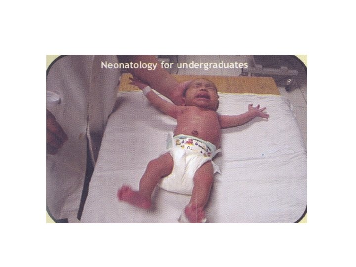
Muscle tone Connvulsions Neonatal reflexes Moro Grasp Tonic Neck Stepping and Placing Rooting &Suckling

• Posture – Term infants normal posture is hips abducted and partially flexed, with knees flexed. – Arms are abducted and flexed at the elbow. – Fists are often clenched, with the fingers covering the thumb • Tone – To test, support the infant with one hand under the chest. Neck extensors should be able to hold head in line for 3 seconds – There should be no more than 10% head lag when moving from supine to sitting positions.

Hypotonia

Neonatal reflexes Also known as developmental, primary, or primitive reflexes. They consist of autonomic behaviors that do not require higher level brain functioning They can provide information about integrity of C. N. S. Their absence indicate C. N. S depression They are often protective and disappear as higher level motor functions emerge.

Moro Reflex Onset: 28 -32 weeks GA Disappearance: 4 - 6 months It is the most important reflex in neonatal period

Moro reflex Stimulus : when baby in supine position elevate his head by your hand then allow head to drop suddenly : Response • Extension of the back • Extension and abduction of the UL • Flexion and adduction of the UL with open fingers • Crying

Significance of Moro Bilateral absence: • CNS depression by narcotics or anesthesia • Brain anoxia and kernicterus • Very Premature baby • Asymmetric response: • Erbs palsy , fracture clavicle or humerus Persistence beyond 6 th month: • CNS damage

Suckling Reflex • When a finger or nipple is placed in the mouth, the normal infant will start to suck vigorously • Appears at 32 w & disappears by 3 – 4 m

Suckling Reflex

Rooting Reflex Well-established: 32 -34 weeks GA Disappears: 3 -4 months Elicited by the examiner stroking the upper lip or corner of the infant’s mouth The infant’s head turns toward the stimulus and opens its mouth

Rooting Reflex

Palmar grasp Well-established: 36 weeks GA Disappears: 4 months Elicited by the examiner placing her finger on the palmar surface of the infant’s hand grasps the finger Attempts to remove the finger result in the infant tightening the grasp

Grasp reflex Technique: put the examiner finger in the baby palm with slight rubbing. Response: the infant grasp the finger firmly Significance: • Absent CNS depression CNS • Persist damage

Stepping Reflex Onset: 35 -36 weeks GA Disappearance: 6 weeks Elicited by touching the top of the infant’s foot to the edge of a table while the infant is held upright. The infant makes movements that resemble stepping

Stepping Hold : baby in upright position then lower him till his sole touch table → stepping movement start.

Placing : When dorsum of the baby foot touches the under surface of the table → flexion then extension to place or put his foot on the table

Placing Reflex
 Evident at 4 weeks PGA Disappearance: 7 months Elicited by")
Tonic neck (Fencing posture) Evident at 4 weeks PGA Disappearance: 7 months Elicited by rotating the infant’s head from midline to one side The infant should respond by extending the arm on the side to which the head is turned and flexing the opposite arm Appearance at birth or persistence beyond 9 m indicate cerebral palsy
")
Tonic neck (Fencing posture)

Gestational Age Assessment Obstetricians - LMP - Ultrasound New Ballard score

Gestational Age Assessment New Ballard Score - Performed within 12 -24 hours - Neuromuscular maturity (6) - Physical maturity (6) Ballard JL, et al. JPediatrics; 1991: 119 (3)

Ballard Score • External Characteristics – Edema – Skin texture, color, and opacity – Lanugo – Plantar creases – Nipples and breasts – Ear form and firmness – Genitals • Neuromuscular Score – – – Posture Square Window Arm recoil Popliteal angle Scarf sign Heel to ear
")
New Ballard Score Ballard JL, et al. JPediatrics; 1991: 119 (3)

- Slides: 130