Neck Space Infections Dr Sheetal Rai Assistant Professor


- Slides: 40

Neck Space Infections Dr. Sheetal Rai Assistant Professor Dept of ENT Yenepoya Medical College Mangalore

Specific Learning Objectives n 1. 2. 3. 4. Outline the anatomy of Submandibular and submental space Parapharyngeal space Retropharyngeal space Peritonsillar space

Specific Learning Objectives n 1. 2. 3. 4. List the causes and outline the clinical features of Ludwig’s angina Parapharyngeal abscess Retropharyngeal abscess Quinsy (Peritonsillar abscess)

Specific Learning Objectives n 1. 2. 3. 4. Outline the management of Ludwig’s angina Parapharyngeal abscess Retropharyngeal abscess Quinsy (Peritonsillar abscess)

Specific Learning Objectives n 1. 2. 3. 4. List the complications of Ludwig’s angina Parapharyngeal abscess Retropharyngeal abscess Quinsy (Peritonsillar abscess)

DEEP NECK SPACE INFECTIONS n Ludwig’s angina n Retropharyngeal abscess n Parapharyngeal abscess n Peritonsillar abscess

Ludwig’s Angina n n Infection of Submandibular space Origin in submaxillary space �sublingual space

SUBMANDIBULAR SPACE n n Divided by mylohyoid into two spacessubmaxillary and sublingual. Contents-

Ludwig’s Angina Criteria for Diagnosis n Rapidly progressing cellulitis; no tendency to form abscess n Involvement of submaxillary & sublingual space n Spread along fascial planes n Involvement of muscle & facsia but not submandibular gland & LN

Aetiology n Importance of mylohyoid line

Clinical features n n n Fever Increasing oral or neck pain & swelling Neck rigidity Trismus, odynophagia Drooling of saliva Dyspnoea & stridor

Clinical features n On examination n Floor of the mouth swollen and inflammed, tense, indurated Tongue displaced upwards & backwards towards the palatal vault Woody induration

X-ray : shows soft tissue edema and airway encroachment n

Treatment n n n Early stage : IV antibiotics, supportive measures, extraction of inciting tooth Advanced stage : Surgery Secure Airway : Tracheostomy under LA

Surgical Drainage : Incision – Median horizontal incision 2 finger breadths below the mandibular margin Drainage : Multiple drains placed & wounds left open.

Complications n n Airway obstruction Spread of infection to carotid sheath or retropharyngeal space �mediastinal extension n Septicaemia n Aspiration pneumonia

Parapharyngeal abscess

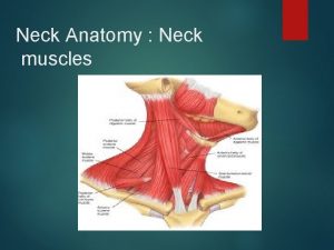
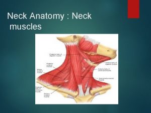
Parapharyngeal space • Inverted pyramidal shape • Extent • Boundaries Compartments • Contents •

Aetiology n Pharynx: acute & chronic tonsillitis, adenoiditis, peritonsillar abscess n Teeth: infn. from lower last molar tooth n Ear: Bezold’s abscess, petrositis n Other spaces n External trauma: penetrating neck injuries

Clinical features ANTERIOR COMPARTMENT n n Anterior & medial displacement of the tonsil POSTERIOR COMPARTMENT n Trismus n n Swelling of the lateral pharyngeal wall behind the posterior pillar External swelling behind the angle of the jaw n HORNER’S SYNDROME Swelling of the parotid region

Clinical features Common to both compartments: n Fever n Odynophagia n Sore throat n Torticollis n Signs of toxemia

Treatment n n n Early stage: IV antibiotics, fluid replacement, close observation Late stage: surgical drainage Incision: submandibular incision two & half finger breadths inferior to mandibular margin from ant. limit of submandibular gland to just past the angle of the mandible

Surgical incision

Complications n n Airway compromise IJV thrombosis Mediastinitis Carotid artery h’ge

Retropharyngeal abscess

Retropharyngeal space n Extent n Boundaries n Content

Aetiology: n Abscessed LNs draining ear, nose & throat. n ü ü ü Regional trauma: FB ingestion oral endotracheal intubation endoscopic procedures

RETROPHARNGEAL ABSCESS n ACUTE Abscessed LNs draining ear, nose & throat. n n n ü ü ü Regional trauma: FB ingestion oral endotracheal intubation endoscopic procedures CHRONIC Caries of cervical spine TB lymphadenitis of retropharyngeal LN

Clinical features n n n Children Irritability, poor appetite, fever Neck rigidity, tenderness, muffled cry, drooling, dyspnoea. n n n Adults Fever, sore throat, odynophagia, neck tenderness. U/L swelling of posterior pharyngeal wall pushing the soft palate forwards. DD: Pott’s abscess. Inv: X ray lat view of neck. CXR: Mediastinal involvement

Management Acute retropharyngeal abscess n Initial: IV antibiotics/ Fluid replacement. n n Surgical: Incision & Drainage per oral (Rose’s position) Inferior extension: Incision along anterior border of SCM from hyoid to cricoid.

Management Chronic retropharyngeal abscess n Treat the underlying cause – Tuberculosis with ATT. n External incision - along anterior border of SCM from hyoid to cricoid. n Intraoral drainge of chronic RP abscess is contraindicated.

Complications n n n Haemorrhage Aspiration Danger space infection

Peritonsillar abscess

Peritonsillar space n n n Between capsule of the tonsil and superior constrictor muscle. Importance –spread of infection to para pharyngeal space, carotid sheath and mediastinum. posterior spread to retropharyngeal space.

Peritonsillar abscess n n n ü ü ü Aetiology : Follows acute tonsillitis Bacteriology: Mixed growth Clinical Features : young adults, Unilateral General symptoms : Fever, chills, malaise Local: Severe throat pain , odynophagia, drooling of saliva, hot potato voice, foul breath, ipsilateral ear ache, trismus

On examination: n Tonsil pillars and soft palate, congested & swollen n Uvula oedematous, pushed to opposite side n Bulging of soft palate & anterior pillar above the tonsil n Tender enlarged JD nodes n Torticollis

Treatment n n n Hospitalisation Aspiration of pus with a wide bore needle Analgesics & IV antibiotics Ø Incision & Drainage Quinsy forceps is used n Interval tonsillectomy n

Complications Ø Ø Ø Respiratory obstruction Parapharyngeal abscess IJV thrombosis Carotid artery H’ge Septicemia Pneumonitis

Summary n n n Deep Fascia of the neck divides the neck into various facial compartments and spaces Anatomy of the deep neck spaces is important to know about the extent and spread of deep neck space infections. Early diagnosis and treatment of deep neck space infection is necessary to prevent dangerous complications.

THANK YOU All images used in this presentation are obtained from a Google image search
 Sheetal rai
 Promotion from assistant to associate professor
 Fok ping kwan
 Dr savur
 Cluster computing definition
 Parapharyngeal space
 Pharengyal
 Neck spaces anatomy
 Chapter 25 sexually transmitted infections and hiv/aids
 Acute gingival infections
 Cryptosporidiose
 Opportunistic infections
 Genital infections
 Understanding the mirai botnet
 Eye infections
 Methotrexate and yeast infections
 Genital infections
 Storch infections
 Postpartum infections
 Opportunistic infections
 Salmonella life cycle
 Bone and joint infections
 Storch infections
 Retroviruses and opportunistic infections
 Balwant rai mehta committee
 El rai de la medusa comentario
 Rai kamaluddin
 Binita rai
 Rai cereal
 Clasificacion rai y binet
 Piattaforma acquisti rai
 Naomi rai
 Sudhendu rai
 Rai koulutus
 Lady rai
 Rai cmh
 Copiii sunt mainile cu care ne prindem de rai
 Deepcluster
 Gvk rao samiti
 Richa rai
 Rai
Rai
