NECK LUMP MIDLINE LUDWIGS ANGINA SUBMENTAL LN SUBLINGUAL

NECK LUMP

MIDLINE � � � � � LUDWIGS ANGINA SUBMENTAL LN SUBLINGUAL DERMOID THYROGLOSSAL CYST SUBHYOID BURSITIS RETEROSTERNAL GOITRE THYMIC SWELLING BONY SWELLING Sebaceouscyst, lipoma, fibroma etc may arise anywhere.

LATERAL SWELLINGS � In the submandibular triangle � LN. � SUBMANDIBULAR SALIVARY GLAND. � PLUNGING RANULA.

LATERAL SWELLINGS � IN THE CAROTID TRIANGLE � ANEURYSM OF CAROTID ARTERY. � CAROTID BODY TUMOR. � BRANCHIAL CYST. � BRANCHIOGENIC CA. � THYROID. � LN.

LATERAL SWELLINGS � IN THE POSTERIOR TRIANGLE � CYSTIC HYGROMA. � PHARYNGEAL POUCH. � SUBCLAVIAN ANEURSYM. � CERVICAL RIB. � COLD ABSCESS.


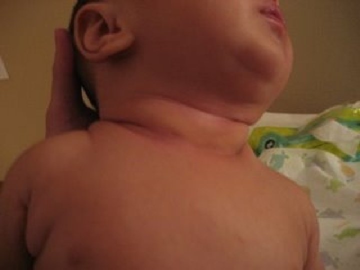
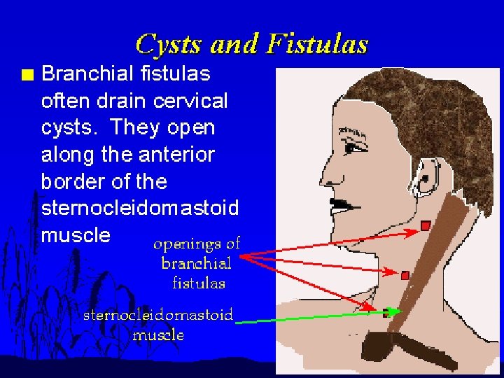
BRANCHIAL CYST

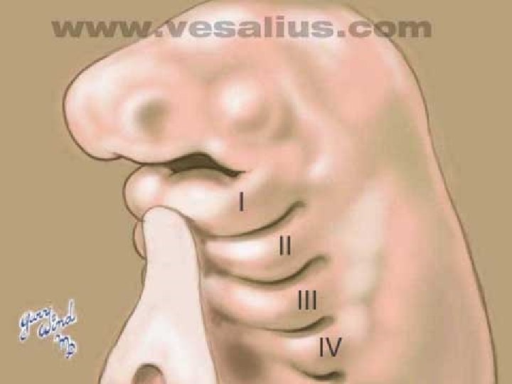
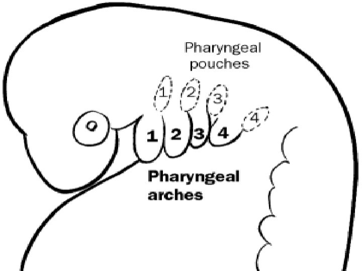
Branchial cleft cysts are congenital epithelial cysts, which arise on the lateral part of the neck from a failure of obliteration of the second branchial cleft in embryonic development. � At the fourth week of embryonic life, the development of 4 branchial (or pharyngeal) clefts results in 5 ridges known as the branchial (or pharyngeal) arches. � The second arch grows caudally and, ultimately, covers the third and fourth arches. � The buried clefts become ectoderm-lined cavities, which normally involute around week 7 of development. If a portion of the cleft fails to involute completely, the entrapped remnant forms an epithelium-lined cyst with or without a sinus tract to the overlying skin. �

S/S � � � A branchial cyst commonly presents as a solitary, painless mass in the neck of a child or a young adult. A history of intermittent swelling and tenderness of the lesion during upper respiratory tract infection may exist. Discharge may be reported if the lesion is associated with a sinus tract. In some instances, patients may present with locally compressive symptoms. A family history may be present. Branchial cysts are smooth, nontender, fluctuant masses, which occur along the lower one third of the anteromedial border of the sternocleidomastoid muscle between the muscle and the overlying skin. The lesion may be tender if secondarily inflamed or infected. When associated with a sinus tract, mucoid or purulent discharge onto the skin or into the pharynx may be present.

TT Medical Care Antibiotics are required to treat infections or abscesses. Surgical Care Surgical excision is definitive treatment for this condition. A series of horizontal incisions, known as a stairstep or stepladder incision, is made to fully dissect out the occasionally tortuous path of the cyst. � Surgery is best delayed until the patient is at least age 3 months. � Definitive surgery should not be attempted during an episode of acute infection or if an abscess is present. � Surgical incision and drainage of abscesses is indicated if present, usually along with concurrent antimicrobial therapy. � � �

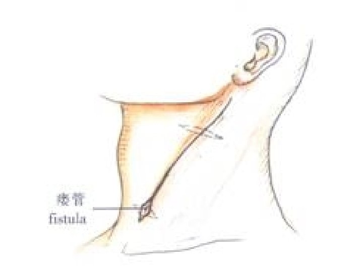
BRANCHIAL FISTULA

Occurs when the cervical sinus of His persists. � The fistula opens externally onto the lower third of neck just anterior to the SCM. � It may be asymptomatic or intermittent mucoid discharge. � Abscess may develop. � If asymptomtic no tt reqd. � Excision. �

CYSTIC HYGROMA
 is a cystic lymphatic lesion that can affect any anatomic subsite")
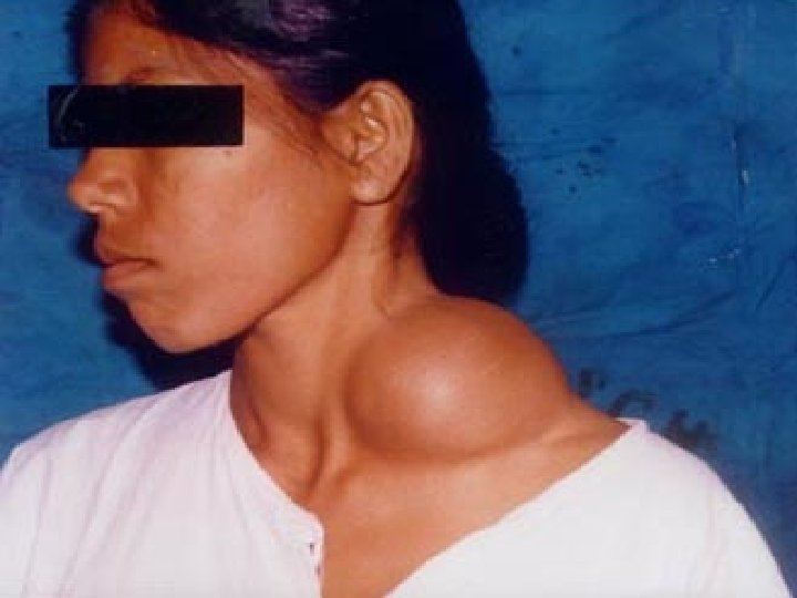
Cystic hygroma (CH) is a cystic lymphatic lesion that can affect any anatomic subsite in the human body. � CH usually affects the head and neck (approximately 75%), with a left-sided predilection. Within the neck, the posterior triangle tends to be most frequently affected. � Approximately 20% of CHs occur in the axilla; more infrequent subsites include the mediastinum, groin, and retroperitoneum. � CH is synonymous with cystic lymphangioma, which is also known as a macrocystic lymphatic malformation and was first described in 1828 by Redenbacker. �

� Lymphangiomas are thought to arise from a combination of the following: a failure of lymphatics to connect to the venous system, abnormal budding of lymphatic tissue, and sequestered lymphatic rests that retain their embryonic growth potential. � In addition to congential development, lymphangiomas can be acquired. They can arise from trauma (including surgery), inflammation, or obstruction of a lymphatic drainage pathway.
 vary depending on the")
The presenting signs and symptoms of the cystic hygroma (CH) vary depending on the lesion's location. � The microcystic form of lymphangioma tends to predominate over CH in the oral cavity and oropharynx. Microcystic lymphangiomas commonly appear as clusters of clear, black, or red vesicles on the buccal mucosa or tongue. � CHs tend to predominate below the mylohyoid muscle and can involve both the anterior and posterior triangles of the neck. � The cysts are typically large and thick walled and have little involvement of surrounding tissue. The overlying skin can take on a bluish hue or may appear normal. �


CHs often present after a sudden increase in size secondary to infection or intralesional bleeding. Spontaneous decompression or shrinkage is uncommon. � Rarely, children with CH display symptoms of newly onset obstructive sleep apnea syndrome (OSAS). This situation may involve children with CH or other space-occupying lesions of the supraglottis or paraglottic region. Suprahyoid lymphangiomas tend to cause more breathing difficulties than infrahyoid lesions. � Potentially life-threatening airway compromise that manifests as noisy breathing (stridor) and cyanosis is a possible symptom of lymphangiomas. � Feeding difficulties, as well as failure to thrive, may alert the clinician to a potential lymphangioma. This is especially true when the lesion affects structures of the upper aerodigestive tract. �
 masses. � A CH typically transilluminates. �")
CHs are typically soft, painless, compressible (doughy) masses. � A CH typically transilluminates. � In children who present with CH of the neck, closely evaluate for tracheal deviation or other evidence of impending airway obstruction. � Closely inspect the tongue, oral cavity, hypopharynx, and larynx because any involvement may lead to airway obstruction. �

� MRI. � CT scanning. � Ultrasonography. � Plain radiography. � Lymphoscintigraphy.

� � Medical Care The medical treatment of CH consists of the administration of sclerosing agents. Sclerosing agents include OK-432 (an inactive strain of group A Streptococcus pyogenes), bleomycin, pure ethanol, bleomycin, sodium tetradecyl sulfate, and doxycycline. � � � Bleomycin: Bleomycin is considered a poor choice because of its toxicity (pulmonary fibrosis) because CH is a benign disease and other treatment options are available. Alcohol: Absolute alcohol as a sclerosing agent has been used with some success in some patients; alcohol works well in vascular malformations. Interferon alfa-2 a: This has been used in the treatment of hemangiomas, and its use has been proposed in lymphangiomas. However, its efficacy has never been documented and it carries a serious side effect profile. Fibrin sealant: The use of a fibrin sealant after aspiration of CH has been reported in the literature. An infected CH should be treated with intravenous antibiotics, and definitive surgery should be performed once the infection has resolved. Incision and drainage or aspiration results in only temporary shrinkage, and subsequent fibrosis can further complicate the resection. Radiotherapy has not been demonstrated to be effective. The preferred treatment of all CH is surgical resection. Only resection can truly offer the potential for cure.

Surgical Care The mainstay of treatment is surgical excision. If acute infection occurs prior to resection, surgery should be delayed at least 3 months. � The surgical team should attempt to completely remove the lymphangioma or to remove as much as possible, sparing all vital neurovascular structures. Complete excision has been estimated to be possible in roughly 40% of cases. � Microcystic lesions are much more difficult to remove because of their intimate association with nearby tissues. Laser therapy is a recent advancement in the treatment of microcystic lesions. � �

Signs of airway obstruction require surgical evaluation at the time of diagnosis. In emergency situations, aspiration with an 18 -gauge or 20 -gauge needle may obviate the need for an emergency tracheostomy. � Radiofrequency ablation has been advocated for use with intraoral lymphatic malformations, especially microcystic lesions. � Magnetic resonance–controlled laser-induced interstitial thermotherapy is a novel therapy that has been proposed for treatment of lymphangiomas. �

SUBLINGUAL DERMOID CYST

Congenital sequestration dermoid. Formd by inclusion of ectoderm at fusion line of first arch. Thin walled cyst lined by squamous epi. Lateral and median variety. Can be supra and inframylohyoid. Usually seen bw 10 -25 years of age. Co of a painless swelling under the tongue or below the chin. � Pain may be asso with infection. � Tranillumintion is –ve. � Tt is excision. � � � �

PLUNGING RANULA

A ranula is a type of mucocele found on the floor of the mouth. � Ranulas present as a swelling of connective tissue consisting of collected mucin from a ruptured salivary gland duct, which is usually caused by local trauma. � The Latin rana means frog, and a ranula is so named because its appearance is sometimes compared to a frog's underbelly. �

� � � The gland that most likely causes a ranula is the sublingual gland An oral ranula is a fluctuant swelling with a bluish translucent color that somewhat resembles the underbelly of a frog. If it is deeper it does not have this bluish appearance. If it is large ( 2 or more cm. ), it may hide the salivary gland affect the location of the tongue. A ranula below the mylohyoid muscle is referred to as a "plunging or cervical ranula", and produces swelling of the neck with or without swelling in the floor of the mouth.

� � � Ranulas are usually asymptomatic, . The overlying skin is usually intact. The mass is not fixed and is also not tender. The mass is not connected to the thyroid gland or lymph nodes. The mass may not be well defined. If it gets large enough it may interfere with swallowing, and cervical ranulas may even interfere with breathing. Some pain may be connected with very large ranulas. Microscopically, ranulas are cystic saliva filled distensions of salivary gland ducts on the floor of the mouth alongside the tongue, and are lined by epithelium. A salivary mucocele, in contrast is not lined by epithelium.

� Treatment of ranulas could involve either a procedure known as "marsupialization" or more often excision of both the gland lesion. � Ranulas are likely to reoccur if the sublingual gland or other gland causing them is not removed with the lesion.

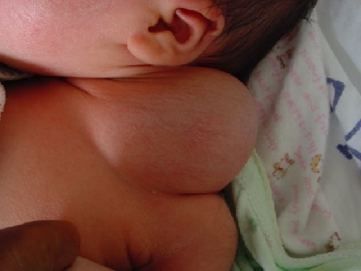
STERNOMASTOID TUMOR

� � � � The sternomastoid "tumor" of infancy is a firm, fibrous mass, appearing at two to three weeks of age, within the substance of the Sternomastoid muscle and appears as a knot. It may or may not be associated with torticollis. Generally, the "tumor" initially grows, then stabilizes, and in about half the cases recedes spontaneously after a few months. It may leave a residual torticollis or may be associated with a facial or cranial asymmetry of a delayed torticollis. The etiology is unknown, a direct cause and effect relationship to birth trauma has been largely disproved although approximately half these children are products of breech deliveries. The treatment is controversial. Approximately half of these "tumors" will resolve spontaneously without sequelae. Progressive torticollis or development of facial asymmetry are considered indications for surgery.

CAROTID BODY TUMOR

Carotid body tumor may also be described as a chemodectoma, glomus tumor or a paraganglioma. It is a slow growing, benign tumor that is usually non-functioning. Aggressive behavior and malignant transformation of the tumor with peri-neural and vascular invasion is known to occur but is rare. Carotid body tumors are neuro-endocrine tumors that arise from the neural crest paraganglionic cells that are located at the level of the carotid bifurcation. These cells line the adventitia of the blood vessel. There may be a familial mode of inheritance. Many patients are asymptomatic or may present with laterally placed, slow-growing, soft masses in the neck. They may transmit the carotid pulsations and a bruit may be heard over the mass. Occasionally a patient may present with a hard and non-pulsatile mass. As the tumor enlarges, it may produce dysphagia/odynophagia or hoarseness. If the tumor is functioning, symptoms due to excessive production of catecholamines occur.

Neck ultrasound with color Doppler is a reliable modality to image the entire extent of carotid body tumors. The treatment of choice is surgical excision. A pre-operative biopsy is usually contraindicated [as these masses are typically very vascular and a biopsy make adequate surgery difficult], and hence the importance of ultrasound in arriving at a diagnosis by demonstrating the classic location of the mass and its internal vascular

COLD ABSCESS
 is a form of tuberculosis, affecting the lymph nodes of")
Scrofula (scrophula or struma) is a form of tuberculosis, affecting the lymph nodes of the neck. � Historically known as the King's Evil, referring to the method of treatment many sufferers used. � In adults it is caused by Mycobacterium tuberculosis and in children by nontuberculous mycobacteria. � The word comes from the Latin scrofula, meaning brood sow. �
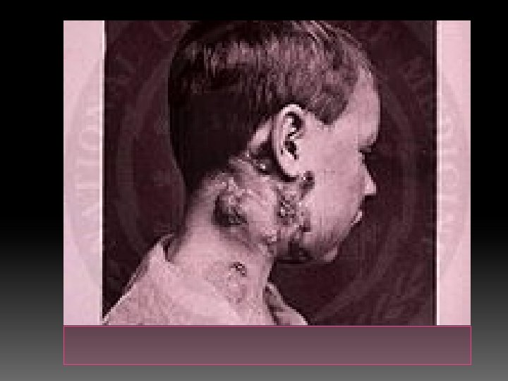

The most usual signs and symptoms are the appearance of a chronic, painless mass in the neck, which is persistent and usually grows with time. � The mass is referred to as a "cold abscess", because there is no accompanying local color or warmth and the overlying skin acquires a violaceous (bluish-purple) color. � NTM infections do not show other notable constitutional symptoms, but scrofula caused by tuberculosis is usually accompanied by other symptoms of the disease, such as fever, chills, malaise and weight loss in about 43% of the patients. � As the lesion progresses, skin becomes adhered to the mass and may rupture, forming a sinus and an open wound. �

� Diagnosis is usually performed by needle aspiration biopsy or excisional biopsy of the mass and the histological demonstration of stainable acid-fast bacteria in the case of infection by M. tuberculosis (Ziehl-Neelsen stain), or the culture of NTM using specific growth and staining techniques.

Treatment approaches are highly dependent on the kind of infection. � Surgical excision of the scrofula does not work well for M. tuberculosis infections, and has a high rate of recurrence and formation of fistulae. Furthermore, surgery may spread the disease to other organs. The best approach then is to use conventional treatment of tuberculosis with antibiotics. � Scrofula caused by NTM, on the other hand, responds well to surgery, but is usually resistant to antibiotics. The affected nodes can be removed either by repeated aspiration, curettage or total excision (with the risk in the latter procedure, however, of causing cosmetically negative effects or damage to the facial nerve, or both). �

Zenker diverticulum

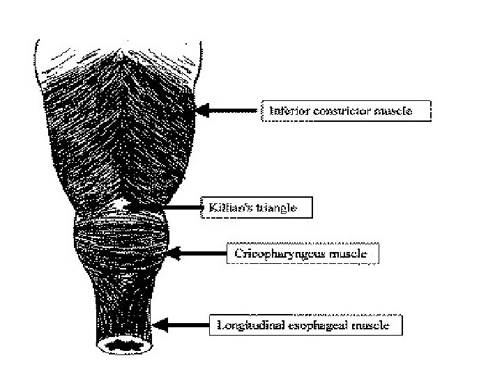
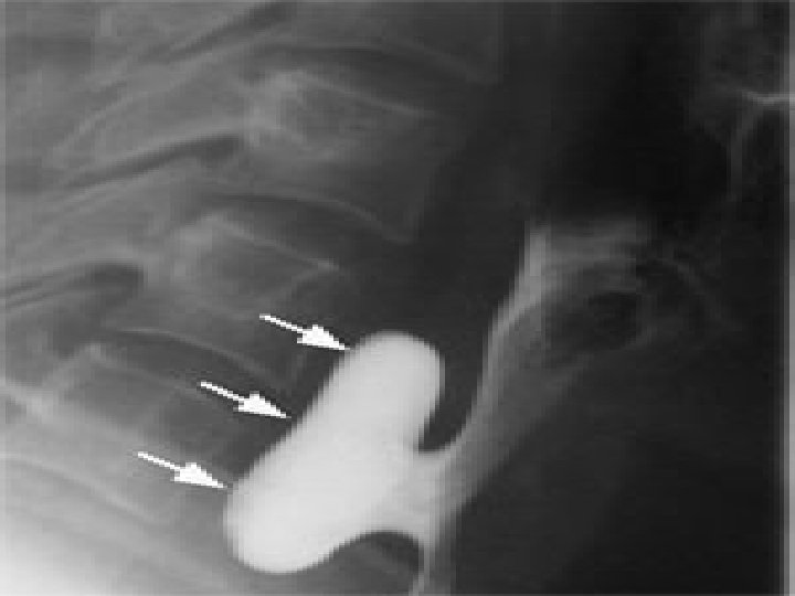
Zenker diverticula occur in a muscular dehiscence that is present most commonly between the oblique muscle fibers of the inferior constrictor muscle and the transverse fibers of the cricopharyngeus (CP) muscle. This area is known as the Killian triangle. � Other areas of muscular dehiscence occur between the oblique and transverse fibers of the CP muscle (ie, Killian-Jamieson area) and between the CP muscle and the esophageal muscles (ie, Laimer triangle). � More inferiorly positioned Zenker diverticula may occur in one of these latter sites �

� Zenker diverticula extend into the left neck 90% of the time. � This is likely due to the slight convexity of the cervical esophagus to the left side and to the more laterally positioned carotid artery on the left side, creating a potential space for the sac.

� � � � Zenker proposed a pulsion mechanism affecting the pharyngeal mucosa above the CP muscle. Hypothetical abnormalities include the following: Abnormal timing of deglutition resulting in closure of the CP muscle when ideally it should be opening Incomplete CP muscle relaxation Elevated resting tone of the entire upper esophageal sphincter (UES) Loss of CP muscle elasticity CP muscle myopathy or denervation atrophy CNS injury with a focal spastic CP muscle spasm in response to gastroesophageal reflux disease (GERD)

� � � The combination of the following symptoms is nearly pathognomonic for Zenker diverticulum: Dysphagia Regurgitation of undigested food hours after eating Sensation of food sticking in the throat Special maneuvers to dislodge food Coughing after eating Aspiration of organic material Unexplained weight loss Fetor ex ore Borborygmi in the neck Symptoms may last from months to years. The most common life-threatening complication is aspiration. Other complications include massive bleeding from the mucosa or from fistulization into a major vessel, esophageal obstruction, and fistulization into the trachea. Squamous cell carcinoma (SCC) within Zenker diverticulum is extremely rare, occurring in 0. 3% of Zenker diverticula worldwide. A Mayo Clinic review suggests an incidence of 0. 48% in the United States. Approximately 50 cases of invasive SCC and carcinoma in situ are reported in the literature. This possibility should be considered when evaluating patients with cervical metastatic SCC with an unknown primary cancer.

Zenker diverticula require intervention only if they produce symptoms. In general, small (ie, <2 cm) lesions found incidentally require no intervention. � Small lesions are satisfactorily treated with a CP myotomy with or without an invagination procedure. � Intermediate and large diverticula (ie, 2 -6 cm) are best managed by open diverticulectomy with CP myotomy or by endoscopic diverticulotomy. � Very large diverticula (ie, >6 cm) are best managed with excision with CP myotomy or a diverticulopexy with CP myotomy, depending on the health of the patient �
. � Rigid or flexible esophagoscopy is essential")
� Flexible endoscopic evaluation of swallowing (FEES). � Rigid or flexible esophagoscopy is essential before surgical management to assess the nature of the mucosa of the Zenker diverticulum and to exclude the presence of SCC or carcinoma in situ. .
 myotomy alone. � Diverticulum invagination or imbrication with CP myotomy. � Diverticulopexy")
Cricopharyngeal (CP) myotomy alone. � Diverticulum invagination or imbrication with CP myotomy. � Diverticulopexy with CP myotomy. � Diverticulectomy with CP myotomy. � Endoscopic diverticulotomy with cautery, laser, or stapler. � Flexible endoscopic diverticulotomy with laser or needle-knife techniques are in the development stage. �

Laryngoceles

Laryngoceles present as lateral neck masses. Most frequently males in the fifth and sixth decade of life. The causative factor of laryngoceles is felt to be either a congenital enlargement of the saccule or acquired by increased, sustained intralaryngeal pressure (i. e. trumpet player). � There are three types of laryngoceles: internal, external, and combined. � The internal laryngocele is found entirely within the larynx and typically extends posterior and superior to the false vocal cord and aryepiglottic fold. � As it is intraluminal, it does not present as a neck mass. � � �

� � � The external laryngocele is seen as a lateral swelling in the neck and passes superiorly through the opening of the thyrohyoid membrane from where the superior laryngeal nerve and vessels pass. The combined laryngocele has features of both internal and external. Symptoms includes lateral neck mass, dysphagia, cough, dyspnea, and occasionally a gurgling sensation as the dilatation releases the contained air. On physical examination, the external and combined laryngoceles may appear as intralaryngeal pressures are elevated and is an easily compressible mass. Additionally, these may present as an acute cervical inflammation if the sac becomes secondarily infected, filled with a purulent fluid, forming a laryngopyocele.

� The management of symptomatic internal and external laryngoceles is surgical, usually through an external approach. � Prior to excision of the laryngocele, it is important that a thorough endoscopic examination is performed to rule-out an underlying carcinoma

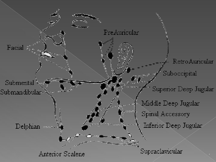
Cervical lymph nodes

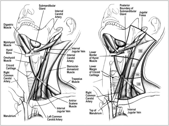
Relevant Anatomy The lymphatic drainage of the head and neck is directed to the lymph nodes located within the fibroadipose tissue that lies between the investing (superficial) layer of the deep fascia superficially and the visceral and prevertebral layers underneath. � In this space, these lymph nodes tend to be aggregated around certain neural and vascular structures such as the internal jugular vein, spinal accessory nerve, and transverse cervical artery. �

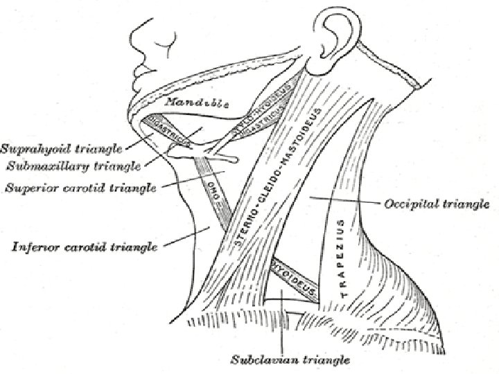
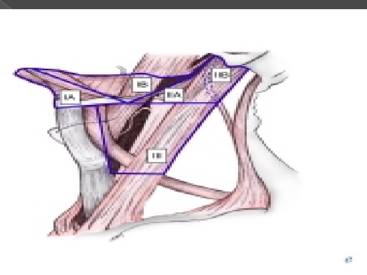
� Lymph nodes in the neck are grouped into levels I-V, corresponding with the submandibular and submental nodes (level I); upper, middle, and lower jugular nodes (levels II, IV); and posterior triangle nodes (level V).

Level I This level is bound by the body of the mandible superiorly, stylohyoid muscle posteriorly, and the anterior belly of the digastric muscle on the contralateral side anteriorly. � This level may be divided into level Ia, which refers to the nodes in the submental triangle (bound by the anterior bellies of the digastric muscles and the hyoid bone), and Ib, which refers to the submandibular triangle nodes. � The nodes of level Ia are at greatest risk of harboring metastasis from cancers that arise from the floor of mouth, anterior tongue, anterior mandibular alveolar ridge, and lower lip, while the nodes of level Ib often receive metastasis from cancers of the oral cavity, anterior nasal cavity, soft tissue structures of the mid face, and submandibular gland. � Closely related, although not strictly a part of the level I group of nodes, are the perifacial nodes, related to the facial vessels above the mandibular margin, and the buccinator nodes, which may become involved with metastasis from tumors in the buccal mucosa, nose, and soft tissues of the cheek and lips. �

Level II lymph nodes are related to the upper third of the jugular vein, extending from the skull base to the inferior border of the hyoid bone. � The anterior border of level II is the stylohyoid muscle, and the posterior border is the posterior border of the sternocleidomastoid muscle. � The spinal accessory nerve, which travels obliquely across this area, is used as a landmark to subdivide this group into IIb, the portion above and behind the nerve, and IIa, the part that lies anteroinferiorly and closer to the internal jugular vein. � The nodes in level II are at greatest risk of harboring metastasis from cancers that arise from the oral cavity, nasal cavity, nasopharynx, oropharynx, hypopharynx, larynx, and parotid gland. �

Level III nodes are located between the hyoid superiorly and a horizontal plane defined by the inferior border of the cricoid cartilage. � The sternohyoid muscle marks the anterior limit of level III, and the posterior border of the sternocleidomastoid muscle is the posterior border. � Level III most commonly receives metastasis from cancers that originate in the oral cavity, nasopharynx, oropharynx, hypopharynx, and larynx. �

Level IV Group of nodes related to the lower third of the jugular vein. � These nodes are located between the inferior border of the cricoid cartilage and the clavicle, and, like level III, the anterior boundary is the sternohyoid muscle, and the posterior border is the posterior border of the sternocleidomastoid muscle. � The nodes of level IV commonly harbor metastasis from cancer that originates in the larynx, hypopharynx, thyroid, and cervical esophagus. �

Level V This refers to the lymph nodes located in the posterior triangle of the neck. These include the spinal accessory, transverse cervical, and supraclavicular group of nodes. � Level V is bound anteriorly by the posterior border of the sternocleidomastoid muscle and posteriorly by the anterior border of the trapezius muscle. � Level V extends from the apex of the convergence of the sternocleidomastoid and trapezius muscle superiorly to the clavicle inferiorly. This level is subdivided by a plane defined by the inferior border of the cricoid cartilage into level Va superiorly and level Vb inferiorly. � Level. Va contains the nodes associated with the spinal accessory nerve, and level Vb contains the transverse cervical and supraclavicular nodes. � The posterior triangle nodes are at greatest risk for harboring metastasis from cancers that arise in the nasopharynx, oropharynx, and skin of the posterior scalp and neck �

Level VI This refers to lymph nodes of the anterior, or central, compartment of the neck. � Defined by the carotid arteries laterally, the hyoid bone superiorly, and the suprasternal notch inferiorly, it is rich in lymphatics that drain the thyroid gland, subglottic larynx, cervical trachea, hypopharynx, and cervical esophagus. � Lymph nodes in this compartment are located in the tracheoesophageal groove (paratracheal nodes), in front of the trachea (pretracheal nodes), around the thyroid gland (parathyroidal nodes), and on the cricothyroid membrane (precricoid or Delphian node). �

Differential Diagnosis for Cervical adenopathy
 › Influenza › Coxacki virus › Cytomegalic virus › Streptococcal")
Infectious Disorders (Specific Agent) › Influenza › Coxacki virus › Cytomegalic virus › Streptococcal pharyngitis, beta type A › Klebsiella › Immune deficiency , acquired (AIDS/HIV) › Lyme disease › Salmonella infection › Staphylococcus aureus infection
 › Bacteroides oralis › Cat-scratch disease › CFS/Fibromyalgia/Chronic fatigue syndrome")
Infectious Disorders (Specific Agent) › Bacteroides oralis › Cat-scratch disease › CFS/Fibromyalgia/Chronic fatigue syndrome › Herpanginia › Herpes simplex › Infectious mononucleosis › Primary HIV/infection syndrome › Tuberculosis › Typhoid fever › Vincent's infection/trench mouth › Atypical mycobacteria
 › Atypical mycobacteria › Diphtheria › Pulmonary anthrax › Tuberculosis,")
Infectious Disorders (Specific Agent) › Atypical mycobacteria › Diphtheria › Pulmonary anthrax › Tuberculosis, disseminated › Anthrax › Atypical mycobacteria, disseminated › Kawasaki disease › Measles (rubeola) › Yersinia enterocolitica infection › Actinomycosis, cervicofacial › Cytomegalic mononucleosis syndrome › Lymphogranuloma venereum › Mycobacteria, atypical pulmonary › Mycobacterium Kansasii
 › Paracoccidiodomycosis (S. A. Blastomyco) › Scrofula/cervical nodes tuberculosis ›")
Infectious Disorders (Specific Agent) › Paracoccidiodomycosis (S. A. Blastomyco) › Scrofula/cervical nodes tuberculosis › Toxoplasmosis, lymphadenitis, acute › Tuberculosis, cavitary pulmonary › Tuberculosis, lymph node › Tularemia › Plague, bubonic › Dengue fever › Monkeypox epizoonosis › Staphylococcus aureus/CA-MRSA virulent PVL gene � Epstein-Barr

Infected organ, Abscesses › Abscess, dentoalveolar › Adenitis/lymph node › Abscess, cervical gland › Tonsillitis/exudative, acute › Cervical adenitis/abscess � Tonsillitis, chronic

Granulomatous, Inflammatory Disorders › Sarcoidosis, pulmonary › Uveoparotid fever/sarcoidosis › Kikuchi's disease

Neoplastic Disorders › Myelogenous/Blastic Leukemia, Acute › Leukemia, acute › Hodgkin's disease › Metastasis, lymph node › Monocytic leukemia, acute › Acute Lymphoblastic/lymphocytic leukemia, › Leukemia › Myelomonocytic, acute leukemia › Adenocarcinoma, parathyroid › tumor, malignant

Neoplastic Disorders › Adenocarcinoma, parathyroid › Adenocarcinoma, thyroid follicular › Burkitt's lymphoma › Carcinoma throat/vallecular › Carcinoma, head and neck › Carcinoma, laryngeal › Carcinoma, medullary, thyroid

Neoplastic Disorders › Carcinoma, nasopharynx › Carcinoma, oral › Carcinoma, papillary, thyroid › Carcinoma, pharynx › Carcinoma, thyroid, anaplastic › Carcinoma, tongue › Hamartoma, angiomatous lymphoid

Neoplastic Disorders › Neuroblastoma, peripheral › Oral carcinoma, floor › Rhabdomyosarcoma › Thyroid lymphoma � Salivary gland tumor, malignant
 › TRAPS/Tumor necrosis")
Hereditary, Familial, Genetic Disorders › Hyper IGD syndrome/HIDS (12 q 24) › TRAPS/Tumor necrosis factor Receptor Periodic Syndrome › PFAPA/Periodic fever (Marshals benign) › Hereditary inflammatory periodic fevers group � Hyperimmunoglobulinemia D-Periodic fever

Neck Dissections

Radical Neck Dissection Originally described by Crile in 1906, this procedure is an en bloc clearance of all fibrofatty tissue from one side of the neck, including the lymph nodes from levels I-V and lymph nodes that surround the tail of the parotid gland, the spinal accessory nerve, the internal jugular vein, and the sternocleidomastoid muscle. � Radical neck dissection does not include the removal of the postauricular, suboccipital, perifacial, buccinator, retropharyngeal, or central compartment nodes. � Previously used for neck disease of any stage, from microscopic to bulky nodal disease, this procedure is now limited to patients with advanced neck disease, recurrent disease after chemoradiation, or gross extracapsular spread to the spinal accessory nerve, sternomastoid muscle, and the internal jugular vein. �

Modified Radical Neck Dissection � This operation involves the removal of the same lymph node groups as those involved in the radical neck dissection (levels I-V) but requires preservation of 1 or more of the following 3 nonlymphatic structures: the spinal accessory nerve, the internal jugular vein, and the sternomastoid muscle. Modified neck dissection is indicated for clinically palpable metastatic neck disease. � Conversion to the radical neck dissection becomes necessary upon gross involvement of the nerve, vein, and muscle, although the involvement of all 3 is unusual, except in very advanced (N 3) disease. �

Selective Neck Dissection This term refers to a type of neck dissection in which one or more lymph node groups normally removed in a radical neck dissection are preserved. � The 1991 classification schema classified selective neck dissections into the following categories: supraomohyoid neck dissection (levels I, III), lateral neck dissection (levels II, III, IV), anterior compartment neck dissection (VI), and posterolateral neck dissection (levels II, IV, V). ). �
- Slides: 92