Nearfield scanning optical microscope NSOM Scanning nearfield optical

")

 • Scanning Near-Field Optical Microscopy (SNOM) • Likely the")
 Break the diffraction limit by working in the near-field")


")


, is")

- Slides: 21
Near-field scanning optical microscope NSOM Scanning near-field optical microscope SNOM
Spatial Resolution of Biological Imaging Techniques • Resolution is diffraction limited. • Abbe (1873) reported that smallest resolvable distance between two points (d) using a conventional microscope may never be smaller than half the wavelength of the imaging light (~200 nm) Ernst Abbe (1840 -1905)
Near-Field Scanning Optical Microscopy (NSOM) • Scanning Near-Field Optical Microscopy (SNOM) • Likely the super-resolution technique with the highest resolution • But only for superficial structures • A form of Scanning Probe Microscopy (SPM)
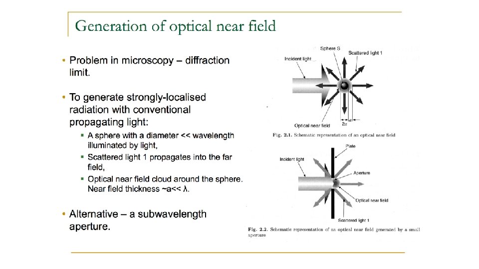
Near-Field Scanning Optical Microscopy (NSOM) Break the diffraction limit by working in the near-field Illuminated “spot” is smaller than diffraction limit (about the size of the tip for a distance equivalent to tip diameter) Launch light through small aperture Near-field = distance of a couple of tip diameters
NSOM working in the near-field • Aperture diameter less than the wavelength of light • In 1993 Eric Betzig and Robert Chichester used NSOM for repetitive single molecule imaging
NSOM working in the near-field • Near-field near surface of object, < λ of light • Near-field consists of light as evanescent wave • Evanescent waves higher frequency, more information • Evanescent waves quantum tunneling phenomenon • Product of Schrödinger wave equations
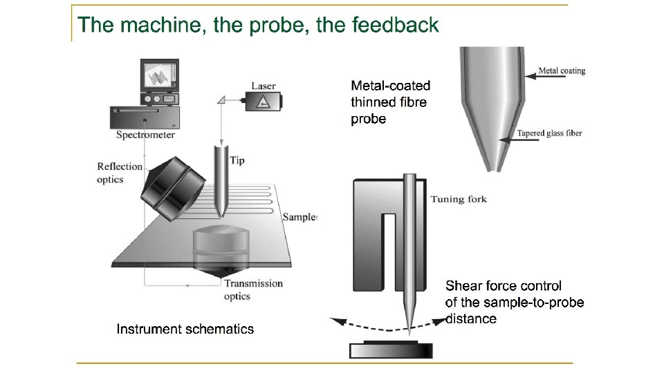
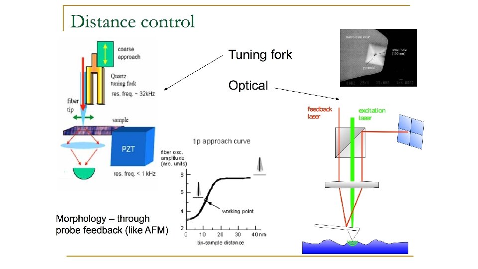
How to move the tip? Steal from AFM Atomic Force Microscopy (AFM)
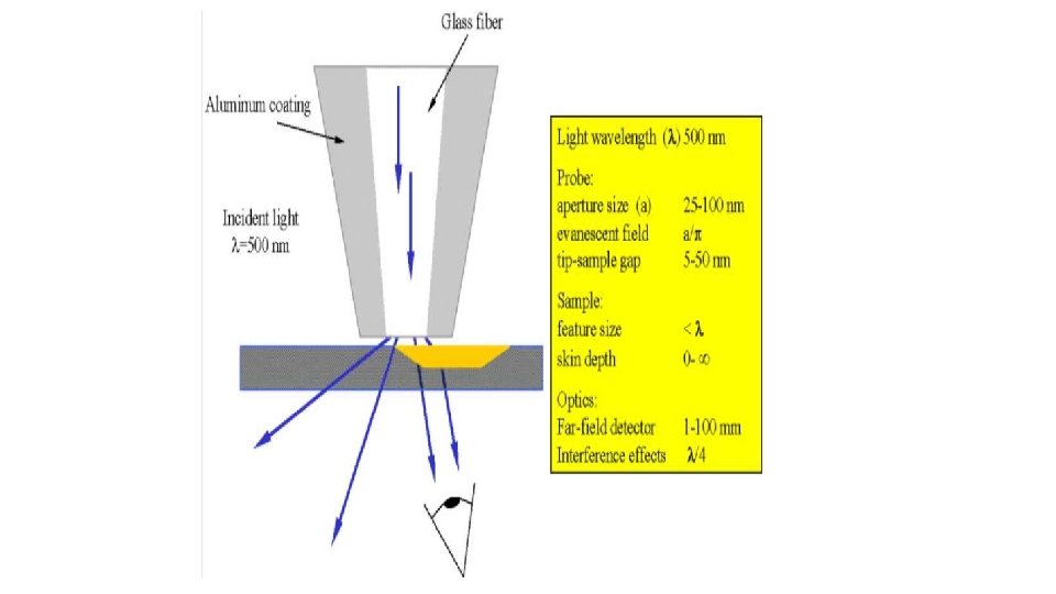
The light source is usually a laser focused into an optical fiber through a polarizer, a beam splitter and a coupler. The scanning tip is usually a pulled or stretched optical fiber coated with metal except at the very tip or just an AFM cantilever with a hole in the center of the pyramidal tip. Standard optical detectors, such as avalanche photodiodes, photomultiplier tubes (PMT) or CCD, can be used.
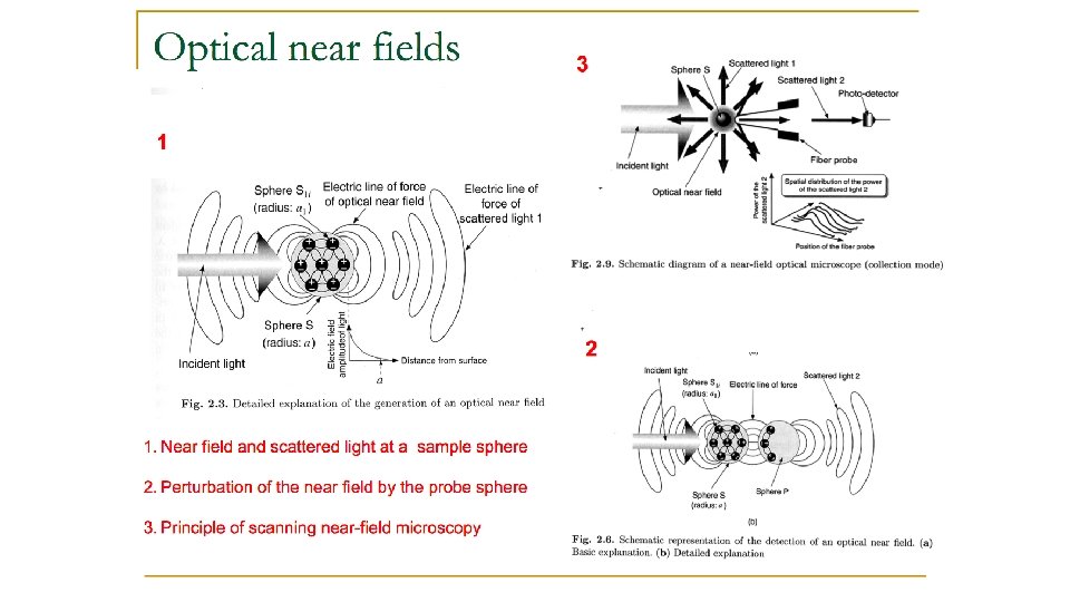
MODES of OPERATION Aperture Mode very popular and could provide very high resolution images Aperture modes include five different operational modes: • • • Illumination Collection Illumination-collection Reflection collection
LIMITATIONS OF SNOM Very short working distance and extremely shallow depth of field Long scan times for large sample areas for high resolution imaging
NSOM images Single molecules of Di. I on glass surface Di. IC 18(3), is a fluorescent lipophilic cationic indocarbocyanine dye
NSOM images