Natural Background Sources of radiation exposure Man made


Natural Background Sources of radiation exposure Man made radiation


 • Space radiation: sun or Cosmic rays: energetic subatomic")
Natural radiation (back ground ) • Space radiation: sun or Cosmic rays: energetic subatomic particles, phrotons(secondary cosmic radiation), helium nuclei, • Exposure : altitude • Each 2000 m: 2 • Airline travel: flight 5 hours : altitude 12 km : 25µsiv, crews us, Europe : radiation workers • Occupational dose limits

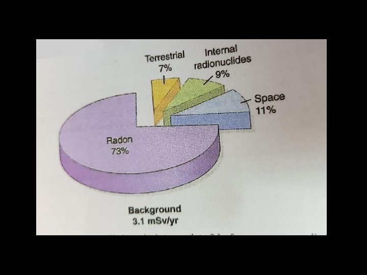
Radon and its progency • Gas, its decays α particles to polonium 218, lead 214 • These decay products become attached to dust particles , inhaled, deposit on bronchial epithelium • 73%

Internal radionuclides • Is ingested • Foods : uranium and thorium and their decay potassium 40 , rubidium 87, carbon 14, tritium and others

Terrestrial sources • External : soil like potassium 40, gama radiation • Internal : radon and other radionuclide that are inhaled or ingested • Radon(52): decay uranium , alfa particles • These decay products become attached to dust particles , inhaled, deposit on bronchial epithelium

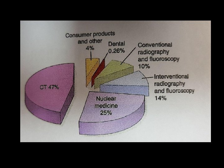
Man –Made radiation • Medical • Industrial • Minor sources
 • Ct")
Medical exposure • 3. 6 billion x-ray examination and nuclear medcine annually(14%dental) • Ct chest and abdomen , cardiac nuclear medicine • Ct older and sicker • Dental exposure : 0. 26% total exposure medical

industrial • • Domestic water supply Tobacco products Combustible fuels Dental porcelain Television receiver Pocket watches Smoke alarms Airport inspection


Dentomaxillofacial radiology: risk and doses • Optimised to produce a diagnostically acceptable image • Less than the threshold needed to cause any deterministic effects • Minimized to keep the risk of stochastic effects within an acceptable range

Estimating cancer risk from diagnostic dentomaxillofacial radiology
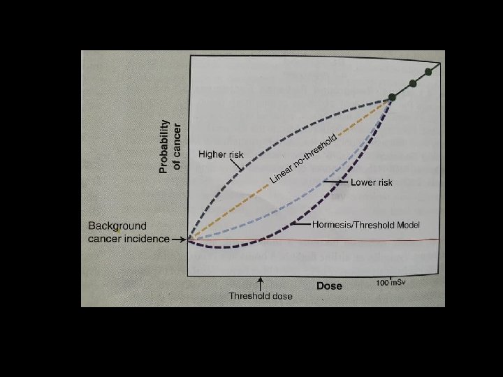
hypothesis • There is a linear relationship between dose and the risk")
LNT(linear nonthreshold )hypothesis • There is a linear relationship between dose and the risk of inducing a new cancer • There is no threshold or safe dose below which there is no added risk.

Ppatient doses from diagnosis dentomaxillofacial radiology

Communication radiation risks to patients

Implementing radiation protection • Justification • Optimization • Dose limitation

2/12/2022 Dr farida Abesi sinus disease 26

Patient protection • Patient selection criteria • Conducting the examination

Reducing Dental Exposure • The most effective approach to reduce unnecessary exposure is to reduce unnecessary radiographic examinations.

Conducting the examination • • • Film and digital imaging Intensifying screens and film or digital imaging Source to skin distance Rectangular collimation Leaded aprons and collars Film and sensor holders Kilovoltage , Mas, film processing, interpreting the images
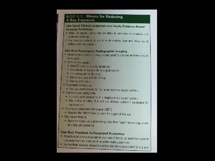

Film and digital imaging • F-speed or digital • Rare –earth intensifying screens are recommended with high speed film of 400 or greater. • Digital system =film in extraoral radiography
 : • Long SOD: less divergent")
Source –to-skin distance • Long distance (40 cm) : • Long SOD: less divergent , volume exposure 10 -25 % exposure tissue
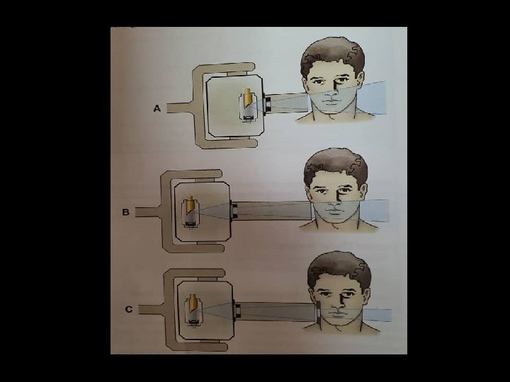

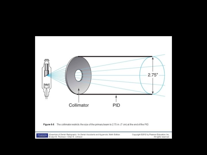


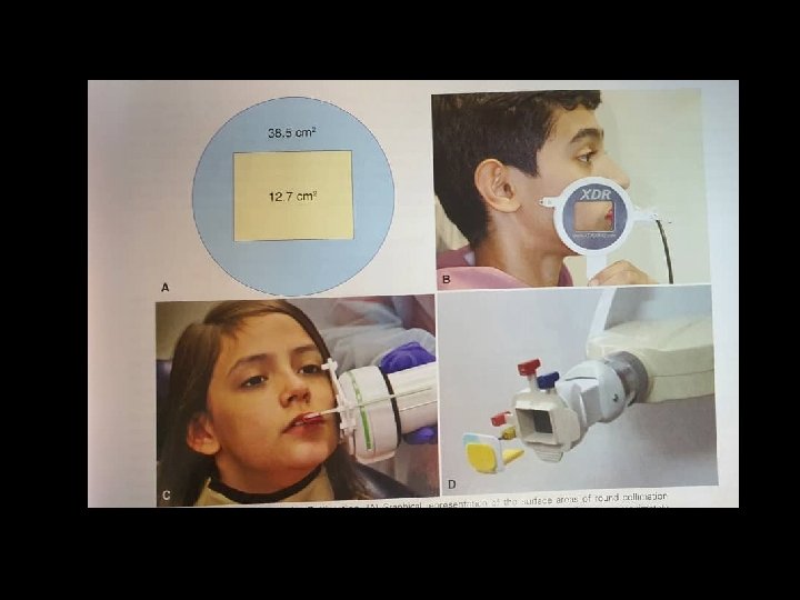
Rectangular Collimation • Rectangular dose 5 round collimation • 60 %

Filtration • Low energy x-ray 20 % • 50 -70 kvp: 1. 5 mm AL • 70: 2. 5 mm AL

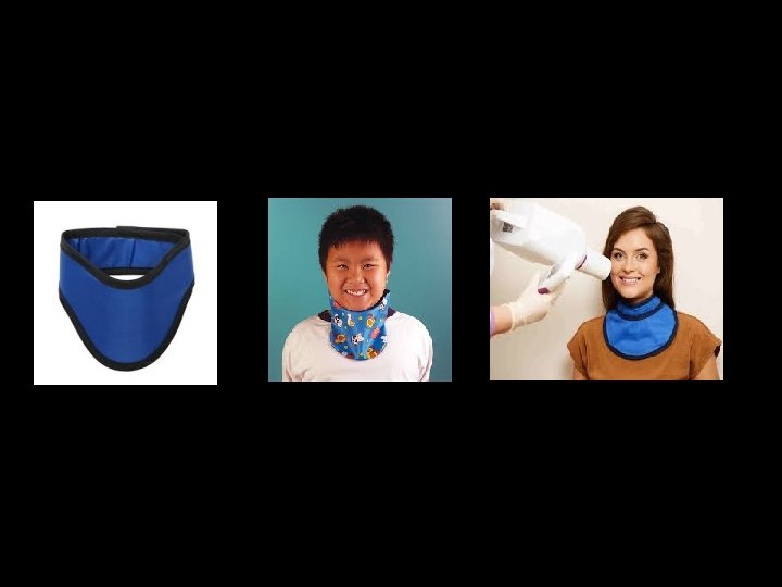
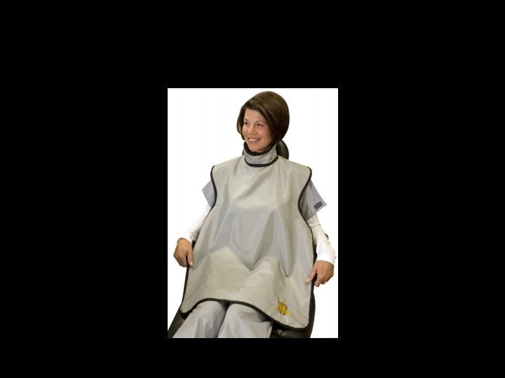
Leaded Aprons and Thyroid Collars • If all of the NCRP are followed , the use of a leaded apron is not required • Thyroid shielding strongly recommended for children • Lead-free a: high A, low density : antimony, tin, tungsten

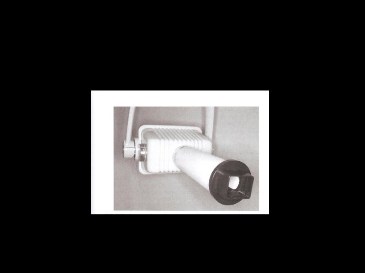
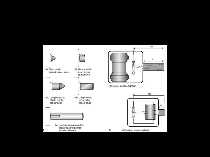
Film and sensor holders • Significant reduction : in unacceptable images • PID: The leadlined cone or cylinder that directs the xray beam during a radiographic exposure.

KVP • KVP greater contrast, enhance diagnosis • Kvp lower contrast, decrease dose • Best balance : 60 -70

m. As • Tube voltage, milliamperage, exposure time • Image density is controlled by m. AS • Radiograph with correct density : very faint soft tissue outlines, gray scale that adequately distinguishes enamel, dentin, cortical bone and trabecular bone.

Film Processing • Time –temperature • Should follow the film manufacturer ‘s recommendation for processing solutions • Automatic film processors : 30 % repeated radiography : processor variability

Interpreting the images • Radiographs : semidarkened room , magnifying glass • Digital images : computer screen, darkened environment

Personnel protection • Operators of radiographic equipement should use barrier protection when possible • 1. Barrier protection : 2 mm pb, (1 m. Gy per year) Sinon position and distance rule • Operator stand 6 feet(2 m), 90 -135 • Never hold films or sensors in place • Neither operator no patient should hold tube housing during exposure • Personnel monitoring

Poposition and distance rule

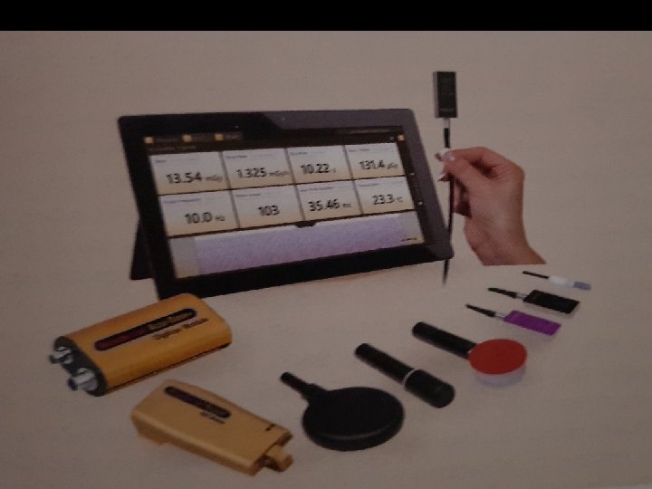
Perssonel Monitoring devices ADA recomends : workers : annual dose >1 msv Personal dosimeter: occupational dose Ffilm badge: 100 micro Gy • Thermoluminescence dosimeter: 50 micro Gy • Optically stimulated luminescence : 10 micro. Gy • Direct ion storage , USB: internet

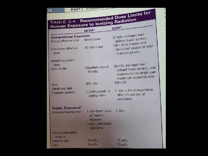
Dose Limits • DLs imply that if received annually , the risk of death would be less than 1 in 10000 • These limits pertain to planned exposure situations (no background , no therapeutic).

Quality assurance Protocols for x-ray tube, image receptor, …… Periodic assessment of the performance of all parts of the radiologic procedure.
- Slides: 59