Nasal CavityII Presented by Dr Sushma Tomar Associate

Nasal Cavity-II Presented by: Dr. Sushma Tomar Associate Professor Department of

Lesson Plan v. Lateral Wall of Nasal Cavity: • Formation • Features • Openings • Arterial Supply • Venous Drainage • Lymphatic Drainage • Nerve Supply • Applied Aspects

Lateral Wall of Nasal Cavity

Bones- Lateral Wall of Nasal Cavity- Formation • Nasal • Maxilla- Frontal process • Lacrimal. • Ethmoid-Superior Concha -Middle Concha -Labyrinth. • Inferior Nasal Concha. • Palatine- Perpendicular plate. • Sphenoid- Medial Pterygoid Plate.

Lateral Wall of Nasal Cavity- Formation contd… Cartilages- • Lateral Nasal Cartilage (Lateral process of Septal Cartilage). • Major Alar Cartilage. • Minor Alar Cartilages (3 -4 in no. ).

Lateral Wall of Nasal Cavity- Features v Lateral wall of nasal cavity is divided into 3 parts: • Anterior part. • Middle part. • Posterior part. Anterior part • Presents a small depressed area, the Vestibule. • It is lined by skin containing short, stiff hair. Middle part • It is known as ‘Atrium of Middle Meatus’. • It is limited above by the agger nasi, which is a faint ridge of mucous membrane. v Limen Nasi- Mucocutaneous junction between the vestibule and atrium of middle meatus. Posterior part • Presents Conchae (Turbinates) and Meatuses.

Conchae v. These are curved bony projections lined by mucosa, directed downwards and medially. v 3 in number: • Superior Nasal Concha. • Middle Nasal Concha. • Inferior Nasal Concha. v. Superior and middle nasal conchae are projections from medial surface of ethmoidal labyrinth. v. Inferior nasal concha is an independent bone. v. Superior concha is smallest and Inferior concha is largest in size.
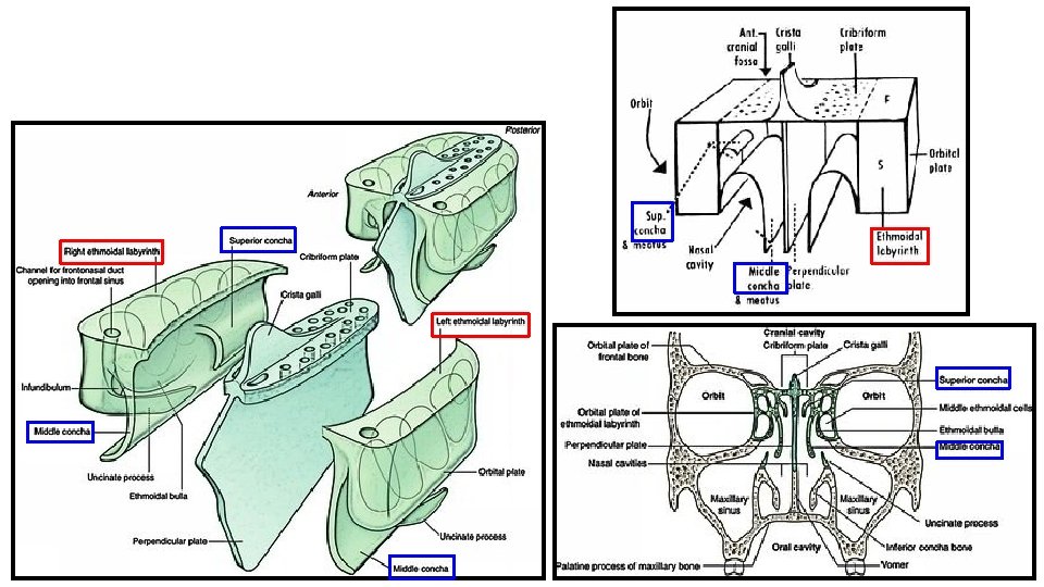

Meatuses- Lateral Wall of Nasal Cavity- Features contd… v. Below and lateral to each concha is a space known as meatus. v 3 in number: • Superior meatus. • Middle meatus. • Inferior meatus. Superior meatus- v. It lies underneath the superior concha. Middle concha (cut) Ethmoidal Bulla Hiatus Semilunaris v. It is smallest. Middle meatus- v. It lies underneath the middle concha. v. It presents the following features: • Ethmoidal bulla (Bulla ethmoidalis). • Hiatus semilunaris. Inferior concha (cut)

Lateral Wall of Nasal Cavity- Features contd… Ethmoidal bulla • A round elevation, produced by underlying middle ethmoidal air sinuses. Hiatus semilunaris • It is a semicircular sulcus below the ethmoidal bulla. Infundibulum • It is a short passage at the anterior end of middle meatus. Inferior meatus • It lies underneath the inferior

Openings in Lateral Wall of Nasal Cavity Site Opening/Openings Spheno-ethmoidal recess Sphenoidal air sinus Superior Meatus Posterior ethmoidal air sinuses Middle Meatus On Ethmoidal Bulla Middle ethmoidal air sinuses In Hiatus semilunaris Anterior part Frontal air sinus Middle part Anterior ethmoidal air sinuses Posterior part Maxillary air sinus Inferior Meatus Nasolacrimal duct (NLD)

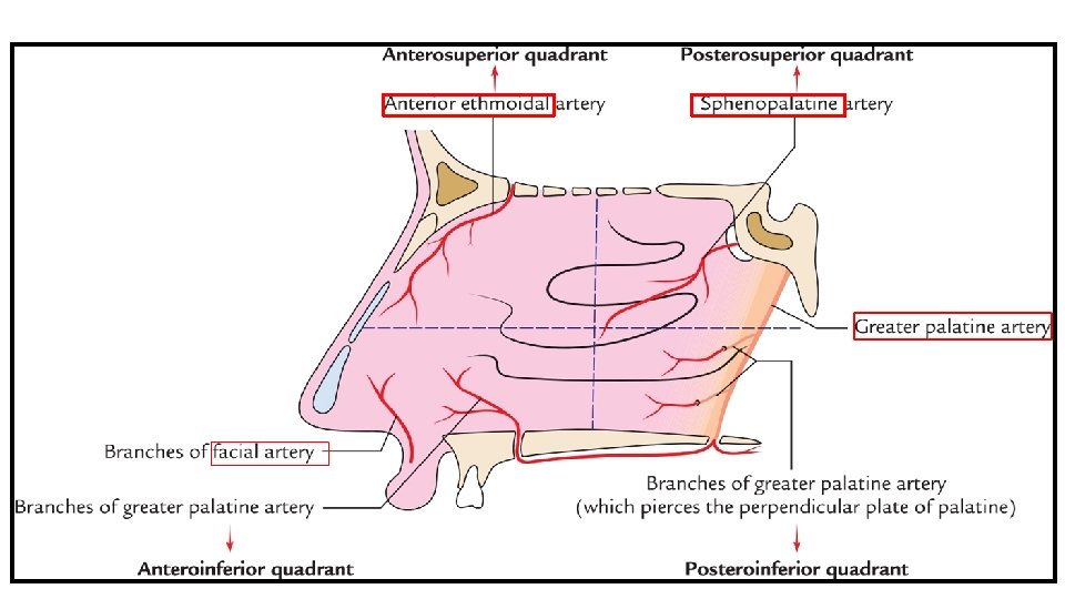
Arterial Supply of Lateral Nasal Wall Anterosuperior Part • Anterior Ethmoidal artery (a branch of Ophthalmic artery). Anteroinferior Part • Lateral Nasal Artery (a branch of Facial artery). • Greater Palatine Artery ( a branch of Maxillary artery). Posterosuperior Part • Sphenopalatine Artery ( a branch of Maxillary artery). Posteroinferior Part • Greater Palatine Artery ( a branch of Maxillary artery).

Venous Drainage of Lateral Nasal Wall v. In lateral wall of nasal cavity, veins accompany the arteries. v. A venous plexus is formed beneath the mucosa. v. Veins drain into: • Facial vein. • Pterygoid venous plexus. • Pharyngeal venous plexus.

Lymphatic Drainage of Lateral Nasal Wall Anterior ½- • Drains into: • Submandibular lymph nodes. Posterior ½- • Drains into: • Retropharyngeal lymph nodes.

Nerve Supply of Lateral Wall of Nasal Cavity Anterosuperior Part • Anterior Ethmoidal Nerve (a branch of Nasociliary nerve). Anteroinferior Part • Anterior Superior Alveolar Nerve (a branch of Infraorbital nerve). Posterosuperior Part • Posterior superior lateral nasal nerves ( branches of Pterygopalatine ganglion). Posteroinferior Part • Nasal branches of Greater Palatine Nerve ( a branch of Pterygopalatine ganglion). Olfactory Part (upper 1/3 rd up to the superior concha)-
![Applied Aspects Rhinoscopy [ Examination of Nasal Cavity] • Anterior Rhinoscopy • Posterior Rhinoscopy](http://slidetodoc.com/presentation_image_h2/12cfb88ffd0dbfe2fea08a474b5fdf43/image-17.jpg "Applied Aspects Rhinoscopy [ Examination of Nasal Cavity] • Anterior Rhinoscopy • Posterior Rhinoscopy")
Applied Aspects Rhinoscopy [ Examination of Nasal Cavity] • Anterior Rhinoscopy • Posterior Rhinoscopy Anterior Rhinoscopy- • Examination of nasal cavity through the nostril. • It is carried out by inserting a nasal speculum through a nostril. Nasal Speculum

Structures seen on Anterior Rhinoscopy • Following structures are seen on Anterior Rhinoscopy: • Middle nasal concha (turbinate). • Inferior nasal concha (turbinate). • Superior meatus. • Middle meatus. • Inferior meatus. • Nasal septum. • Floor of nasal cavity.

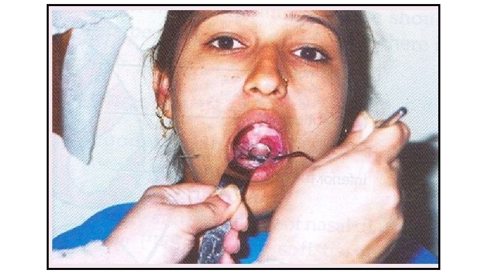
Posterior Rhinoscopy • Examination of nasal cavity through the pharynx. • It is carried out by inserting a mirror into the pharynx. Structures seen on Posterior Rhinoscopy: • Choanae. • Conchae. • Posterior border of nasal septum. Posterior Rhinoscopic Mirror

- Slides: 21