n n n n a subfalcial cingulate herniation

 subfalcial (cingulate) herniation ; 镰下疝 b) uncal herniation")
示意图 n n n n a) subfalcial (cingulate) herniation ; 镰下疝 b) uncal herniation ; 钩疝 c) downward (central, transtentorial) herniation ; 下行性小脑幕疝 d) external herniation ; 颅外 疝 e) tonsillar herniation. 扁桃体 疝 f) ascending transtentorial herniation (reversed tentorial)上行性小脑幕疝 g) sphenoid herniation蝶骨 嵴疝


解剖关系 F F CC CC P O Sy s T lv Sp lv s Mb 3 v Qc O

解剖关系 F F s T d T P Mb Ce s 4 th V Ce

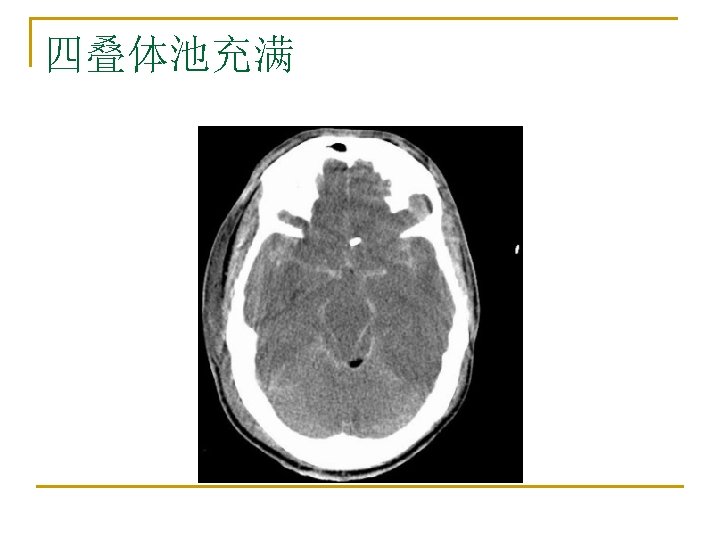
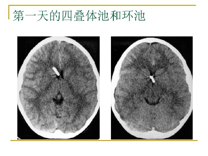
The suprasellar cistern & the quadrigeminal cistern n The left and center images show the suprasellar cistern. Its anterior borders are formed by the frontal lobes (F). Its lateral borders are formed by the uncus (U) of the temporal lobes. The left image shows the 5 -pointed star appearance of the suprasellar cistern where the posterior border is formed by the pons (Po). The black arrow points to the fourth ventricle. The center image shows a higher cut where the suprasellar cistern has a 6 -pointed star appearance since the posterior border is n formed by the cerebral peduncles (P) which have a central cleft. The right image shows the quadrigeminal cistern (black arrow). Note the "baby's bottom" appearance of its anterior border. When ICP is increased, the quadrigeminal cistern space is compressed or obliterated.

The suprasellar cistern & the quadrigeminal cistern. n The midline sagittal MRI scan shows the levels of the axial diagrams. The quadrigeminal cistern is located above (anterior to) the "Q" in the highest cut shown (number 9). The anterior border of the quadrigeminal cistern is formed by the superior colliculi (c). Image 8 (lower cut) also shows the quadrigeminal cistern. In this case, its anterior border is formed by the inferior colliculi (c). This gives the anterior border of the quadrigeminal cistern the appearance of a "baby's bottom". The quadrigeminal plate is comprised of the superior and inferior colliculi. The quadrigeminal cistern is posterior to this quadrigeminal plate, thus its anterior border may be formed by the inferior or superior colliculi.
 Transtentorial herniation n n The suprasellar cistern (left image) is")
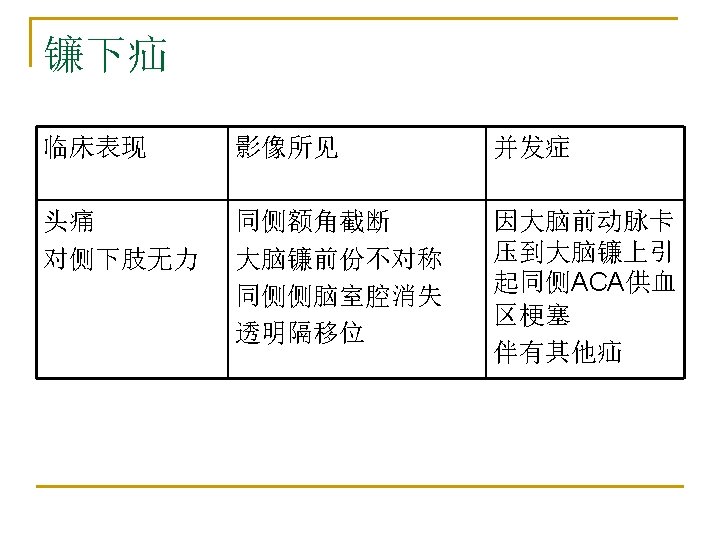
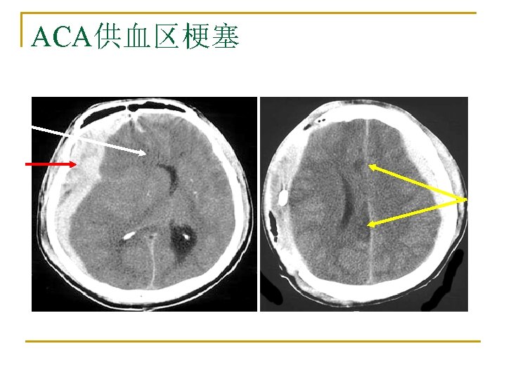
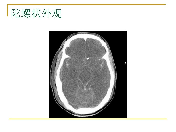
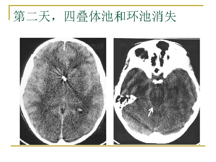
Subfalcine herniation (cingulate herniation) Transtentorial herniation n n The suprasellar cistern (left image) is obliterated. The quadrigeminal cistern is very compressed and pushed posteriorly (center image). A subdural hematoma with a midline shift is noted. There is central transtentorial and subfalcine herniation.

Uncal herniation
 and the quadrigeminal cistern")
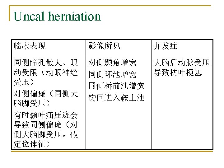
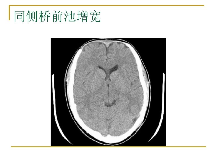
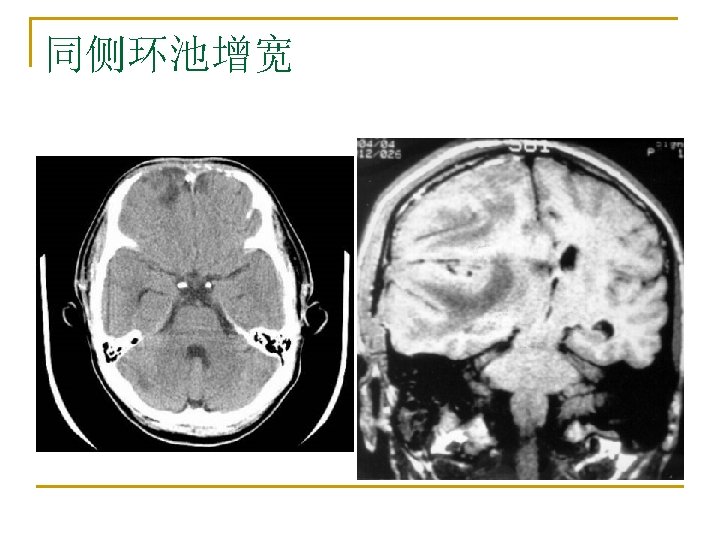
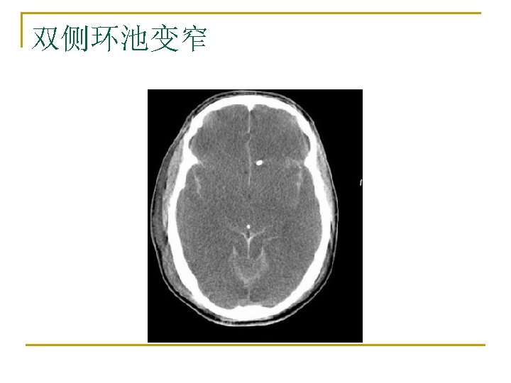
Uncal herniation n obliteration of the suprasellar cistern (red arrow) and the quadrigeminal cistern (green arrow)

Uncal herniation n The ipsilateral ventricle, sulci, fissures are compressed and obliterated, isappeared. n obliteration of the suprasellar cistern(s) and quadrigeminal cistern(q)

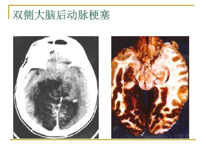
Uncal herniation s q n n Acute infarction 1 st day n n Acute infarction 4 th day

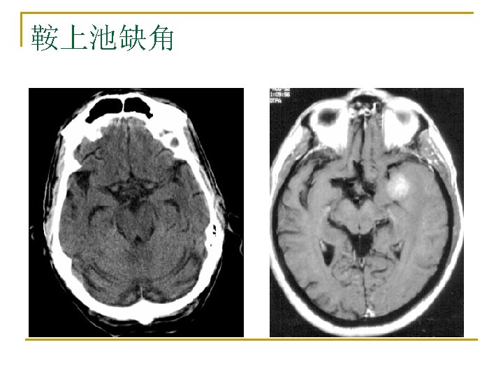
Uncal herniation n n Before surgery, a big GBM in the left temporal lobe with uncal herniation. After surgery, the GBM was removed, the suprasellar cistern and quadrigeminal cisterns are normal.
, this is a complication")
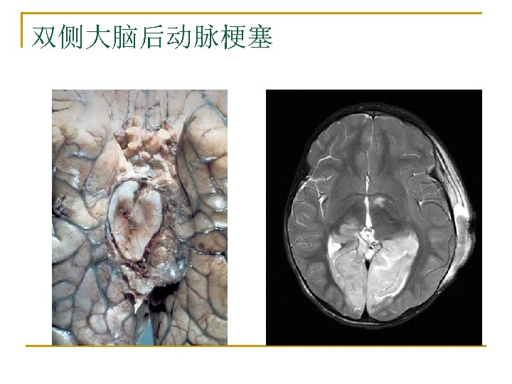
Uncal herniation n Acute infarction of right posterior artery (PCA), this is a complication of uncal/transtentorial herniation, because the PCA was compressed by brain herniation.

Durette hemorrhage

Durette hemorrhage

Kernohan’s notch颞叶疝压迹

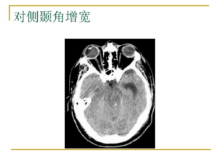
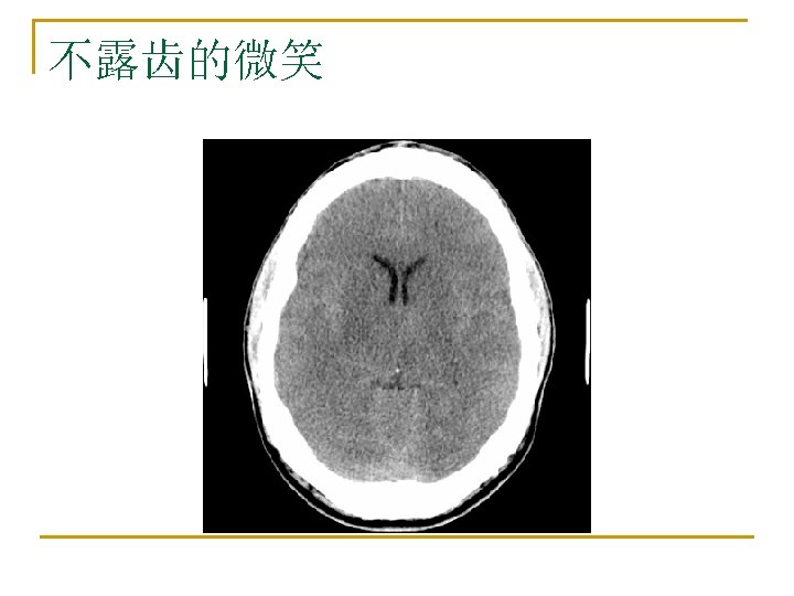
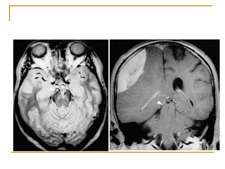
Uncal herniation n When mass effects within or adjacent to the temporal lobe occur, the medial portion of the temporal lobe (uncus) is forced medially and downward over the tentorium. There is ipsilateral pupillary dilation. The uncus is pushed medially into the suprasellar cistern. There is bilateral uncal herniation. The suprasellar cistern is obliterated.

early uncal herniation n The right uncus is pushing into the suprasellar cistern; early right uncal herniation.
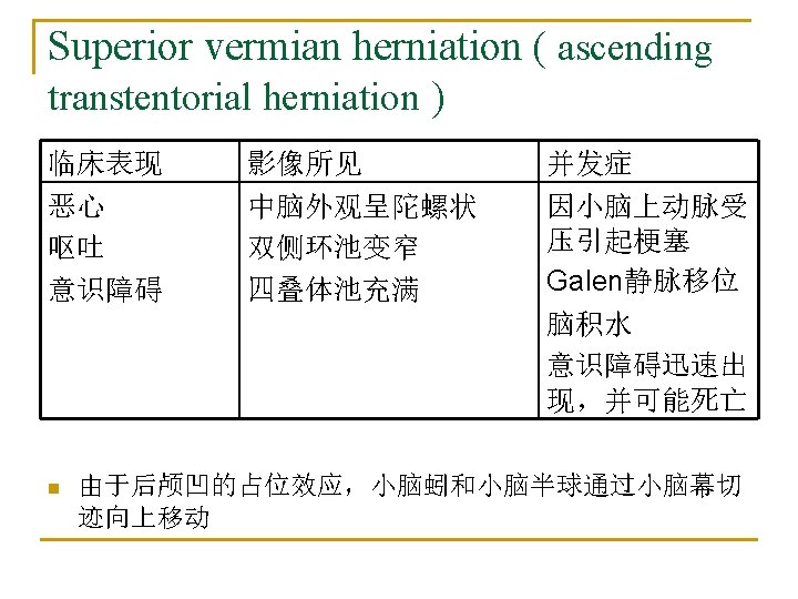

ascending transtentorial herniation
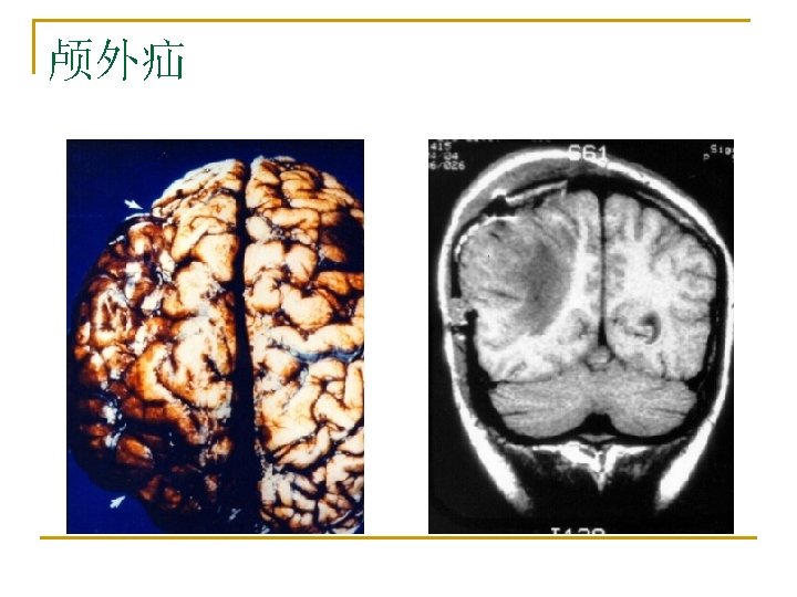
, a mass effect in the posterior fossa")
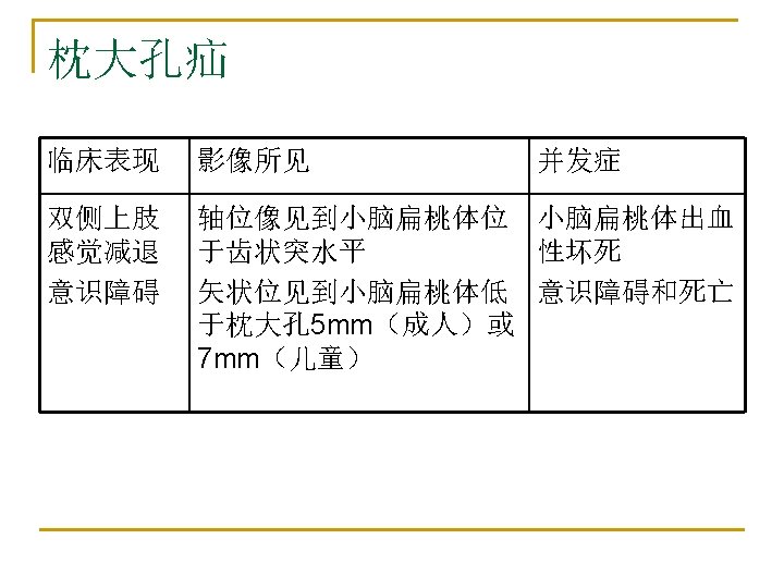
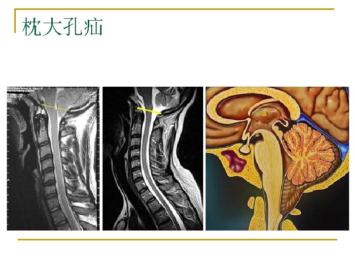
Tonsillar herniation n In tonsillar herniation (rare), a mass effect in the posterior fossa causes the cerebellar tonsils to herniate inferiorly through the foramen magnum compressing the medulla and upper cervical spinal cord. Conscious patients complain of neck pain and vomiting. They may have nystagmus, pupillary dilatation, bradycardia, hypertension and respiratory depression. Early tonsillar herniation is difficult to recognize in an unconscious patient. It may not be evident on CT scan since axial views cannot see the pathology well. It is best seen on sagittal MRI. Clinically changes in vital signs may be the only clinical clue in an unconscious patient.

Tonsillar herniation

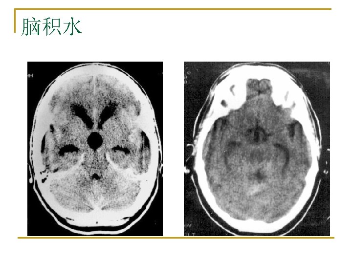
a male patient in his 30's who died of brain stem herniation after completing a marathon. n The CT shows (A) loss of the rostral cerebral sulci suggesting increase in ICP, (B) and (C) a large hydrocephalus with widening of both temporal horns. The grey matter can still be differentiated from the white matter, but all sulci are lost. This suggests that the brain oedema is of relative recent onset and massive tissue ischaemia has not yet occurred. (D) Compression of the fourth ventricle with dilatation of the third ventricle and the caudal aspect of both temporal horns. This is observed with considerable brain oedema and obstructive hydrocephalus. (E) Herniation of the medulla and pons into the foramen magnum. (F) The tonsils are located at the level of the dens which is a good indicator foramen magnum herniation.
 The disc shows florid hemorrhages with relatively little swelling, indicating a rapid,")
n (A) The disc shows florid hemorrhages with relatively little swelling, indicating a rapid, dramatic increase in CSF pressure. Progressive changes of optic disc oedema are seen in a patient with an intracranial tumour who declined treatment (B-D). (B) Early nerve fiber dilatation is seen particularly superiorly, inferiorly and nasally. (C) This increases and venous engorgement develops. (D) Temporal nerve fiber dilatation and swelling of the disc increases and hemorrhages appear. (E) In gross chronic disc oedema the normal retinal vasculature is masked and dilated superficial capillaries are observed. (F) In atrophic optic disc oedema nerve fibers are eventually destroyed and the optic disc without viable nerve fibers does not swell. This patient had longstanding benign intracranial hypertension. Retinochoroidal venous collaterals are present (black arrowhead).

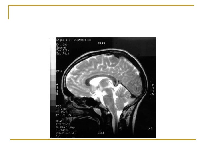
核磁选择 n n 1. Subfalcine herniation. This is best seen on coronal MR images. 2. Descending transtentorial herniation (uncal herniation, hippocampal herniation). best seen on coronal images, but the compression of the brainstem is best observed on axial T 2 -WI. 3. Ascending transtentorial herniation. The sagittal imaging plane is preferred. 4. Cerebellar tonsillar herniation. Sagittal and coronal imaging planes are preferred.
- Slides: 57