n low dose spiral CT autofluorescence bronchoscopy associated

n low dose spiral CT, autofluorescence bronchoscopy associated protocols
 Pure GGO, Mixed GGO (extent of solid")
1. LDCT protocol -GGO pattern - size(mm) Pure GGO, Mixed GGO (extent of solid portion ≤ 50%) Mixed GGO (extent of solid portion > 50%) ≤ 5 mm F/U CT at 12 months If no change, F/U CT annually F/U CT at 6 months, If no change, F/U CT annually > 5 mm, ≤ 10 mm F/U CT at 6 months If no change, F/U CT annually F/U CT at 6 months, If no change, VATS ≥ 10 mm F/U CT at 6 months If no change, F/U CT annually F/U CT at 6 months, If no change, PCNB or VATS If size increased , PCNB or VATS recommanded, F/U: follow up PCNB: percutaneous needle biopsy, VATS: video assisted thoracic surgery
 ≤ 5 mm F/U CT at 12")
LDCT protocol -solid Nodule - Nodule Size(mm) ≤ 5 mm F/U CT at 12 months If no change, no further FU > 5 mm, ≤ 10 mm Initial F/U CT at 3~6 months, Second F/U CT at 6 months, Third, fourth F/U CT at 12 months If no change, no further FU ≥ 10 mm <Option 1> Initial F/U CT at 3 months, Second F/U CT at 6 months, Third, fourth F/U CT at 12 months If no change, no further FU <Option 2> PCNB or VATS If size increased , PCNB or VATS recommanded, F/U: follow up PCNB: percutaneous needle biopsy, VATS: video assisted thoracic surgery

2. Autofluorescence Bronchoscopy Protocol AFB : Abnormal lesion detected Biopsy Carcinoma In Situ and more Dysplasia Moderate to Severe Metaplasia Mild Operation, PDT, F/U AFB electrocautary etc at 3 month at 6~12 month
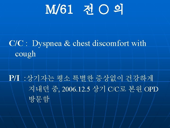
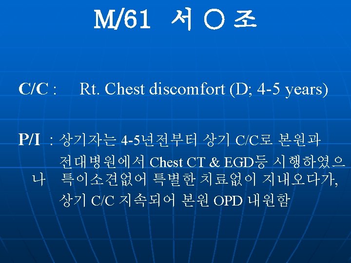
 Operation Hx (-) Pul. Tbc(+) ; 40 yrs-ago")
n P/H : Admission Hx (-) Operation Hx (-) Pul. Tbc(+) ; 40 yrs-ago DM/ HTN / Hepatitis - / - n S/H : Alcohol (+) ; Social Smoking (+) ; 100 PYs(2 yrs-ago, quit) n F/H : N-S
")
Chest PA (06. 12. 5)

Chest LDCT

ONCO-LIFE
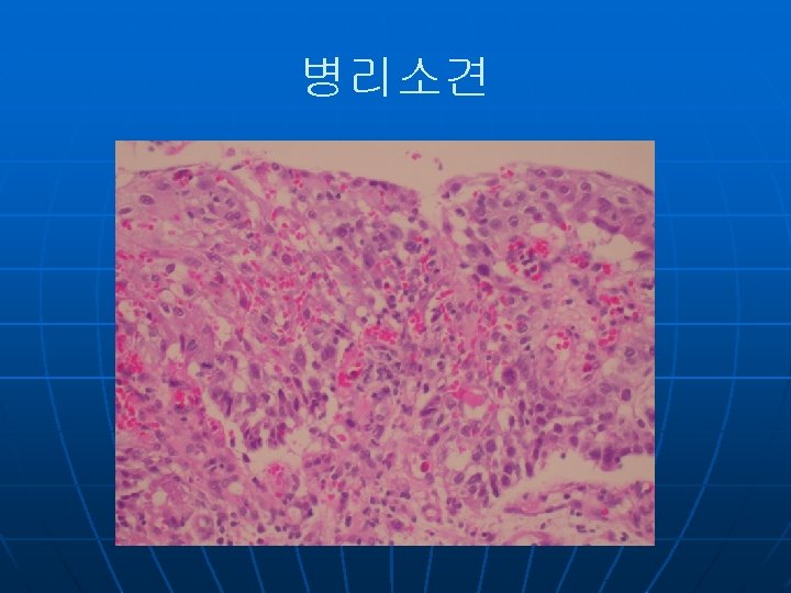

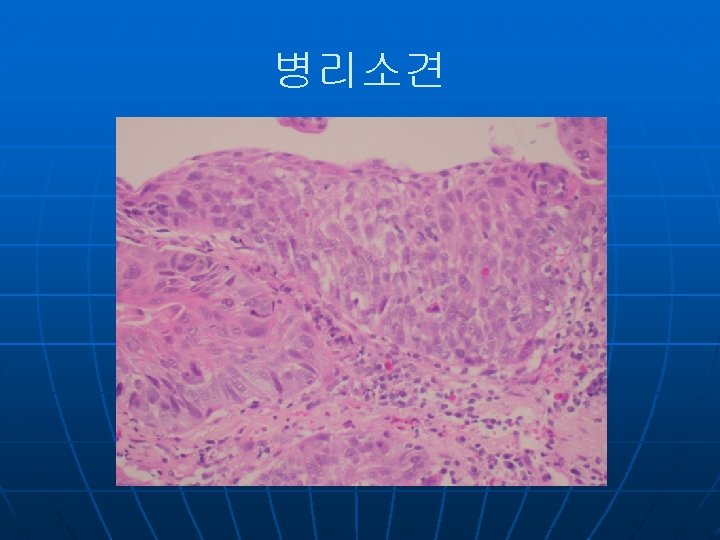
Final Diagnosis 1. Severe dysplasia in Lt. lingular segment 2. Old pulmonary Tbc.
 Operation Hx (-) DM/ HTN / Pul. Tbc")
n P/H : Admission Hx (-) Operation Hx (-) DM/ HTN / Pul. Tbc / Hepatitis - /- / n S/H : Alcohol (+) ; Social Smoking (+) ; 40 PYs n F/H : N-S
")
Chest PA (06. 12. 18)
")
Chest LDCT (06. 12. 18)
")
ONCO-LIFE (06. 12. 19)

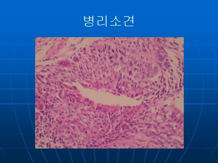
Final Diagnosis 1. Carcinoma in situ in RUL post. segment 2. Bullae in RUL
 n n C/C : Rt. Chest pain, coughing, blood tinged sputum,")
문 O 기(M/66) n n C/C : Rt. Chest pain, coughing, blood tinged sputum, mild resting dyspnea (O: 20 days ago) P/H : 20여년 전 glaucoma Tx. 2년 전 DM, CVA Dx. at 본원 No known Hx. Of HTN, pul. Tbc, Hepatitis No Operation Hx.

n n S/H : Alcohol : None Smoking : None P/I : 상기 환자는 20일 전부터 발생한 Rt. Chest pain 및 cough, blood tinged sputum, mild resting dyspnea있어 Home observation 중 Sx. Aggravation되어 local 들려 check한 chest X-ray 상 pneumonia 소견보여 further evaluation위해 본 원 OPD visit
 4. Bronchoscopy")
Diagnosis plan 1. Chest X-ray 2. Laboratory 3. Chest CT(enhance) 4. Bronchoscopy

Chest PA/Lt. Lat
")
Chest CT(enhance)

Onco-Life

bronchoscopy

Bronchoscopy
")
Chest PA (F/U)


n P/H : No known Hx of DM, Hypertension, Hepatitis, Pul. Tbc n S/H : Alcohol - None Smoking - 30 pys (1갑 x 30년) n F/H : Non-Specific
")
Chest PA & Lt. lateral(01. 09)
")
HRCT ( 08. 01. 09 )
")
Bronchoscopy( 08. 01. 09 )

S 08 -157

LCA

Chromogranin

PET-CT
")
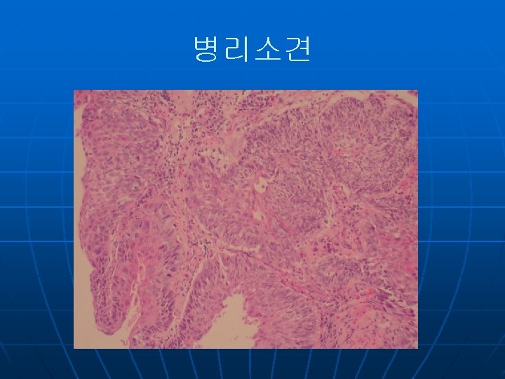
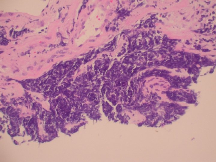
Final Diagnosis n Small cell lung carcinoma (stage 1)
- Slides: 45