n Drawing illustrates the traditional tricompartment model of


 Axial CT image shows an enlarged pancreatic")


 Axial CT image shows thickening of the")


- Slides: 38
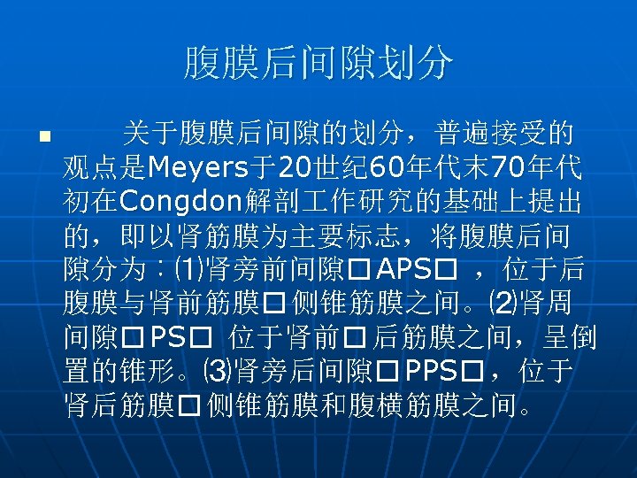
n Drawing illustrates the traditional tricompartment model of the retroperitoneum, which is accordingly divided into the anterior pararenal space (APS), perirenal space (PS), and posterior pararenal space (PPS). The anterior renal fascia (ARF), posterior renal fascia (PRF), and lateroconal fascia (LCF) divide the spaces.
n Drawing illustrates the recently modified tricompartment model, which reflects the understanding that the perirenal fascia is laminar and variably fused and there are interfascial connections between the spaces. The retromesenteric plane (RMP), retrorenal space (RRS), and lateroconal space are potential interfascial communications. Perinephric septa run between the renal capsule and the perinephric fascia, allowing subcapsular fluid to communicate with the retrorenal space or retromesenteric plane. APS = anterior pararenal space, PPS = posterior pararenal space, PS = perirenal space (, ).
十二指肠壁内血肿 n Traumatic duodenal intramural hematoma in a 26 -year-old man who had sustained a seat belt injury in a high-speed motor vehicle collision. Abdominal CT scan obtained with oral and intravenous contrast material shows wall thickening of the third and fourth portions of the duodenum (arrows). No extraluminal air (a finding that would have suggested perforation) was seen. The patient was treated conservatively and recovered without intervention
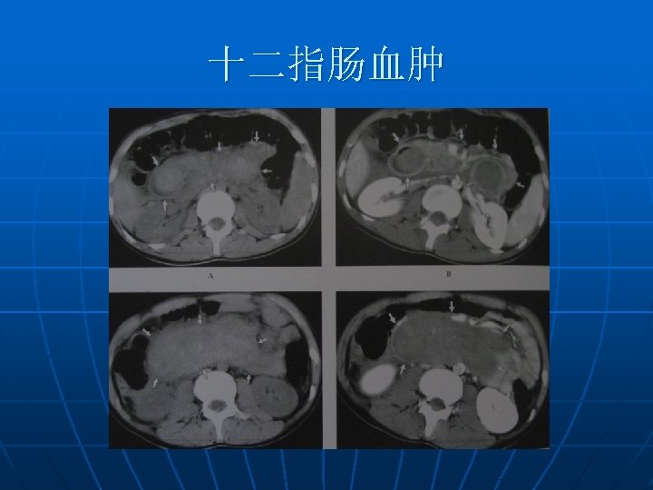
十二指肠血肿 n Large traumatic duodenal hematoma in a 49 -year-old man who was involved in a motor vehicle collision. The patient was also taking anticoagulants. (a) Abdominal CT scan obtained with oral and intravenous contrast material shows a large hematoma (arrowheads) displacing the second portion of the duodenum (arrow) anteromedially and narrowing the duodenal lumen. (b) Coronal reformatted CT image depicts the full extent of the duodenal hematoma (arrowheads).
十二指肠降部挫伤 n Grade I duodenal injury. Axial CT image shows thickening of the duodenal wall (arrow) in the descending part without evidence of free air. There is stranding of the peripancreatic fat.
十二指肠水肿 n Duodenal hematoma in an 11 -year-old boy who had sustained a bicycle handlebar injury. On an abdominal CT scan obtained with oral and intravenous contrast material, the third portion of the duodenum is thickened and edematous (arrowheads). No extraluminal air could be identified to suggest perforation. The patient was treated conservatively and recovered completely.
十二指肠降部破裂 n Grade II duodenal injury. (a) Axial CT image shows an enlarged pancreatic head with mild edema (arrow) (grade I lesion). (b) CT image obtained at a lower level shows thickening of the duodenal wall in the descending part (black arrow). Adjacent to the duodenum is a small collection of extraluminal air (white arrow), which indicates a small grade II laceration of the wall.
十二指肠穿孔 n Duodenal perforation in a 28 -year-old man who sustained blunt trauma in a motor vehicle collision. (a) Abdominal CT scan obtained with intravenous and oral contrast material shows extraluminal air (arrows) adjacent to the duodenum (D). Cholecystectomy clips are also present. (b) Coronal reformatted CT image shows a large amount of fluid in the right anterior pararenal space with a small focus of extraluminal air (arrow), findings that are consistent with a duodenal perforation. The perforation was confirmed and repaired at surgery. D = duodenum.
十二指肠水平部破裂 n Grade II duodenal injury. Axial CT image shows a grade II injury of the horizontal part of the duodenum with small collections of extraluminal air (arrows). A subcapsular hematoma is present at the lower pole of the right liver lobe (arrowhead).
十二指肠破裂 n Grade III duodenal injury. (a) Axial CT image shows thickening of the duodenal wall in the descending part (black arrow). At the transition zone to the horizontal part, there is disruption of the wall (white arrow). Additional findings include a retroperitoneal hematoma and hypoperfusion of the right kidney due to right renal artery occlusion. (b) CT image obtained at a lower level shows the disruption (black arrow) with a large surrounding extraluminal hematoma (white arrow).
n Ruptured duodenum in a 27 -year-old female victim of a motor vehicle accident. CT scan shows fluid in the duodenum and leakage of fluid into the right anterior pararenal space (arrow).
消化内镜胃窦及胰头活检术后 n Axial CT images in patient with perforation of the second portion of the duodenum 1 day after gastroduodenoscopy with endoscopic pancreatic fine-needle aspiration biopsy and gastric biopsy. (a) Unenhanced CT image shows air in anterior pararenal space (white arrow) and right perirenal space (black arrow). (b) A more caudal image shows a focus of discontinuity (short black arrow) in the wall of the second portion of the duodenum, with extraluminal extravasation of oral contrast material (long black arrow). Air is also demonstrated in the right posterior pararenal space (long white arrow), right properitoneal compartment (short white arrow), and peritoneal cavity (arrowhead). (c) Contrast-enhanced CT image obtained 2 years later shows the right anterior renal fascia (white arrow) to extend to the second portion of the duodenum (arrowhead). The cystic lesion in the pancreatic head is an intraductal papillary mucinous neoplasm (black arrow).
消化内镜胰头活检术后 n Contrast-enhanced axial CT images in patient with perforation of the second portion of the duodenum after endoscopic pancreatic fineneedle aspiration biopsy 1 week earlier. The amount of air in the right perirenal space exceeds that in the right anterior pararenal space.
内镜检查后 n Duodenal perforation after endoscopy in a 51 -yearold man. CT scan shows a thick-walled, contracted duodenum with air in the adjacent retroperitone um (arrow).
空肠破裂 n Jejunal perforation in a 66 -year-old woman after a motor vehicle accident. Axial CT image shows hypervascular thickened jejunum with a suspicious defect (curved arrow) and with focal fluid, fat stranding, and extraluminal air (straight arrow) adjacent to jejunal loops. The patient later underwent resection of a 20 -cm segment of the small bowel. No mesenteric injury was found at surgery.
十二指肠溃疡穿孔 n Abdominal pain and a perforated duodenal ulcer in a 79 -year-old man. CT scan obtained with oral contrast material shows intraperitoneal extravasation of contrast material from the lateral portion of the duodenum (white arrow) and leakage of contrast material around the liver (black arrow).
膀胱破裂 n Ruptured bladder in a 43 -year-old female pedestrian who was struck by a car. (a) Axial abdominal CT image shows intraperitoneal areas of free contrast material (straight arrows) and free air (curved arrow). (b) Axial CT image shows a retroperitoneal area of extraluminal contrast enhancement (arrow). These features mimic those found in bowel injury but, instead, are secondary to a bladder rupture, which was found at surgery.
胰腺炎血肿进入十二指壁 n Acute pancreatitis and hemorrhage into the lateral duodenal wall, which caused mass effect and narrowing of the duodenal lumen, in a 46 -year-old man. CT scan shows extensive stranding of the peripancreatic fat secondary to pancreatitis. Massive enlargement of the lateral wall of the duodenum is accompanied by a focal area of increased attenuation at the site of the bleeding (black arrow). The duodenal lumen, which contains low-attenuation fluid, is narrowed and displaced medially (white arrow).