N Cem FICICIOGLU MD Ph D AA MBA

N. Cem FICICIOGLU, MD, Ph. D, . AA, MBA Prof in OB&GYN

The differentiation of the gonad- Y chromosome, SRY gene Gonad is not testis- the absence of antimüllerian hormone-the development of paramesonephric (müllerian) ducts The formation, fusion and resorption of müllerian ducts

Sertoli cell AMH The regression of mullerian duct Leydig cell Testosteron The development of mesonephric Ducts 5 α reductase DHT The differantion of external genitalia
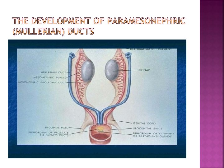
 ducts and mesonephric ducts in fetus, up to eight")
Both ducts, paramesonephric (müllerian) ducts and mesonephric ducts in fetus, up to eight weeks; bipotent period One of them disappears in 12 th week The differantion occurs by AMH secreted by sertoli cell and testosterone secreted by leydig cell In the absence of AMH, uterus, fallopian tubes and 1/3 upper vagina develop from müllerian ducts


Sporadic abnormalities Multifactoriel, Poligenic Familial Uriner system abnormalities accompany with vagina, cervix, fallopian tubes

%5, 5, in selected population %8, in infertile population %13, 3, pregnancy loss %24, 5, reccurent pregnancy loss

Congenital Acquired Myomas Polyps Intrauterine adhesions
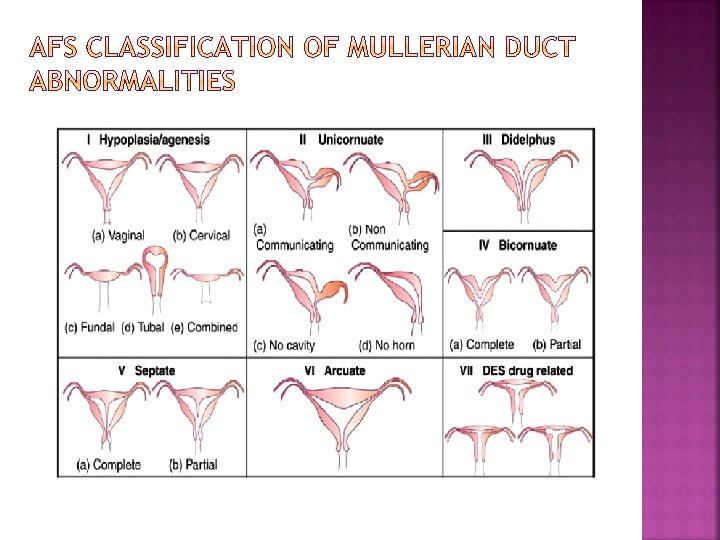

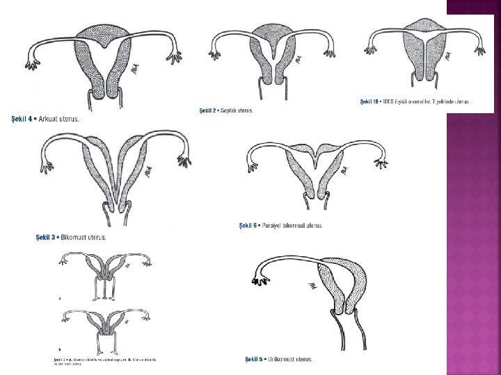
Complete failure of fusion of the paired Mullerian ducts results in duplication of the uterine corpus and cervix called uterus didelphys. A longitudinal vaginal septum is present in most women with a didelphys uterus, and may facilitate the early diagnosis when identified on routine speculum examination

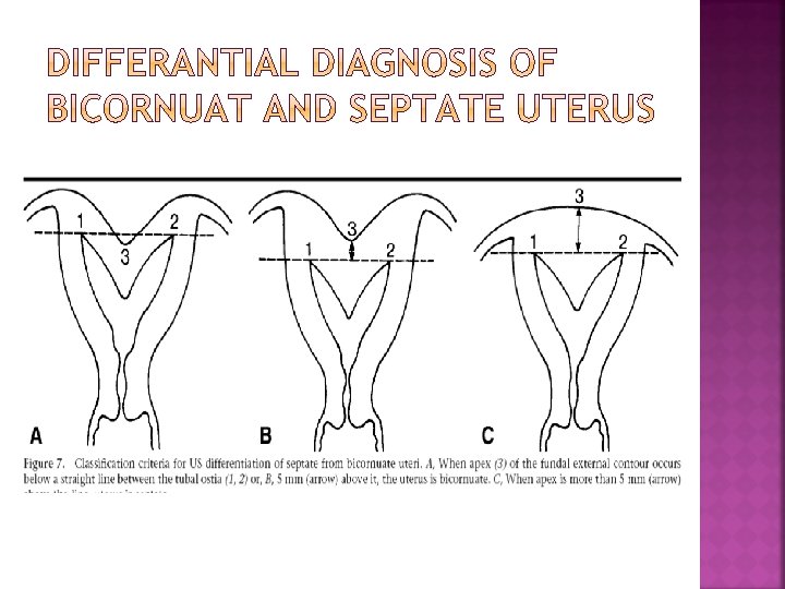
A results from failure of the Mullerian ducts to completely fuse. The central myometrium may extend to the level of the internal cervical os (bicornuate unicollis) or external cervical os (bicornuate bicollis). The latter is distinguished from uterus didelphys because it demonstrate some degree of fusion between the two horns, whereas in classic uterus didelphys the two horns and cervices are separated completely.

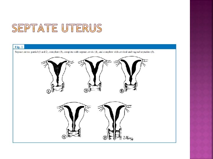
The partition between the ducts is thought to resorb in a caudal to cranial direction Failure of complete resorption results in a fibromuscular septum that can be partial or complete—dividing the uterine cavity and cervical canal into two parts

A unicornuate uterus represents a uterine malformation where the uterus is formed from one only of the paired Mullerian ducts while the other Mullerian duct does not develop or only in a rudimentary fashion. The sometimes called hemi-uterus has a single horn linked to the ipsilateral fallopian tubes that faces its ovary


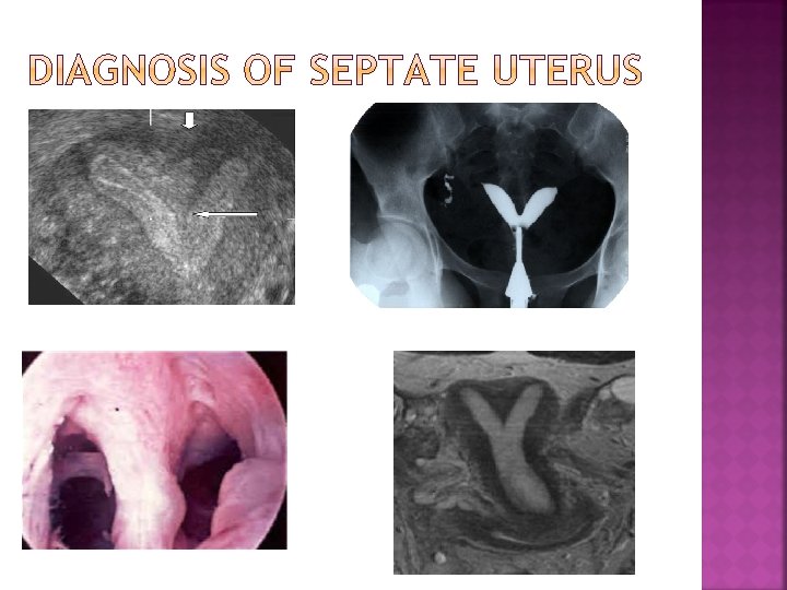
Physical examination Ultrasonography, 2 D and 3 D HSG MRI Laparoscopy

Symptoms depend on the severity and type of the defect Dysmenorrhea, chronic pelvic pain, dyspareunia, hematocolpos, hematometra in adolescent Treccurent pregnancy loss Preterm labor Abnormal fetal presentation Infertility
- Slides: 23