n Anatomy Physiology L 3 Integumentary System Integumentary


 1. protective covering 2. regulate body temp 3.")
 of avascular stratified squamous tissue")
")


 Mechanical injury")


 n n n Subcutaneous")
EX: vaccinations 2. Sub. Q (subcutaneously) EX:")


")

 n n n 1. 2. Apocrine: respond to emotional")


 3.")


 burn")

 burn")
 burn")

 burn")

 Skin disorders of the feet")


")


- Slides: 67
n Anatomy & Physiology L 3 Integumentary System
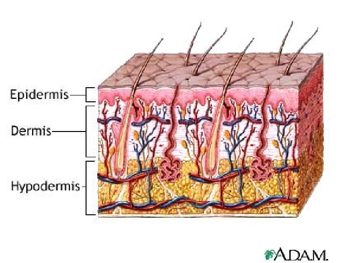
Integumentary System n I. Overview: General structure: composed of epidermis and a dermis, separated by a basement membrane with a subcutaneous layer underneath
n n n General Function: (5) 1. protective covering 2. regulate body temp 3. houses sensory receptors 4. synthesizes various chemicals 5. excretes wastes
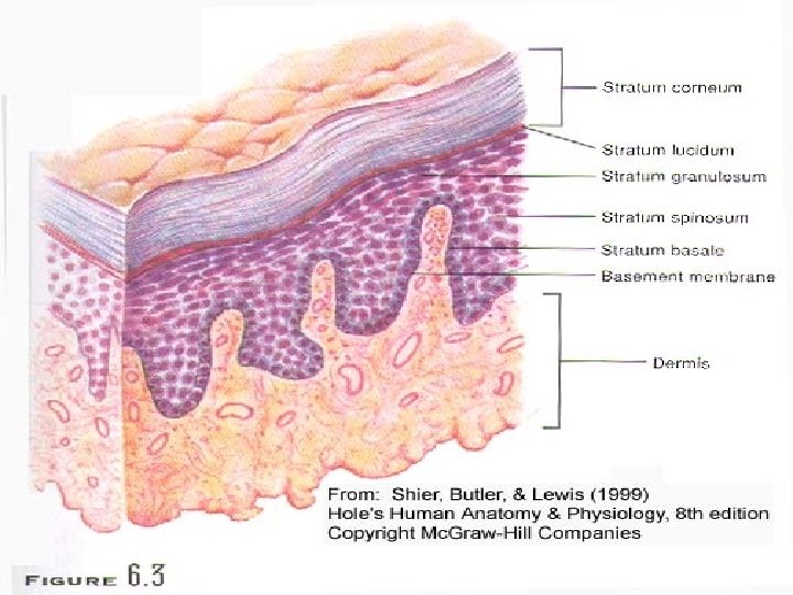
II. Epidermis layer n Layers (5) of avascular stratified squamous tissue
n n 1. stratum corneum: composed of dead epidermal cells, fully keratanized (most superficial) 2. stratum lucidium: in the thickened skin of palms and soles. Contains transparent protein resembling keratin.
n n n 3. stratum granulosum: contain granules in the cell’s cytoplasm which die and become keratinized. 4. stratum spinosum: a thick layer of tiny fibers in the cytoplasm (spiny when pulled apart) 5. stratum basale: (basal layer) cells undergoing mitosis
Keratinization: n Strands of tough, fibrous, waterproof keratin proteins are synthesized and stored in cells causing hardening.
n n epidermal cells undergo process: keratinization, as they are “pushed” towards the surface Mitotic rate is normally EQUAL to that of those cells lost
n 1. 2. Mitotic rate INCREASES when friction occurs causing: calluses on hands and feet corns (keratinized masses on toes)
n n Overall Fxn: Protect underlying tissues against: Water loss (*Wrinkly fingers) Mechanical injury Affects of harmful chemicals / pathogens
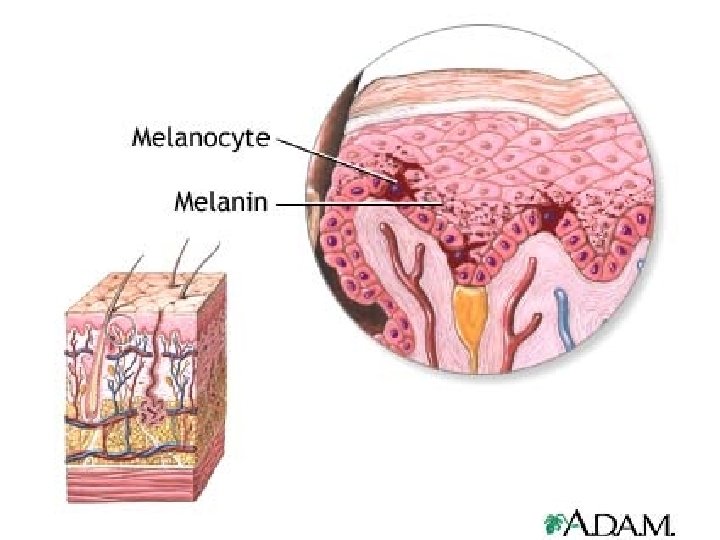
Melanocytes n : Melanocytes n n n Structures lying deep in the epidermis Produce melanin: fxn is to protect underlying cells from UV light Cytocrine secretion: the transfer of granules of melanin into nearby epidermal cells.
Melanocytes
III Dermal layer n n n Layer of loose connective tissue binding epidermis to underlying tissues, such as muscles, adipose, etc Composed mainly of collagen & elastin: Avg. thickness: 1 -2 mm. 0. 5 on eyelids, 3 mm soles of feet Contains: muscle fibers, nerve endings, and blood supply
n n n Basic fxn: 1. blood vessels supply nutrients to all skin cells 2. regulate body temp.
n Nervous tissue scattered throughout the dermis n n Some carrying impulses to muscles / glands of the skin Some associated with various sensory receptors in the skin
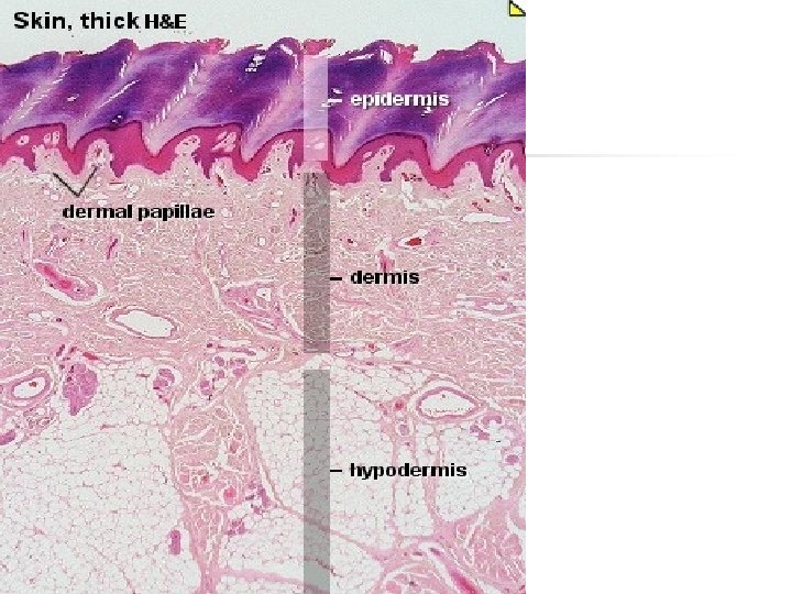
Fingerprints n n n Specialized structure: Dermal papillae Uneven ridges formed during fetal development NO TWO sets are exactly alike!
IV Extension of Dermis, also called hypodermis/ 3 rd layer) n n n Subcutaneous layer composed mainly of loose connective and adipose Layer containing majority of dermal blood vessels Therefore fxn: insulates= conserves body heat and impedes entrance of heat from outside
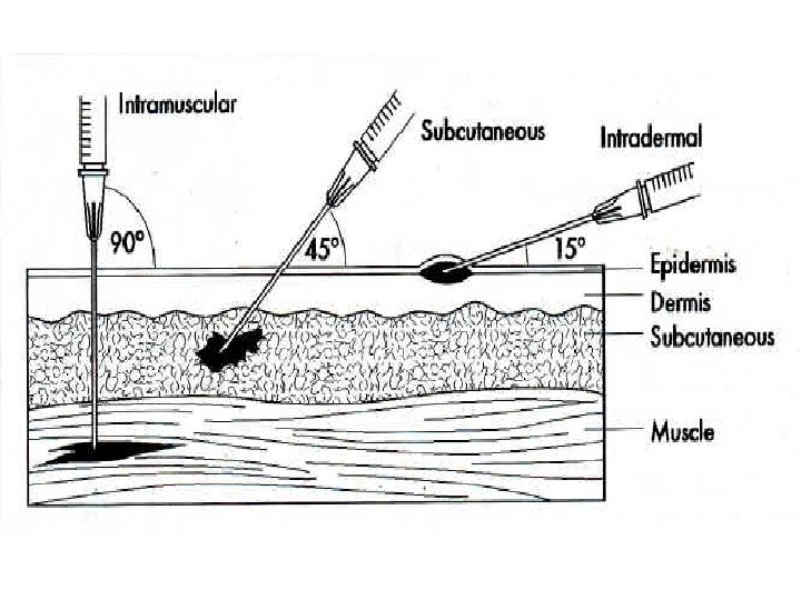
Types of injections: n n 1. IM (Intramuscularly)EX: vaccinations 2. Sub. Q (subcutaneously) EX: insulin 3. Intradermally EX: TB test 4. Transdermally EX: birth control, smoking patch, pain patches
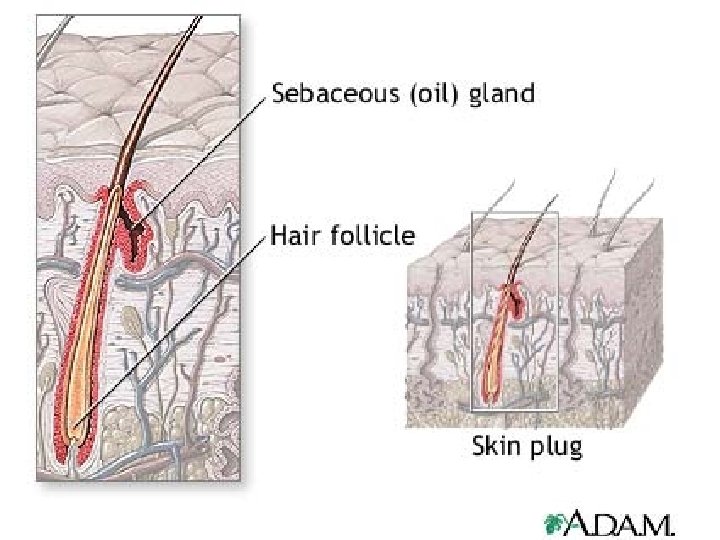
V Accessory organs of the skin n n 1. Hair follicles: hair found almost all areas of the body each hair develops at base called: follicle as newly formed cells develop and grow, older cells “pushed” towards surface and keratinize: called hair shaft
n n hair color genetically determined bundle of smooth muscle and one or more sebaceous glands attached to each hair follicle: arrector pili muscle
2. Sebacious glands: n n n glands that secrete sebum, helping to keep skin and hair soft and waterproof In some regions, open directly to the skin surface: 1. lips 2. corners of mouth 3. external reproductive parts
None found on n n Palms Soles
3. Nails: n n -protective covers on the ends of fingers and toes (tactile) -undergo keratinization
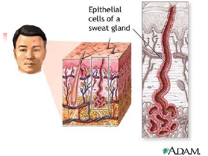
4. Sweat glands: primary fx=cool body n n Excretes wastes located in nearly all areas of the skin Each gland consists of a coiled tube, lined with sweat secreting epithelial cells Sweat is primarily composed of water, also containing salt and urea
Types of sweat glands (3) n n n 1. 2. Apocrine: respond to emotional stress Eccrine: respond to elevated body temp. Specialized apocrine: ceruminous gland-secretes earwax Mammary glands-secretes milk
V. Regulation of Body Temperature n n n Normal or core body temp, is about 98. 6 F, 37 C) Heat Production and loss Heat is by- product of cellular respiration
Heat n Lost to outside environment by the following ways: n n n Evaporation Conduction Convection Radiation*#1 way to cool body Hyperthermia vs. Hypothermia
Problems with temperature regulation n n Humidity due to saturation EX: hyperthermia- sweat glands activated but cannot evaporate sweat r/t hot humid day. Body temp. rises
n 1. 2. 3. At risk age groups for Temperature related condition Very young Very old Very thin
VI. Skin Color n n Melanin: Primary skin pigment. All humans have same concentration of melanocytes. Skin color is due to amt. of melanin sent to the epidermis. Genetics: each person inherits genes for melanin production.
n n Environment: sunlight, uv light, x-ray Causes: existing melanin to darken and stimulate additional melanin production
VII. Physiological factors n n n 1. Oxygen content in blood. Normal / to high: red blood causes pinkish look to skin Insufficient (Too low): ↓ 02= bluish (cyanosis)
n n 2. Additional skin pigmentation carotene: in subcut. Causes yellowish look (Asian) 3. Variety of diseases EX: Jaundice- infants or adults with liver disease.
VIII. Response to injuries n n Inflammation: Is a response to injury in which: Blood vessels dilate Membrane permeability increases Benefits of Inflammation are: aid in healing by providing additional nutrients and 02 to tissue
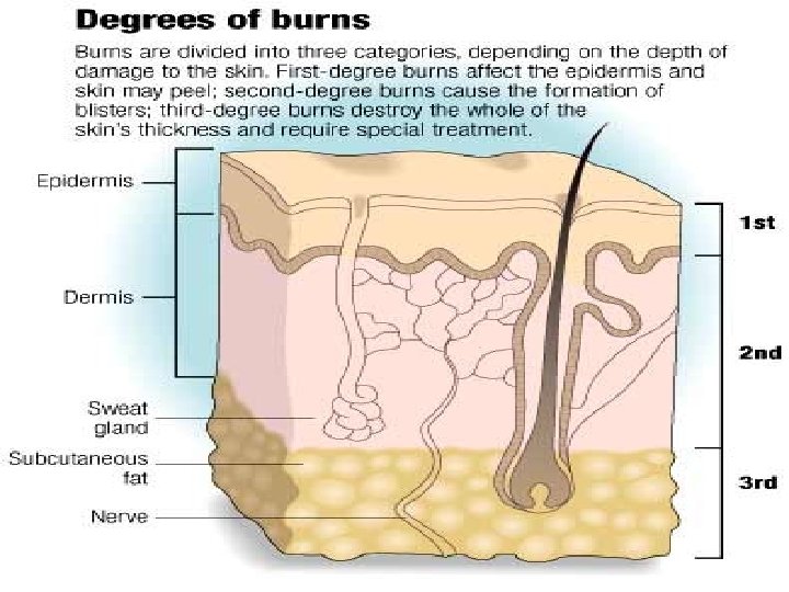
n IX. Burns
1 st degree burn Epidermis ONLY affected n Redness and burning n EX: sunburn n
Epidermal (1 st degree) burn
2 nd degree burn Damage to both the epidermis and dermis n Damage not enough to PREVENT healing n
Dermal (2 nd degree) burn
Deep dermal (2 nd degree) burn
3 rd Degree burn ENTIRE epidermis, and accessory organs destroyed n Tissue death n
Sub-dermal (3 rd degree) burn
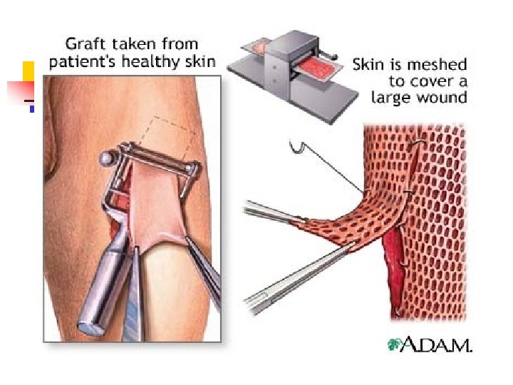
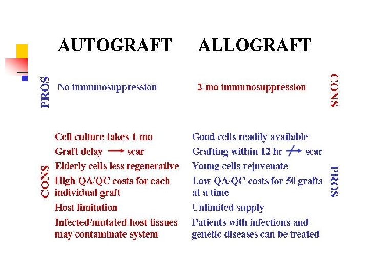
Skin graft
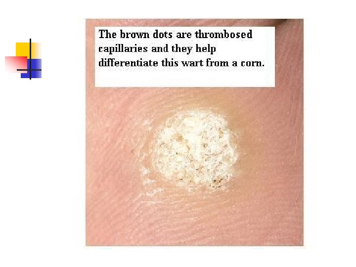
Common disorders of the Feet Athlete's Foot (right down) Skin disorders of the feet such as Callus, Athlete's foot and Plantar Warts
Tinea pedis
Acne
Contact dermatitis
Moles are harmless skin growths that may be flat or protruding. They vary in color from pink flesh tones to dark brown or black.
Pediculosis
Decubitus pressure ulcer (Bed sore)
Where do pressure ulcers form? n n Where bone causes the greatest force on the skin and tissue. For bedridden persons, most pressure ulcers form on the lower back below the waist (sacrum), the hip bone (trochanter), and heels.
Diabetic Ulcer
Pathophysiology: diabetic foot ulceration Neuropathy Motor Sensory Autonomic Abnormal foot biomechanics Loss of protective sensation Reduced skin compliance and lubrication Ulceration Vascular insufficiency Infection
Eczema
Quick Quiz: 2 pts each n n n 1. Name 3 of the, at least 8 fx’s of integ. system 2. List and describe the 3 layers of the skin 3. Name/describe top 2 ways we lose body heat 4. Describe keratin and keratinocytes 5. Describe the relationship between skin color and melanin
Quick Quiz: cont n n n 6. Describe what a sebacious gland is/does 7. Describe what a sweat gland is/does 8. What does Vit D have to do with skin? 9. Discuss the differences in 1 st, 2 nd, 3 rd degree burns 10. Discuss two factors that can influence diabetic ulcers and pressure ulcers