
MYOPATHIES Dr Mohammad Ali Kukaswadia M D WHAT


- Slides: 111
MYOPATHIES Dr Mohammad Ali Kukaswadia, M. D.
WHAT WE NEED TO LEARN TODAY • Muscle structure & physiology • Clinical manifestations • Diagnostic approach • Individual myopathies
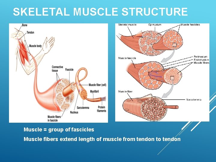
SKELETAL MUSCLE STRUCTURE Muscle = group of fascicles Muscle fibers extend length of muscle from tendon to tendon
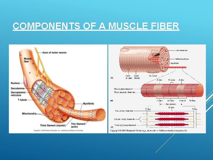
COMPONENTS OF A MUSCLE FIBER
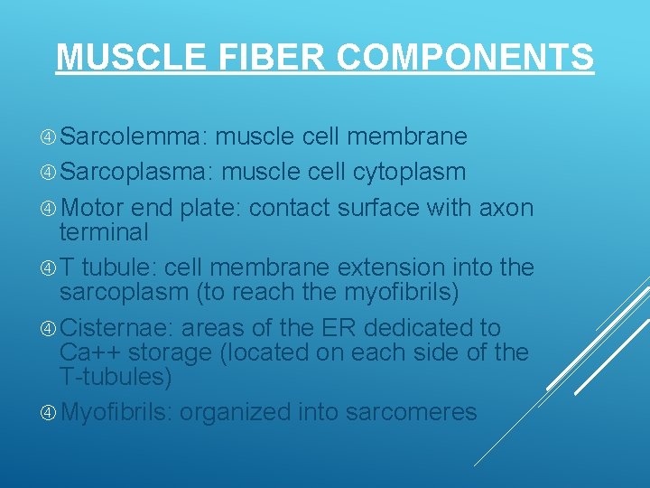
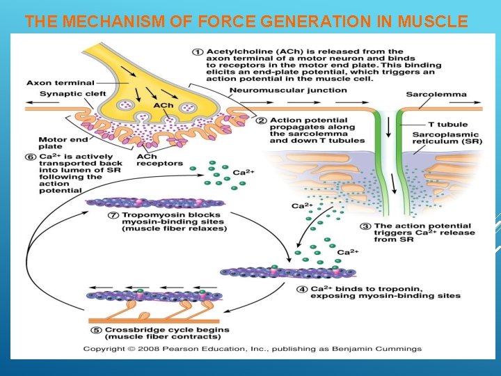
MUSCLE FIBER COMPONENTS Sarcolemma: muscle cell membrane Sarcoplasma: muscle cell cytoplasm Motor end plate: contact surface with axon terminal T tubule: cell membrane extension into the sarcoplasm (to reach the myofibrils) Cisternae: areas of the ER dedicated to Ca++ storage (located on each side of the T-tubules) Myofibrils: organized into sarcomeres
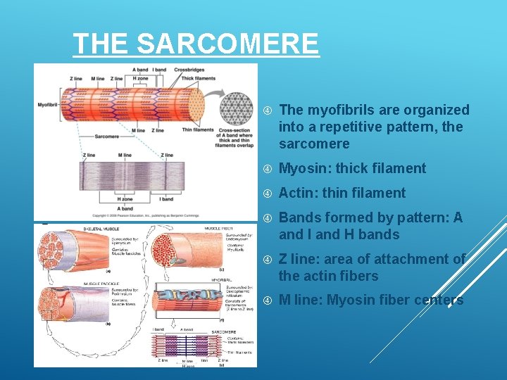
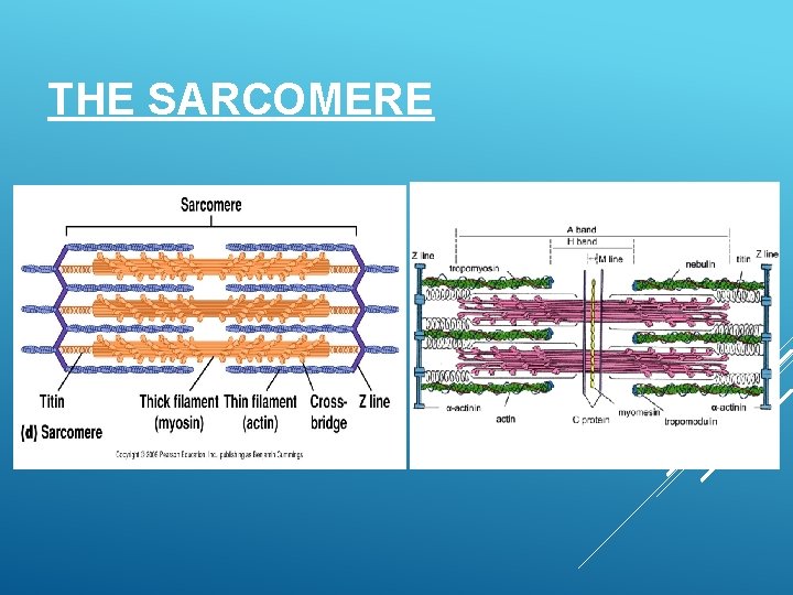
THE SARCOMERE The myofibrils are organized into a repetitive pattern, the sarcomere Myosin: thick filament Actin: thin filament Bands formed by pattern: A and I and H bands Z line: area of attachment of the actin fibers M line: Myosin fiber centers
THE SARCOMERE
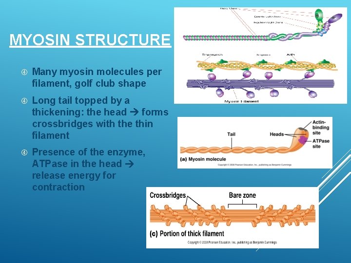
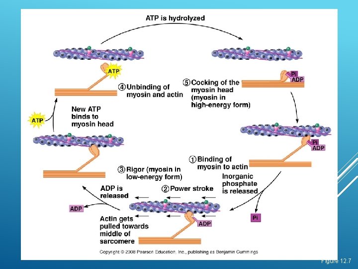
MYOSIN STRUCTURE Many myosin molecules per filament, golf club shape Long tail topped by a thickening: the head forms crossbridges with the thin filament Presence of the enzyme, ATPase in the head release energy for contraction
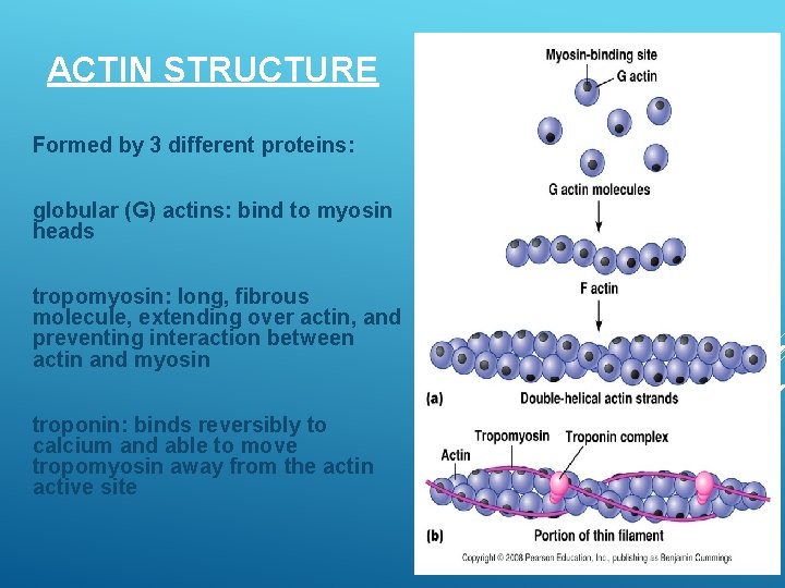
ACTIN STRUCTURE Formed by 3 different proteins: globular (G) actins: bind to myosin heads tropomyosin: long, fibrous molecule, extending over actin, and preventing interaction between actin and myosin troponin: binds reversibly to calcium and able to move tropomyosin away from the actin active site
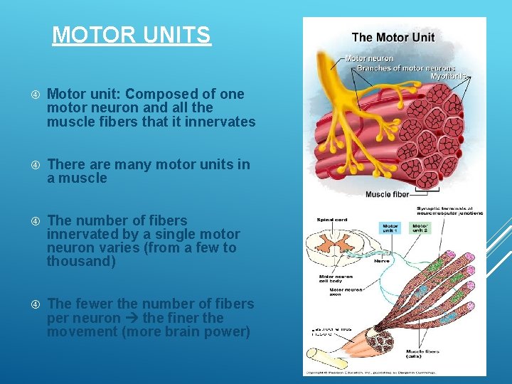
MOTOR UNITS Motor unit: Composed of one motor neuron and all the muscle fibers that it innervates There are many motor units in a muscle The number of fibers innervated by a single motor neuron varies (from a few to thousand) The fewer the number of fibers per neuron the finer the movement (more brain power)
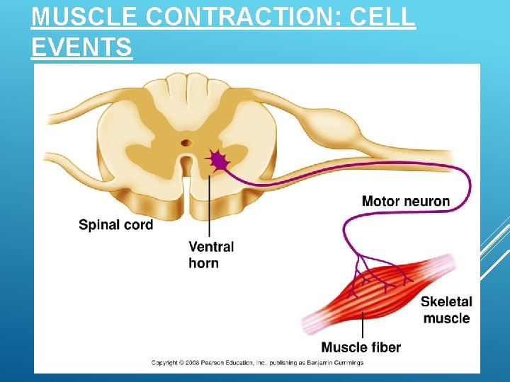
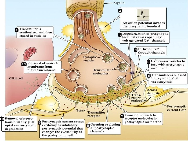
MUSCLE CONTRACTION: CELL EVENTS
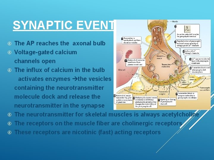
SYNAPTIC EVENTS The AP reaches the axonal bulb Voltage-gated calcium channels open The influx of calcium in the bulb activates enzymes the vesicles containing the neurotransmitter molecule dock and release the neurotransmitter in the synapse The neurotransmitter for skeletal muscles is always acetylcholine The receptors on the muscle fiber are cholinergic receptors These receptors are nicotinic (fast) acting receptors
THE MECHANISM OF FORCE GENERATION IN MUSCLE
Figure 12. 7
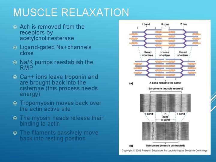
MUSCLE RELAXATION Ach is removed from the receptors by acetylcholinesterase Ligand-gated Na+channels close Na/K pumps reestablish the RMP Ca++ ions leave troponin and are brought back into the cisternae (this process needs energy) Tropomyosin moves back over the actin active site The myosin heads release their binding to actin The filaments passively move back into resting position
Myopathies are disorders in which there is a primary functional or structural impairment of skeletal muscle. The myopathies are subdivided into acquired and hereditary disorders
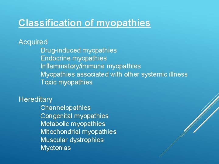
Classification of myopathies Acquired Drug-induced myopathies Endocrine myopathies Inflammatory/immune myopathies Myopathies associated with other systemic illness Toxic myopathies Hereditary Channelopathies Congenital myopathies Metabolic myopathies Mitochondrial myopathies Muscular dystrophies Myotonias
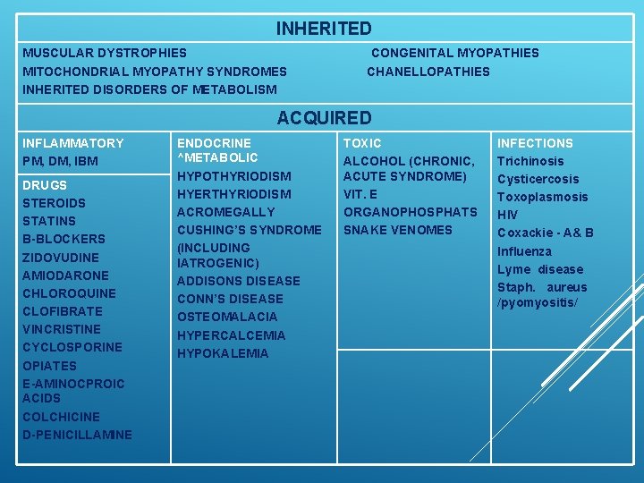
INHERITED MUSCULAR DYSTROPHIES MITOCHONDRIAL MYOPATHY SYNDROMES INHERITED DISORDERS OF METABOLISM CONGENITAL MYOPATHIES CHANELLOPATHIES ACQUIRED INFLAMMATORY PM, DM, IBM DRUGS STEROIDS STATINS B-BLOCKERS ZIDOVUDINE AMIODARONE CHLOROQUINE CLOFIBRATE VINCRISTINE CYCLOSPORINE OPIATES E-AMINOCPROIC ACIDS COLCHICINE D-PENICILLAMINE ENDOCRINE ^METABOLIC HYPOTHYRIODISM HYERTHYRIODISM ACROMEGALLY CUSHING’S SYNDROME (INCLUDING IATROGENIC) ADDISONS DISEASE CONN’S DISEASE OSTEOMALACIA HYPERCALCEMIA HYPOKALEMIA TOXIC ALCOHOL (CHRONIC, ACUTE SYNDROME) VIT. E ORGANOPHOSPHATS SNAKE VENOMES INFECTIONS Trichinosis Cysticercosis Toxoplasmosis HIV Coxackie - A& B Influenza Lyme disease Staph. aureus /pyomyositis/
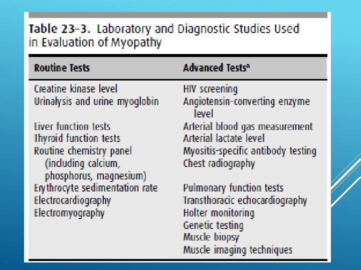
In practice, patients mainly seek neurological advice because of muscular symptoms and signs, accidentally discovered CK elevation genetic counseling.
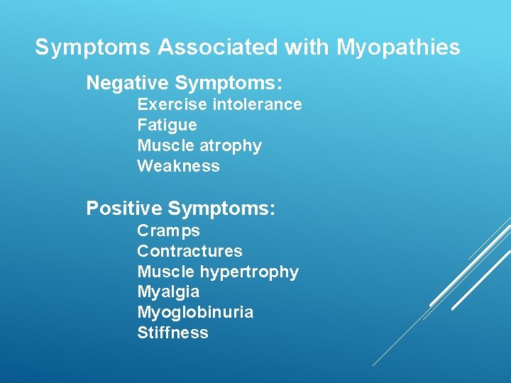
Symptoms Associated with Myopathies Negative Symptoms: Exercise intolerance Fatigue Muscle atrophy Weakness Positive Symptoms: Cramps Contractures Muscle hypertrophy Myalgia Myoglobinuria Stiffness
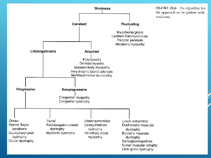
Approach to the patient with muscle weakness: The evaluation of the patient presenting with a complaint of “weakness” involves three steps: Distinguishing true muscle weakness from asthenia or motor impairment not due to loss of muscle power Localizing, within the neuromuscular system, the site of the lesion that is producing weakness Determining the cause of the lesion Asthenia - is a medical term referring to a condition in which the body lacks or has lost strength either as a whole or in any of its parts
DISTINGUISHING TRUE MUSCLE WEAKNESS FROM ASTHENIA Many patients who complain of weakness are not objectively weak when muscle strength is formally tested. A careful history and physical examination will permit the distinction History — systemic disorders like cardiopulmonary disease, joint disease, anemia, cachexia from malignancy or chronic infectious or inflammatory disease, and/or depression. Patients with any of these conditions may be functionally limited but not truly weak - patient is limited by shortness of breath, chest pain, joint pain, fatigue, poor exercise tolerance, paresthesias, or spasticity rather than a true decrease in muscle power.
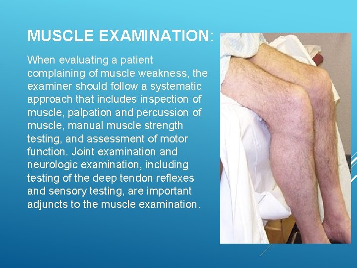
MUSCLE EXAMINATION: When evaluating a patient complaining of muscle weakness, the examiner should follow a systematic approach that includes inspection of muscle, palpation and percussion of muscle, manual muscle strength testing, and assessment of motor function. Joint examination and neurologic examination, including testing of the deep tendon reflexes and sensory testing, are important adjuncts to the muscle examination.
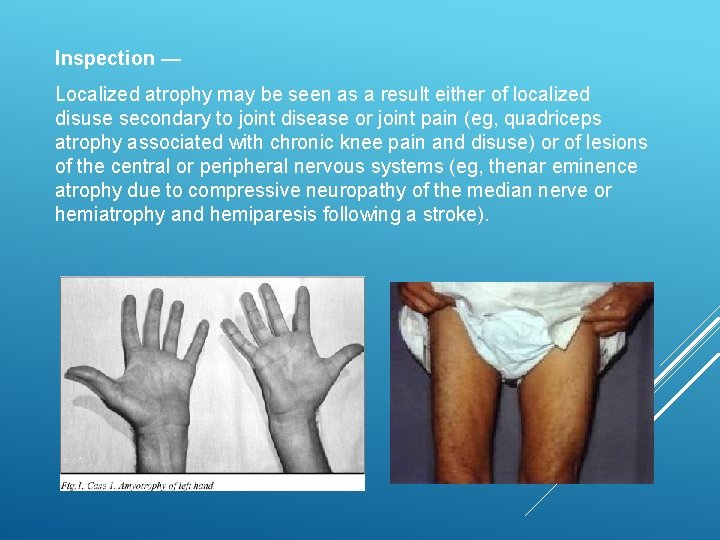
Inspection — Localized atrophy may be seen as a result either of localized disuse secondary to joint disease or joint pain (eg, quadriceps atrophy associated with chronic knee pain and disuse) or of lesions of the central or peripheral nervous systems (eg, thenar eminence atrophy due to compressive neuropathy of the median nerve or hemiatrophy and hemiparesis following a stroke).
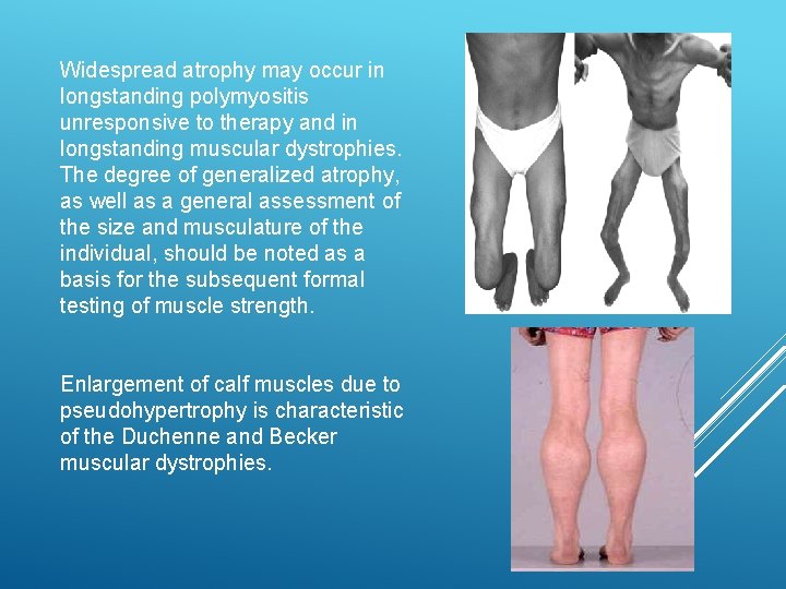
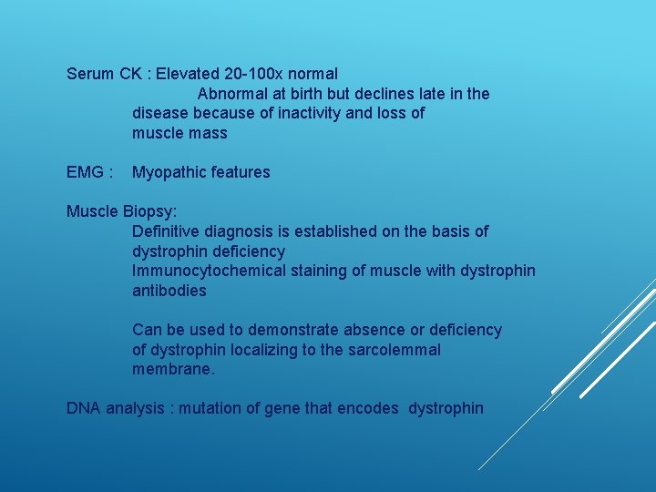
Widespread atrophy may occur in longstanding polymyositis unresponsive to therapy and in longstanding muscular dystrophies. The degree of generalized atrophy, as well as a general assessment of the size and musculature of the individual, should be noted as a basis for the subsequent formal testing of muscle strength. Enlargement of calf muscles due to pseudohypertrophy is characteristic of the Duchenne and Becker muscular dystrophies.
Palpation and percussion: Muscle tenderness suggests an infectious etiology or a metabolic myopathy such as Mc. Ardle’s disease. In comparison, tenderness is an unusual finding in the idiopathic inflammatory myopathies such as polymyositis or dermatomyositis. Increased muscle tone or rigidity is present in Parkinson disease, Increased tone also occurs in upper motor neuron lesions Myotonia describes abnormal lack of relaxation after muscle contraction and is typical of myotonic dystrophy.
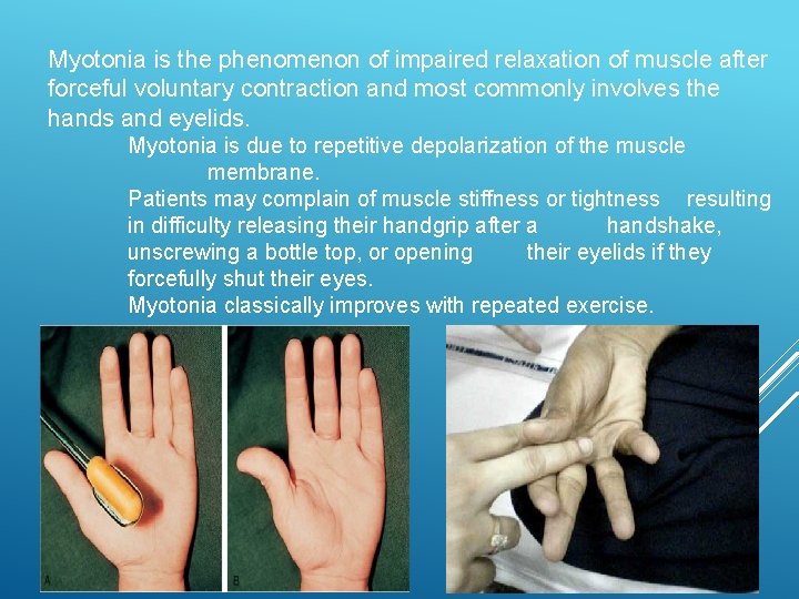
Myotonia is the phenomenon of impaired relaxation of muscle after forceful voluntary contraction and most commonly involves the hands and eyelids. Myotonia is due to repetitive depolarization of the muscle membrane. Patients may complain of muscle stiffness or tightness resulting in difficulty releasing their handgrip after a handshake, unscrewing a bottle top, or opening their eyelids if they forcefully shut their eyes. Myotonia classically improves with repeated exercise.
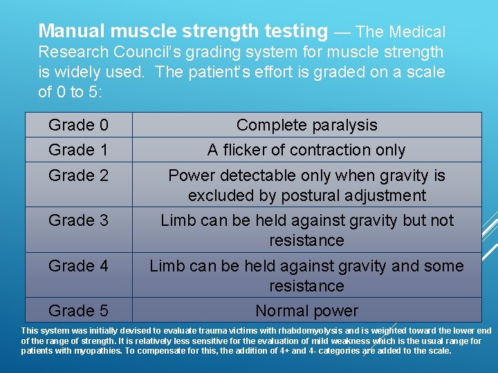
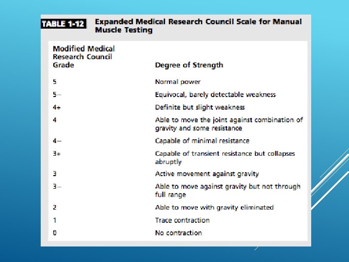
Manual muscle strength testing — The Medical Research Council’s grading system for muscle strength is widely used. The patient’s effort is graded on a scale of 0 to 5: Grade 0 Complete paralysis Grade 1 A flicker of contraction only Grade 2 Power detectable only when gravity is excluded by postural adjustment Grade 3 Limb can be held against gravity but not resistance Grade 4 Limb can be held against gravity and some resistance Grade 5 Normal power This system was initially devised to evaluate trauma victims with rhabdomyolysis and is weighted toward the lower end of the range of strength. It is relatively less sensitive for the evaluation of mild weakness which is the usual range for patients with myopathies. To compensate for this, the addition of 4+ and 4 - categories are added to the scale.
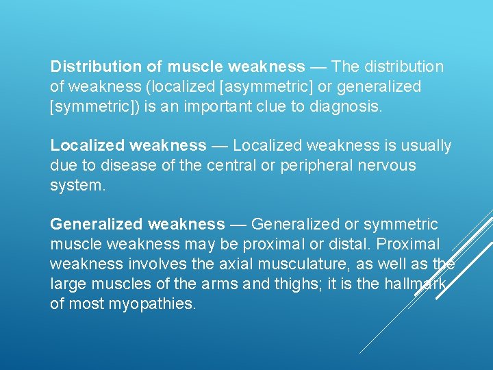
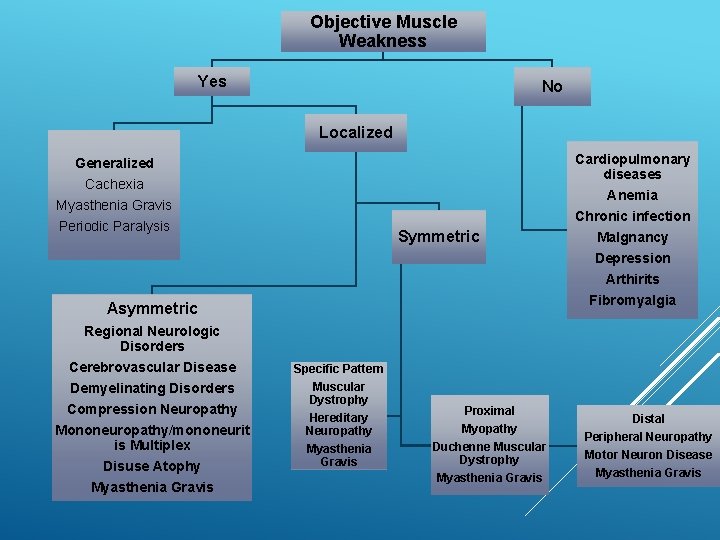
Distribution of muscle weakness — The distribution of weakness (localized [asymmetric] or generalized [symmetric]) is an important clue to diagnosis. Localized weakness — Localized weakness is usually due to disease of the central or peripheral nervous system. Generalized weakness — Generalized or symmetric muscle weakness may be proximal or distal. Proximal weakness involves the axial musculature, as well as the large muscles of the arms and thighs; it is the hallmark of most myopathies.
Objective Muscle Weakness Yes No Localized Generalized Cachexia Myasthenia Gravis Periodic Paralysis Symmetric Arthirits Fibromyalgia Asymmetric Regional Neurologic Disorders Cerebrovascular Disease Demyelinating Disorders Compression Neuropathy Mononeuropathy/mononeurit is Multiplex Disuse Atophy Myasthenia Gravis Cardiopulmonary diseases Anemia Chronic infection Malgnancy Depression Specific Pattern Muscular Dystrophy Hereditary Neuropathy Myasthenia Gravis Proximal Myopathy Duchenne Muscular Dystrophy Myasthenia Gravis Distal Peripheral Neuropathy Motor Neuron Disease Myasthenia Gravis
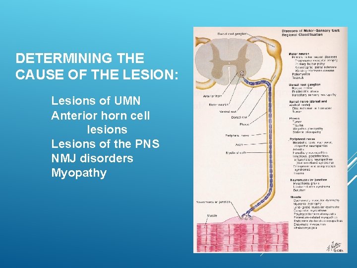
DETERMINING THE CAUSE OF THE LESION: Lesions of UMN Anterior horn cell lesions Lesions of the PNS NMJ disorders Myopathy
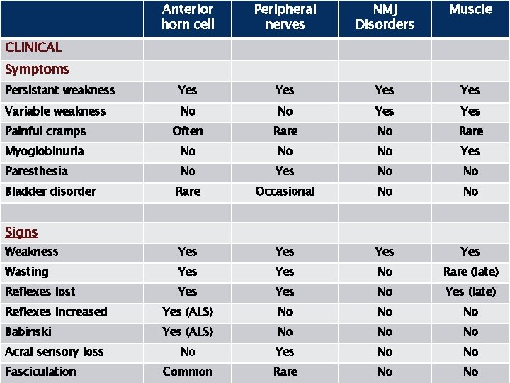
Anterior horn cell Peripheral nerves NMJ Disorders Muscle Persistant weakness Yes Yes Variable weakness No No Yes Painful cramps Often Rare No Rare Myoglobinuria No No No Yes Paresthesia No Yes No No Rare Occasional No No Weakness Yes Yes Wasting Yes No Rare (late) Reflexes lost Yes No Yes (late) Reflexes increased Yes (ALS) No No No Babinski Yes (ALS) No No Yes No No Common Rare No No CLINICAL Symptoms Bladder disorder Signs Acral sensory loss Fasciculation
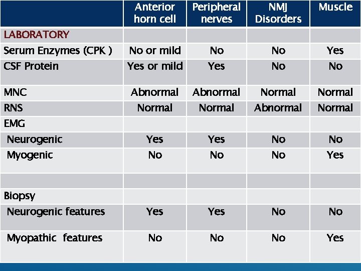
Anterior horn cell Peripheral nerves NMJ Disorders Muscle Serum Enzymes (CPK ) No or mild No No Yes CSF Protein Yes or mild Yes No No MNC Abnormal Normal RNS Normal Abnormal Neurogenic Yes No No Myogenic No No No Yes Neurogenic features Yes No No Myopathic features No No No Yes LABORATORY EMG Biopsy
Which negative and/or positive symptoms and signs does the patient demonstrate? What is the distribution of weakness? Family history Are associated systemic symptoms or signs present? History What is the temporal evolution? Are there precipitating factors that trigger episodic weakness or myotonia?
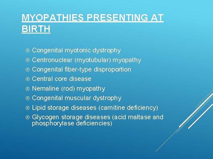
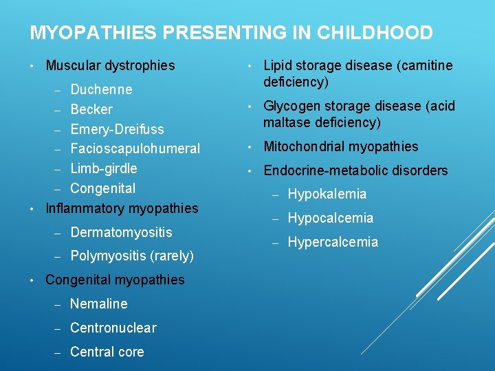
MYOPATHIES PRESENTING AT BIRTH Congenital myotonic dystrophy Centronuclear (myotubular) myopathy Congenital fiber-type disproportion Central core disease Nemaline (rod) myopathy Congenital muscular dystrophy Lipid storage diseases (carnitine deficiency) Glycogen storage diseases (acid maltase and phosphorylase deficiencies)
MYOPATHIES PRESENTING IN CHILDHOOD • Muscular dystrophies Duchenne – Becker – Emery-Dreifuss – Facioscapulohumeral – Limb-girdle – Congenital • Inflammatory myopathies • Lipid storage disease (carnitine deficiency) • Glycogen storage disease (acid maltase deficiency) • Mitochondrial myopathies • Endocrine-metabolic disorders – • – Dermatomyositis – Polymyositis (rarely) Congenital myopathies – Nemaline – Centronuclear – Central core – Hypokalemia – Hypocalcemia – Hypercalcemia
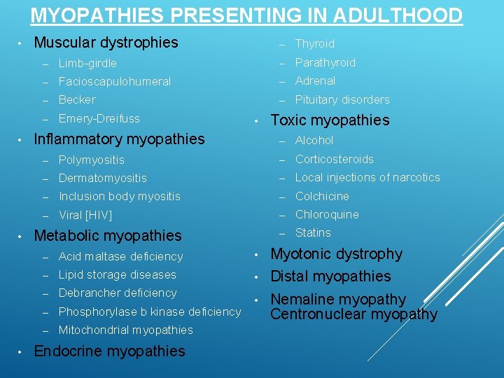
MYOPATHIES PRESENTING IN ADULTHOOD • • Muscular dystrophies – Thyroid – Limb-girdle – Parathyroid – Facioscapulohumeral – Adrenal – Becker – Pituitary disorders – Emery-Dreifuss • Inflammatory myopathies Toxic myopathies – Alcohol – Polymyositis – Corticosteroids – Dermatomyositis – Local injections of narcotics – Inclusion body myositis – Colchicine – Viral [HIV] – Chloroquine – Statins Metabolic myopathies – Acid maltase deficiency – Lipid storage diseases – Debrancher deficiency – Phosphorylase b kinase deficiency – Mitochondrial myopathies Endocrine myopathies Myotonic dystrophy • Distal myopathies • Nemaline myopathy Centronuclear myopathy •
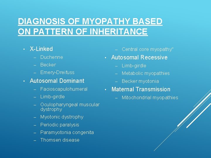
DIAGNOSIS OF MYOPATHY BASED ON PATTERN OF INHERITANCE • • X-Linked – Central core myopathy" Autosomal Recessive – Duchenne – Becker – Limb-girdle – Emery-Dreifuss – Metabolic myopathies Autosomal Dominant – Becker myotonia – Facioscapulohumeral – Limb-girdle – Oculopharyngeal muscular dystrophy – Myotonic dystrophy – Periodic paralysis – Paramyotonia congenita – Thomsen disease • • Maternal Transmission – Mitochondrial myopathies
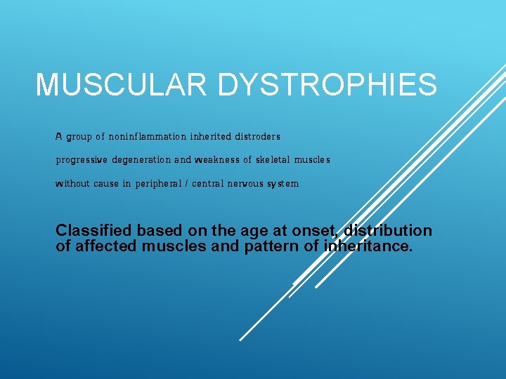
MUSCULAR DYSTROPHIES A group of noninflammation inherited distroders progressive degeneration and weakness of skeletal muscles without cause in peripheral / central nervous system Classified based on the age at onset, distribution of affected muscles and pattern of inheritance.
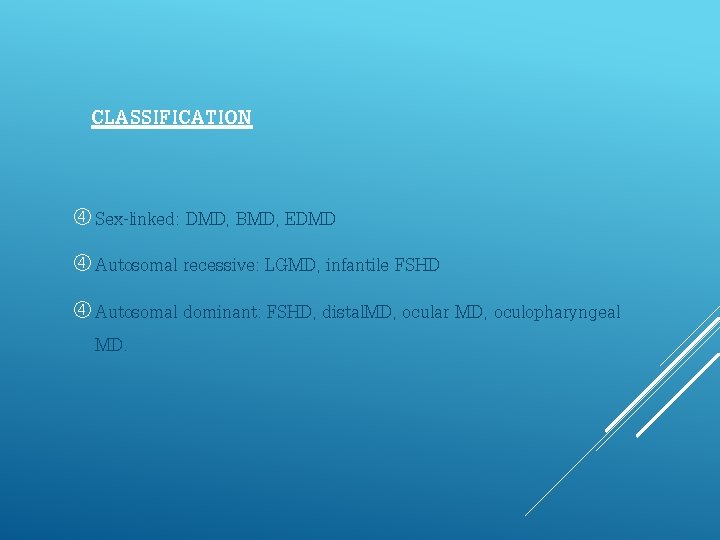
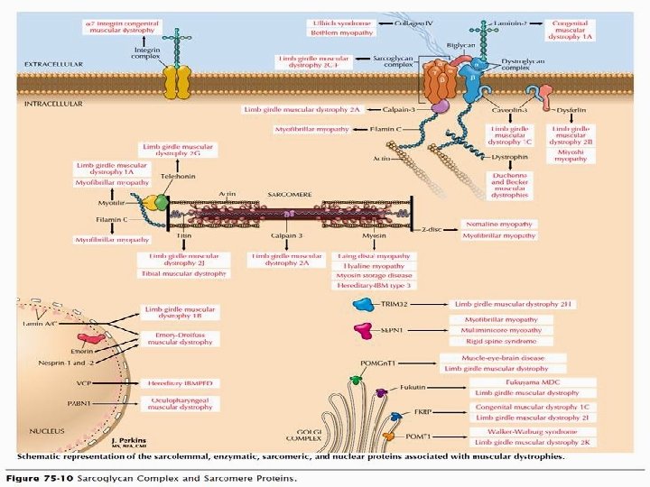
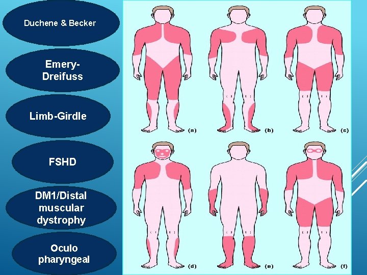
CLASSIFICATION Sex-linked: DMD, BMD, EDMD Autosomal recessive: LGMD, infantile FSHD Autosomal dominant: FSHD, distal. MD, ocular MD, oculopharyngeal MD.
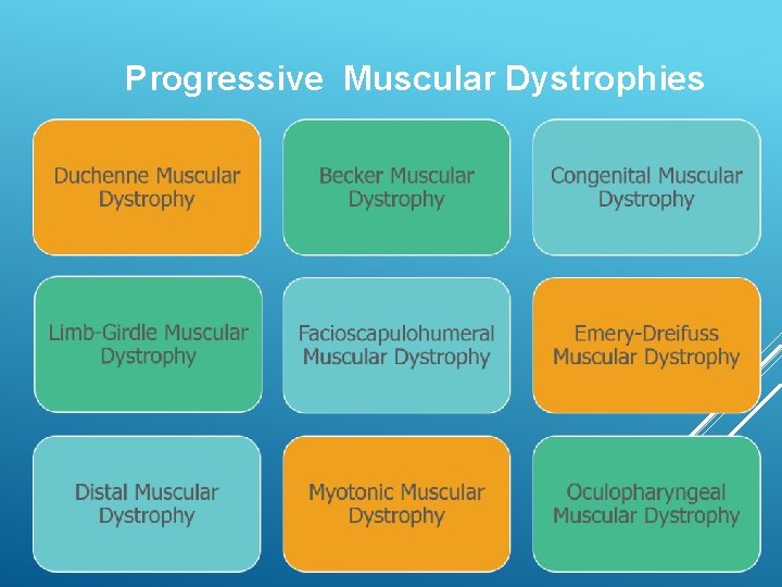
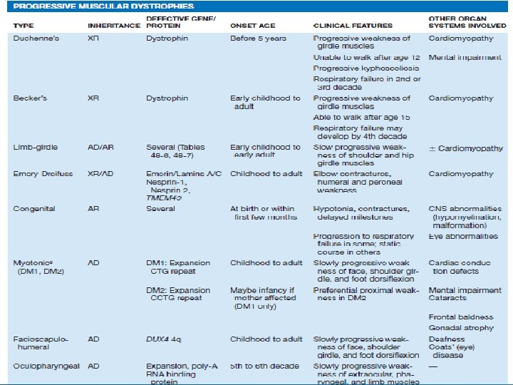
Progressive Muscular Dystrophies
Duchene & Becker Emery. Dreifuss Limb-Girdle FSHD DM 1/Distal muscular dystrophy Oculo pharyngeal
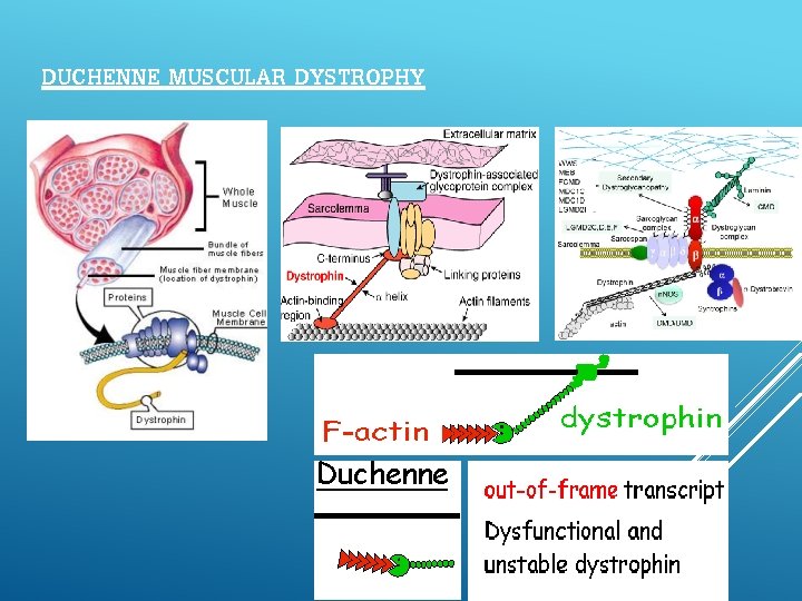
DUCHENNE MUSCULAR DYSTROPHY
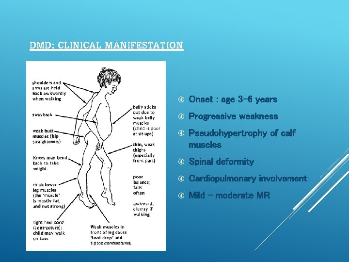
DMD: CLINICAL MANIFESTATION Onset : age 3 -6 years Progressive weakness Pseudohypertrophy of calf muscles Spinal deformity Cardiopulmonary involvement Mild - moderate MR
Serum CK : Elevated 20 -100 x normal Abnormal at birth but declines late in the disease because of inactivity and loss of muscle mass EMG : Myopathic features Muscle Biopsy: Definitive diagnosis is established on the basis of dystrophin deficiency Immunocytochemical staining of muscle with dystrophin antibodies Can be used to demonstrate absence or deficiency of dystrophin localizing to the sarcolemmal membrane. DNA analysis : mutation of gene that encodes dystrophin
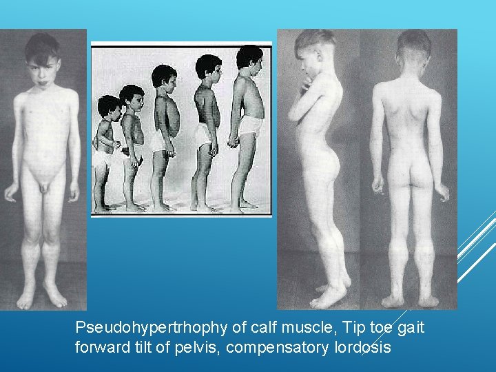
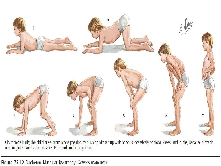
Pseudohypertrhophy of calf muscle, Tip toe gait forward tilt of pelvis, compensatory lordosis
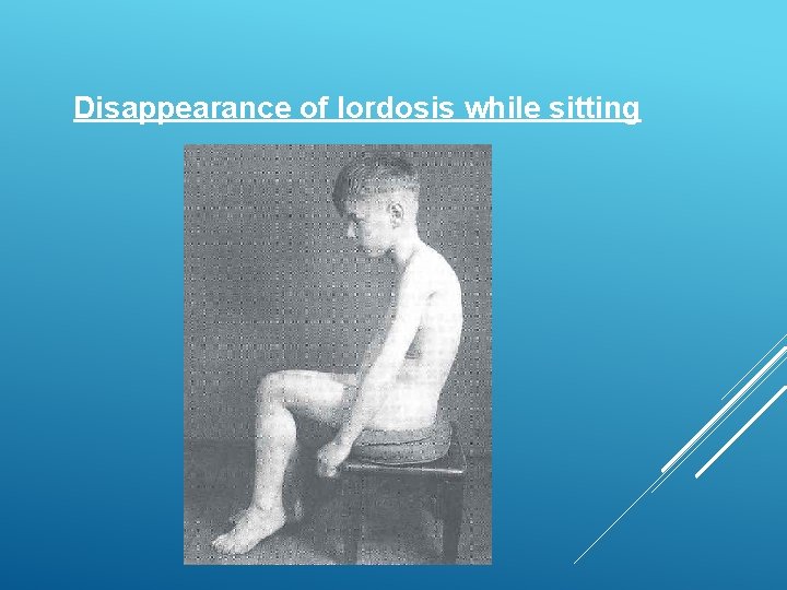
Disappearance of lordosis while sitting
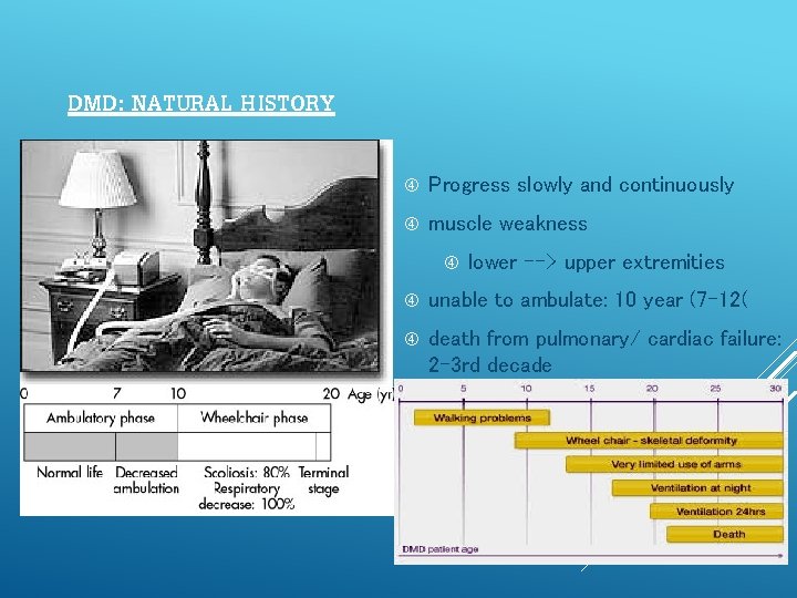
DMD: NATURAL HISTORY Progress slowly and continuously muscle weakness lower --> upper extremities unable to ambulate: 10 year (7 -12( death from pulmonary/ cardiac failure: 2 -3 rd decade
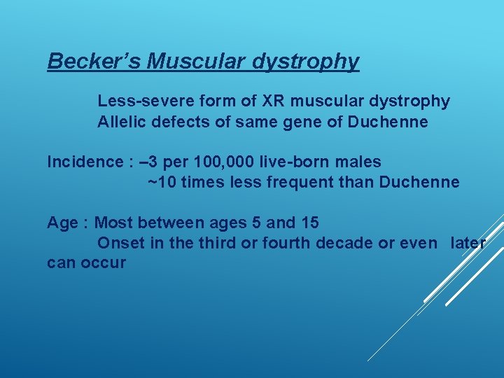
Becker’s Muscular dystrophy Less-severe form of XR muscular dystrophy Allelic defects of same gene of Duchenne Incidence : – 3 per 100, 000 live-born males ~10 times less frequent than Duchenne Age : Most between ages 5 and 15 Onset in the third or fourth decade or even later can occur
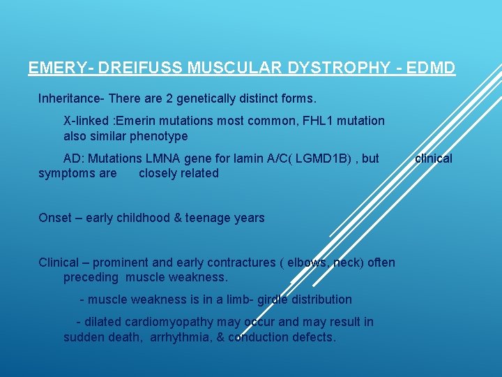
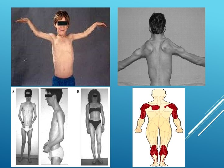
EMERY- DREIFUSS MUSCULAR DYSTROPHY - EDMD Inheritance- There are 2 genetically distinct forms. X-linked : Emerin mutations most common, FHL 1 mutation also similar phenotype AD: Mutations LMNA gene for lamin A/C( LGMD 1 B) , but symptoms are closely related Onset – early childhood & teenage years Clinical – prominent and early contractures ( elbows, neck) often preceding muscle weakness. - muscle weakness is in a limb- girdle distribution - dilated cardiomyopathy may occur and may result in sudden death, arrhythmia, & conduction defects. clinical
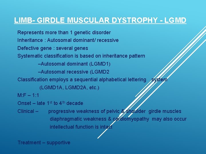
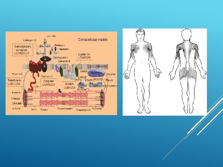
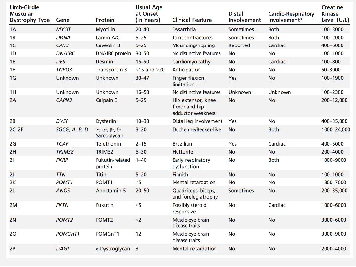
LIMB- GIRDLE MUSCULAR DYSTROPHY - LGMD Represents more than 1 genetic disorder Inheritance : Autosomal dominant/ recessive Defective gene : several genes Systematic classification is based on inheritance pattern –Autosomal dominant (LGMD 1) –Autosomal recessive (LGMD 2 Classification employs a sequential alphabetical lettering system (LGMD 1 A, LGMD 2 A, etc. ) M: F – 1: 1 Onset – late 1 st to 4 th decade Clinical – progressive weakness of pelvic & shoulder girdle muscles diaphragmatic weakness & cardiomyopathy may also occur intellectual function is intact Treatment – supportive
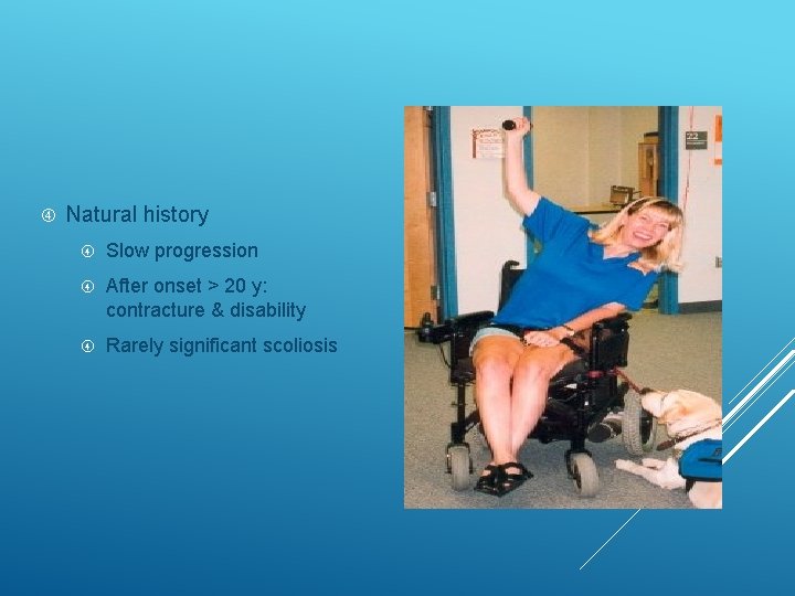
Natural history Slow progression After onset > 20 y: contracture & disability Rarely significant scoliosis
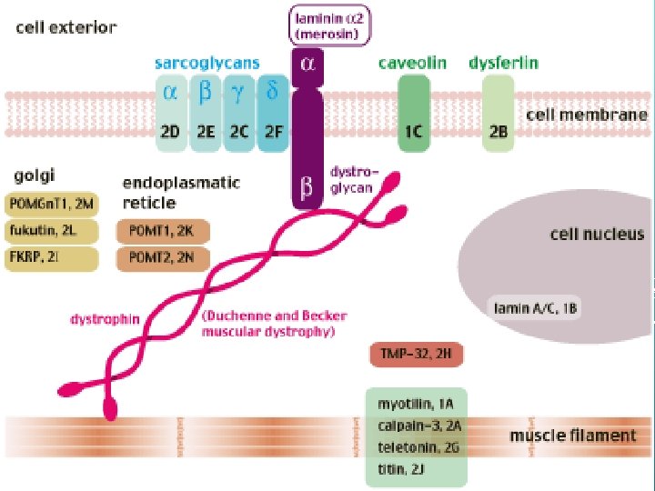
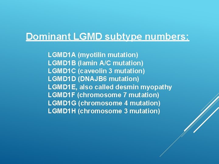
Dominant LGMD subtype numbers: LGMD 1 A (myotilin mutation) LGMD 1 B (lamin A/C mutation) LGMD 1 C (caveolin 3 mutation) LGMD 1 D (DNAJB 6 mutation) LGMD 1 E, also called desmin myopathy LGMD 1 F (chromosome 7 mutation) LGMD 1 G (chromosome 4 mutation) LGMD 1 H (chromosome 3 mutation)
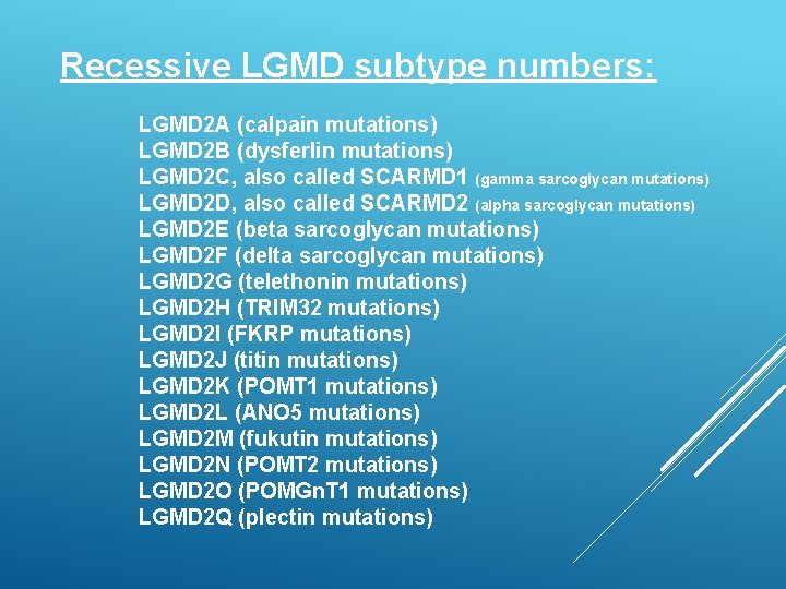
Recessive LGMD subtype numbers: LGMD 2 A (calpain mutations) LGMD 2 B (dysferlin mutations) LGMD 2 C, also called SCARMD 1 (gamma sarcoglycan mutations) LGMD 2 D, also called SCARMD 2 (alpha sarcoglycan mutations) LGMD 2 E (beta sarcoglycan mutations) LGMD 2 F (delta sarcoglycan mutations) LGMD 2 G (telethonin mutations) LGMD 2 H (TRIM 32 mutations) LGMD 2 I (FKRP mutations) LGMD 2 J (titin mutations) LGMD 2 K (POMT 1 mutations) LGMD 2 L (ANO 5 mutations) LGMD 2 M (fukutin mutations) LGMD 2 N (POMT 2 mutations) LGMD 2 O (POMGn. T 1 mutations) LGMD 2 Q (plectin mutations)
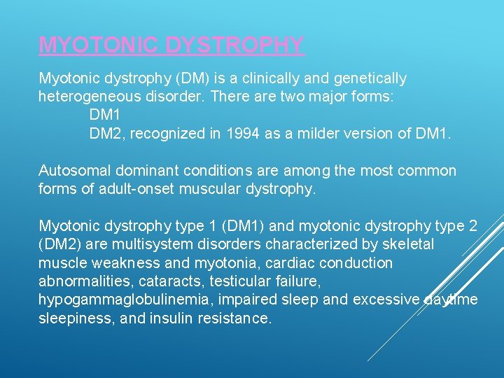
MYOTONIC DYSTROPHY Myotonic dystrophy (DM) is a clinically and genetically heterogeneous disorder. There are two major forms: DM 1 DM 2, recognized in 1994 as a milder version of DM 1. Autosomal dominant conditions are among the most common forms of adult-onset muscular dystrophy. Myotonic dystrophy type 1 (DM 1) and myotonic dystrophy type 2 (DM 2) are multisystem disorders characterized by skeletal muscle weakness and myotonia, cardiac conduction abnormalities, cataracts, testicular failure, hypogammaglobulinemia, impaired sleep and excessive daytime sleepiness, and insulin resistance.
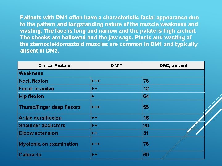
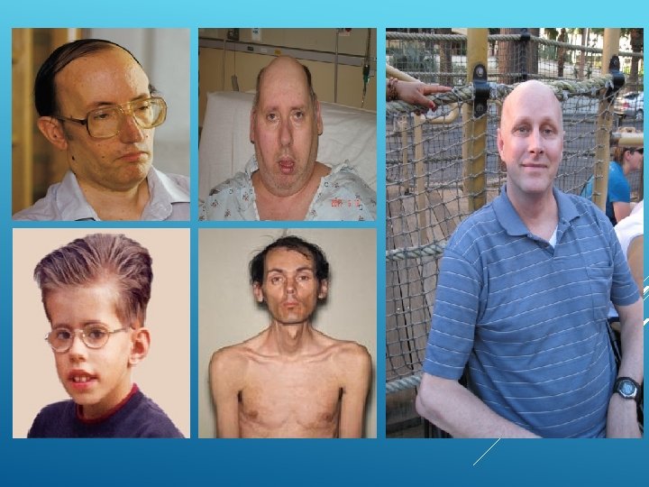
Patients with DM 1 often have a characteristic facial appearance due to the pattern and longstanding nature of the muscle weakness and wasting. The face is long and narrow and the palate is high arched. The cheeks are hollowed and the jaw sags. Ptosis and wasting of the sternocleidomastoid muscles are common in DM 1 and typically absent in DM 2. Clinical Feature DM 1* DM 2, percent Weakness Neck flexion +++ 75 Facial muscles ++ 12 Hip flexion + 64 Thumb/finger deep flexors +++ 55 Ankle dorsiflexion ++ 16 Shoulder abductors ++ 20 Elbow extension ++ 31 Myotonia on examination +++ 75 Cataracts ++ 60
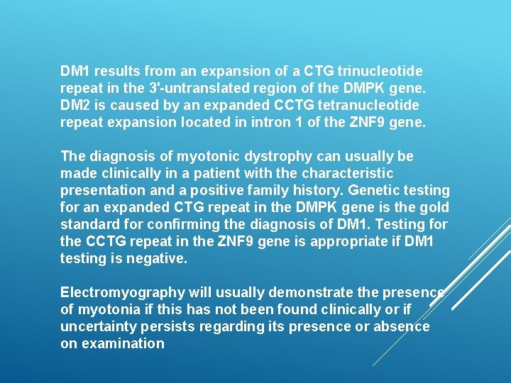
DM 1 results from an expansion of a CTG trinucleotide repeat in the 3'-untranslated region of the DMPK gene. DM 2 is caused by an expanded CCTG tetranucleotide repeat expansion located in intron 1 of the ZNF 9 gene. The diagnosis of myotonic dystrophy can usually be made clinically in a patient with the characteristic presentation and a positive family history. Genetic testing for an expanded CTG repeat in the DMPK gene is the gold standard for confirming the diagnosis of DM 1. Testing for the CCTG repeat in the ZNF 9 gene is appropriate if DM 1 testing is negative. Electromyography will usually demonstrate the presence of myotonia if this has not been found clinically or if uncertainty persists regarding its presence or absence on examination
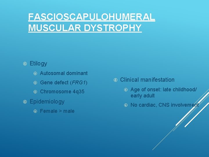
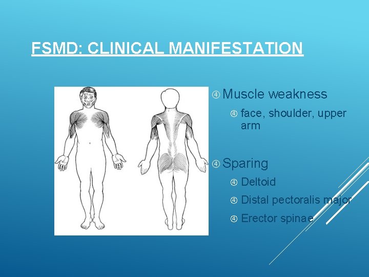
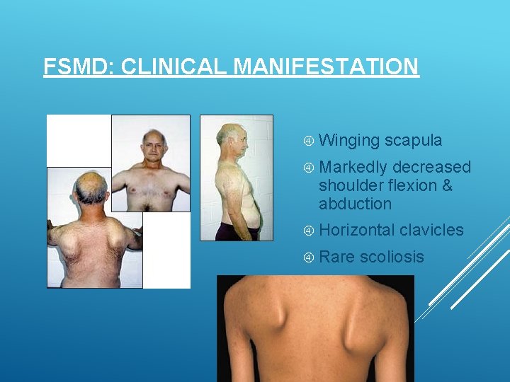
FASCIOSCAPULOHUMERAL MUSCULAR DYSTROPHY Etilogy Autosomal dominant Gene defect (FRG 1) Chromosome 4 q 35 Epidemiology Female > male Clinical manifestation Age of onset: late childhood/ early adult No cardiac, CNS involvement
FSMD: CLINICAL MANIFESTATION Muscle weakness face, shoulder, upper arm Sparing Deltoid Distal pectoralis major Erector spinae
FSMD: CLINICAL MANIFESTATION Winging scapula Markedly decreased shoulder flexion & abduction Horizontal Rare clavicles scoliosis
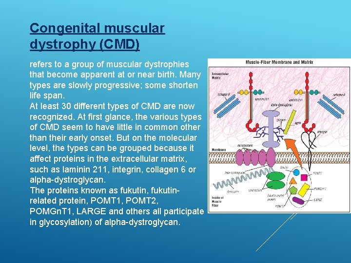
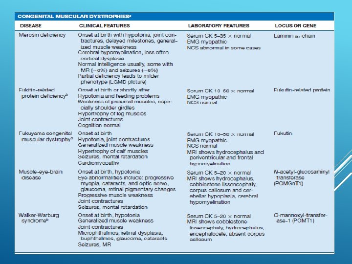
Congenital muscular dystrophy (CMD) refers to a group of muscular dystrophies that become apparent at or near birth. Many types are slowly progressive; some shorten life span. At least 30 different types of CMD are now recognized. At first glance, the various types of CMD seem to have little in common other than their early onset. But on the molecular level, the types can be grouped because it affect proteins in the extracellular matrix, such as laminin 211, integrin, collagen 6 or alpha-dystroglycan. The proteins known as fukutin, fukutinrelated protein, POMT 1, POMT 2, POMGn. T 1, LARGE and others all participate in glycosylation) of alpha-dystroglycan.
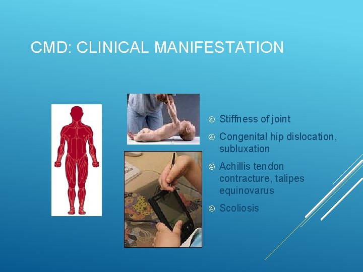
CMD: CLINICAL MANIFESTATION Stiffness of joint Congenital hip dislocation, subluxation Achillis tendon contracture, talipes equinovarus Scoliosis
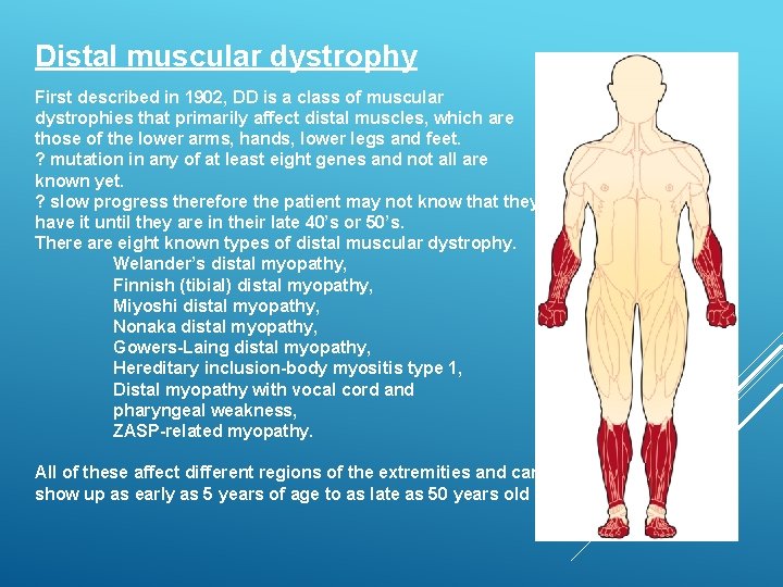
Distal muscular dystrophy First described in 1902, DD is a class of muscular dystrophies that primarily affect distal muscles, which are those of the lower arms, hands, lower legs and feet. ? mutation in any of at least eight genes and not all are known yet. ? slow progress therefore the patient may not know that they have it until they are in their late 40’s or 50’s. There are eight known types of distal muscular dystrophy. Welander’s distal myopathy, Finnish (tibial) distal myopathy, Miyoshi distal myopathy, Nonaka distal myopathy, Gowers-Laing distal myopathy, Hereditary inclusion-body myositis type 1, Distal myopathy with vocal cord and pharyngeal weakness, ZASP-related myopathy. All of these affect different regions of the extremities and can show up as early as 5 years of age to as late as 50 years old
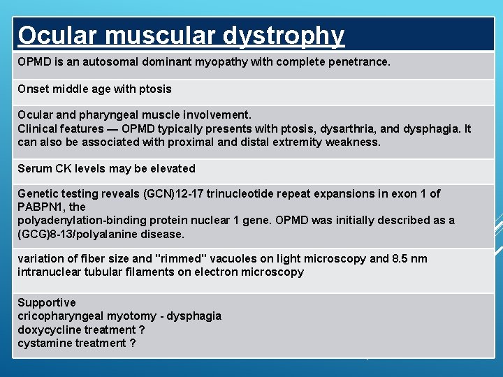
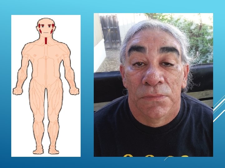
Ocular muscular dystrophy OPMD is an autosomal dominant myopathy with complete penetrance. Onset middle age with ptosis Ocular and pharyngeal muscle involvement. Clinical features — OPMD typically presents with ptosis, dysarthria, and dysphagia. It can also be associated with proximal and distal extremity weakness. Serum CK levels may be elevated Genetic testing reveals (GCN)12 -17 trinucleotide repeat expansions in exon 1 of PABPN 1, the polyadenylation-binding protein nuclear 1 gene. OPMD was initially described as a (GCG)8 -13/polyalanine disease. variation of fiber size and "rimmed" vacuoles on light microscopy and 8. 5 nm intranuclear tubular filaments on electron microscopy Supportive cricopharyngeal myotomy - dysphagia doxycycline treatment ? cystamine treatment ?
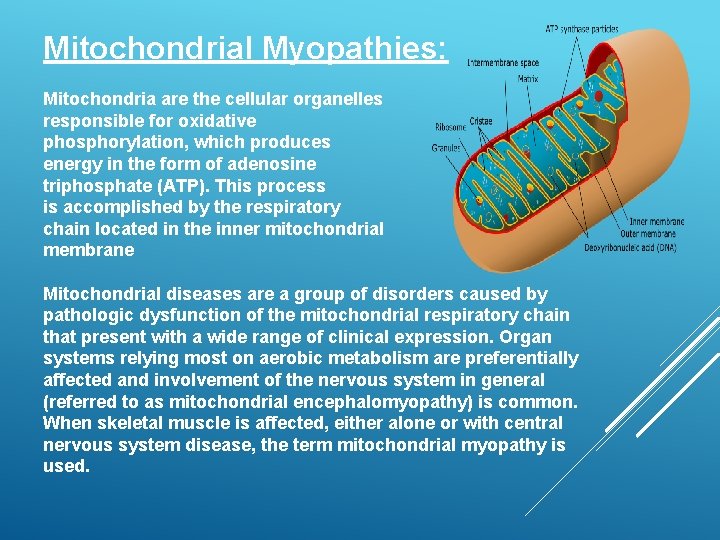
Mitochondrial Myopathies: Mitochondria are the cellular organelles responsible for oxidative phosphorylation, which produces energy in the form of adenosine triphosphate (ATP). This process is accomplished by the respiratory chain located in the inner mitochondrial membrane Mitochondrial diseases are a group of disorders caused by pathologic dysfunction of the mitochondrial respiratory chain that present with a wide range of clinical expression. Organ systems relying most on aerobic metabolism are preferentially affected and involvement of the nervous system in general (referred to as mitochondrial encephalomyopathy) is common. When skeletal muscle is affected, either alone or with central nervous system disease, the term mitochondrial myopathy is used.
Accumulating evidence suggests that mitochondrial disorders are among the most common inherited metabolic diseases. Muscle is frequently affected, although in some cases the severity is mild or subclinical The phenotypic expression of mitochondrial encephalomyopathies is extremely variable. The following groups illustrate the different ways mitochondrial myopathies can present clinically: As chronic progressive external ophthalmoplegia (with or without mild proximal muscle weakness) or Kearns. Sayre syndrome As an isolated myopathy with or without exercise intolerance ● and/or myalgia As a severe myopathy or encephalomyopathy of infancy and As a predominantly multisystem disease with myopathy (eg, MELAS and MERRF)
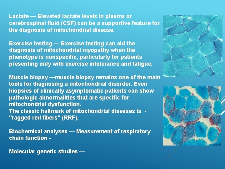
Lactate — Elevated lactate levels in plasma or cerebrospinal fluid (CSF) can be a supportive feature for the diagnosis of mitochondrial disease. Exercise testing — Exercise testing can aid the diagnosis of mitochondrial myopathy when the phenotype is nonspecific, particularly for patients presenting only with exercise intolerance and fatigue. Muscle biopsy —muscle biopsy remains one of the main tools for diagnosing a mitochondrial disorder. Even biopsies of clinically asymptomatic patients can show pathologic abnormalities that are specific for mitochondrial dysfunction. The classic hallmark of mitochondrial diseases is "ragged red fibers" (RRF). Biochemical analyses — Measurement of respiratory chain function Molecular genetic studies —
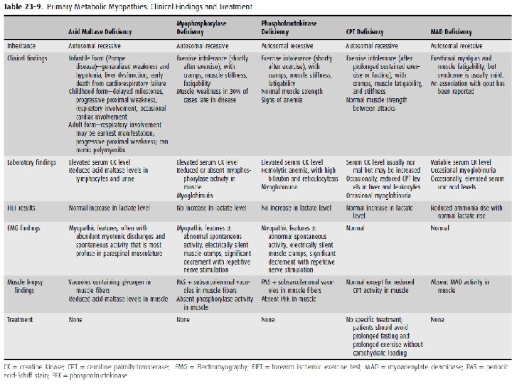
PRIMARY METABOLIC MYOPATHIES
Drug-induced myopathies Drug-induced myopathy is among the most common causes of muscle disease. It ranges from mild myalgias with or without mild weakness to chronic myopathy with severe weakness and to massive rhabdomyolysis with acute renal failure Drug-induced myopathy may result from several different mechanisms, including direct myotoxicity, immunologically induced inflammatory myopathy, indirect muscle damage, or a combination of multiple mechanisms.
Drugs that may cause direct myotoxicity include: Alcohol Cocaine Glucocorticoids Lipid-lowering drugs Antimalarials Colchicine Zidovudine Drugs that may cause an immunologically induced inflammatory myopathy include Interferon alpha D-penicillamine Indirect muscle damage — drug-induced coma with subsequent ischemic muscle compression, drug-induced hypokalemia (eg, diuretics), drug-induced hyperkinetic states (eg, delirium tremens or seizures secondary to alcohol), dystonic states associated with phenothiazines, hyperthermia related to cocaine use, and the neuroleptic malignant syndrome.
Glucocorticoid-induced myopathy Myopathy, a well-recognized side effect of glucocorticoid (corticosteroid) therapy, can occur with any of the glucocorticoid preparations. The risk may be increased in older or malnourished patients and in those with cancer. Glucocorticoid-induced myopathy can occur with the initiation of systemic therapy as well as in chronic maintenance therapy when the dose is increased. Affected patients typically present with gradual onset over several weeks of proximal muscle weakness accompanied by muscle wasting. A common manifestation is difficulty getting up from a chair or climbing stairs. Myalgias and muscle tenderness are not observed.
Glucocorticoids have a direct catabolic effect on skeletal muscle via effects on intermediary metabolism that provide amino acids as a substrate for gluconeogenesis. Activation of the glucocorticoid receptor appears to be involved, since myopathy can be prevented by a glucocorticoid receptor antagonist. An additional mechanism in critical illness was suggested in an experimental model. Glucocorticoid therapy interfered with insulin-like growth factor-I (IGF-I) signaling, leading to increased myocyte apoptosis.
Onset of weakness – dose and duration of weakness - Daily doses in excess of 40 to 60 mg/day can induce clinically important weakness within two weeks and almost always result in some degree of muscle weakness when continued for more than one month. unusual in patients treated with less than 10 mg/day of prednisone or its equivalent. Inhaled glucocorticoids are rarely associated with muscle weakness. The diagnosis of glucocorticoid-induced myopathy is one of exclusion, absence of other causes of myopathy. demonstrating improved strength within three to four weeks after appropriate dose reduction. Muscle strength begins to improve within three to four weeks after appropriate dose reduction and eventually resolves in virtually all patients if glucocorticoid therapy can be discontinued.
Statin associated adverse muscle events are variable: Myalgia: A symptom of musclediscomfort, including muscle aches, soreness, stiffness, tenderness, or cramps with or soon after exercise, with a normal creatine kinase (CK) level. Myopathy: Muscle weakness (not due to pain), with or without an elevation in CK level. Myositis: Muscle inflammation. Myonecrosis: Elevation in muscle enzymes compared with either baseline CK levels (while not on statin therapy) or the upper limit of normal that has been adjusted for age, race, and sex: Mild: Threefold to 10 fold elevation in CK Moderate: Tenfold to 50 fold elevation in CK Severe: Fiftyfold or greater elevation in CK Clinical rhabdomyolysis: myonecrosis with myoglobinuria or acute renal failure (an increase in serum creatinine of least 0. 5 mg/d. L [44 micromol/L]).
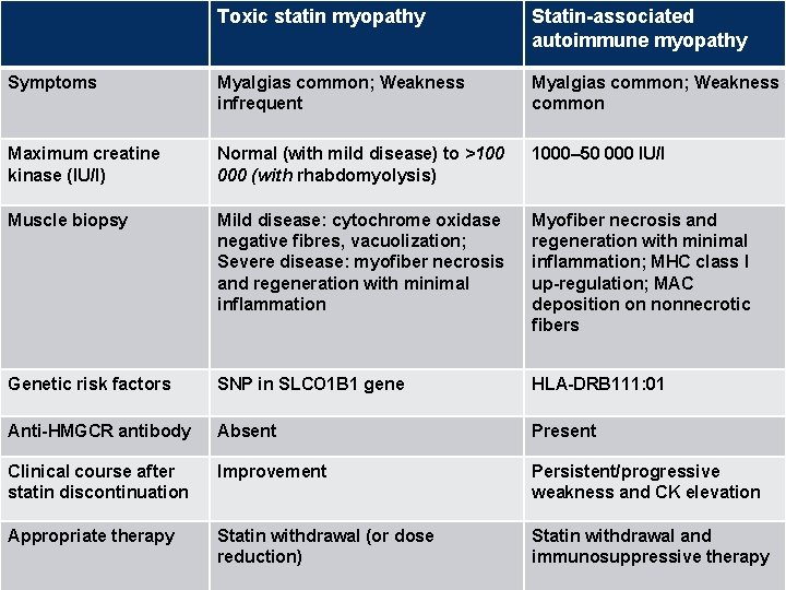
Toxic statin myopathy Statin-associated autoimmune myopathy Symptoms Myalgias common; Weakness infrequent Myalgias common; Weakness common Maximum creatine kinase (IU/l) Normal (with mild disease) to >100 000 (with rhabdomyolysis) 1000– 50 000 IU/l Muscle biopsy Mild disease: cytochrome oxidase negative fibres, vacuolization; Severe disease: myofiber necrosis and regeneration with minimal inflammation Myofiber necrosis and regeneration with minimal inflammation; MHC class I up-regulation; MAC deposition on nonnecrotic fibers Genetic risk factors SNP in SLCO 1 B 1 gene HLA-DRB 111: 01 Anti-HMGCR antibody Absent Present Clinical course after statin discontinuation Improvement Persistent/progressive weakness and CK elevation Appropriate therapy Statin withdrawal (or dose reduction) Statin withdrawal and immunosuppressive therapy
Endocrine Myopathies: Major categories of endocrine myopathy include those associated with (1) adrenal dysfunction (as in Cushing disease or steroid myopathy); (2) thyroid dysfunction (as in myxedema coma or thyrotoxic myopathy); (3) parathyroid dysfunction (as in multiple endocrine neoplasia); (4) pituitary dysfunction; (5) islands of Langerhans dysfunction (as in diabetic myopathy from ischemic infarction of the femoral muscles). Steroid myopathy is the most common endocrine myopathy
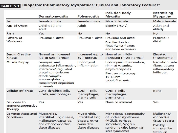
IDIOPATHIC INFLAMMATORY MYOPATHIES
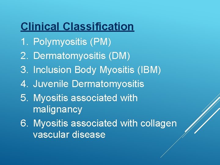
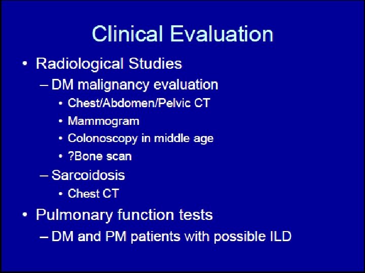
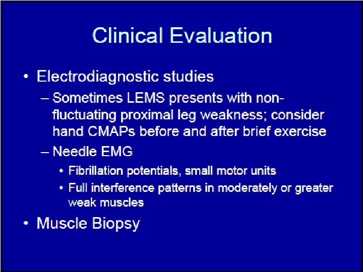
Clinical Classification 1. 2. 3. 4. 5. Polymyositis (PM) Dermatomyositis (DM) Inclusion Body Myositis (IBM) Juvenile Dermatomyositis Myositis associated with malignancy 6. Myositis associated with collagen vascular disease
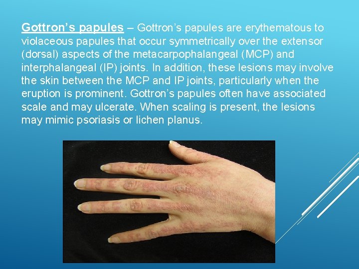
Gottron’s papules – Gottron’s papules are erythematous to violaceous papules that occur symmetrically over the extensor (dorsal) aspects of the metacarpophalangeal (MCP) and interphalangeal (IP) joints. In addition, these lesions may involve the skin between the MCP and IP joints, particularly when the eruption is prominent. Gottron’s papules often have associated scale and may ulcerate. When scaling is present, the lesions may mimic psoriasis or lichen planus.
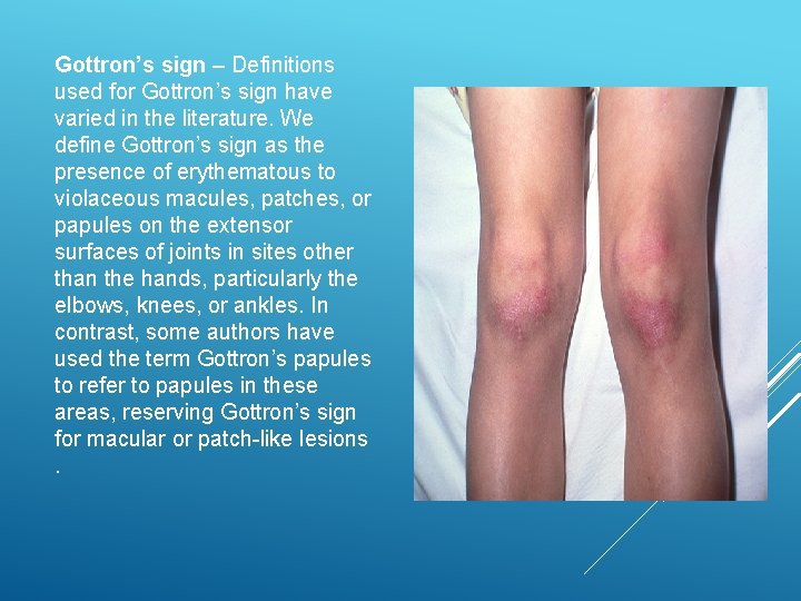
Gottron’s sign – Definitions used for Gottron’s sign have varied in the literature. We define Gottron’s sign as the presence of erythematous to violaceous macules, patches, or papules on the extensor surfaces of joints in sites other than the hands, particularly the elbows, knees, or ankles. In contrast, some authors have used the term Gottron’s papules to refer to papules in these areas, reserving Gottron’s sign for macular or patch-like lesions.
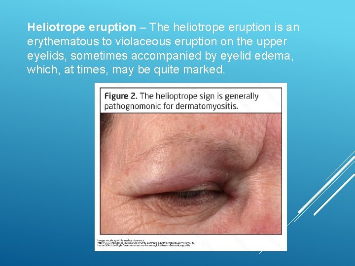
Heliotrope eruption – The heliotrope eruption is an erythematous to violaceous eruption on the upper eyelids, sometimes accompanied by eyelid edema, which, at times, may be quite marked.
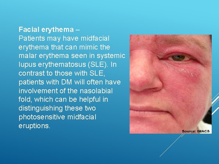
Facial erythema – Patients may have midfacial erythema that can mimic the malar erythema seen in systemic lupus erythematosus (SLE). In contrast to those with SLE, patients with DM will often have involvement of the nasolabial fold, which can be helpful in distinguishing these two photosensitive midfacial eruptions.
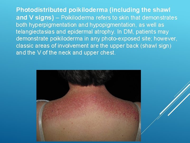
Photodistributed poikiloderma (including the shawl and V signs) – Poikiloderma refers to skin that demonstrates both hyperpigmentation and hypopigmentation, as well as telangiectasias and epidermal atrophy. In DM, patients may demonstrate poikiloderma in any photo-exposed site; however, classic areas of involvement are the upper back (shawl sign) and the V of the neck and upper chest.
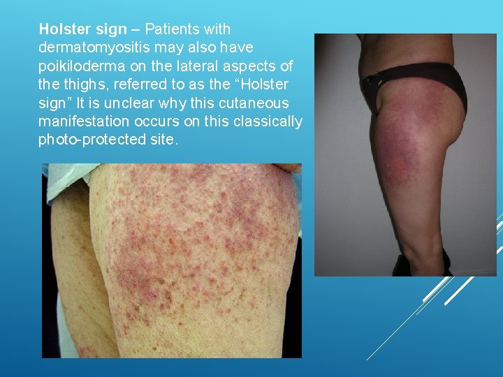
Holster sign – Patients with dermatomyositis may also have poikiloderma on the lateral aspects of the thighs, referred to as the “Holster sign” It is unclear why this cutaneous manifestation occurs on this classically photo-protected site.
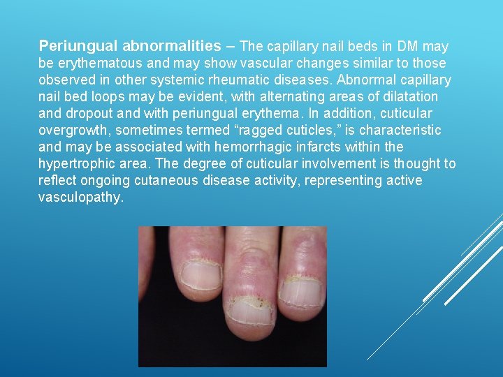
Periungual abnormalities – The capillary nail beds in DM may be erythematous and may show vascular changes similar to those observed in other systemic rheumatic diseases. Abnormal capillary nail bed loops may be evident, with alternating areas of dilatation and dropout and with periungual erythema. In addition, cuticular overgrowth, sometimes termed “ragged cuticles, ” is characteristic and may be associated with hemorrhagic infarcts within the hypertrophic area. The degree of cuticular involvement is thought to reflect ongoing cutaneous disease activity, representing active vasculopathy.
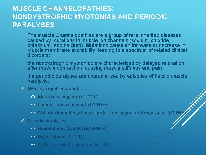
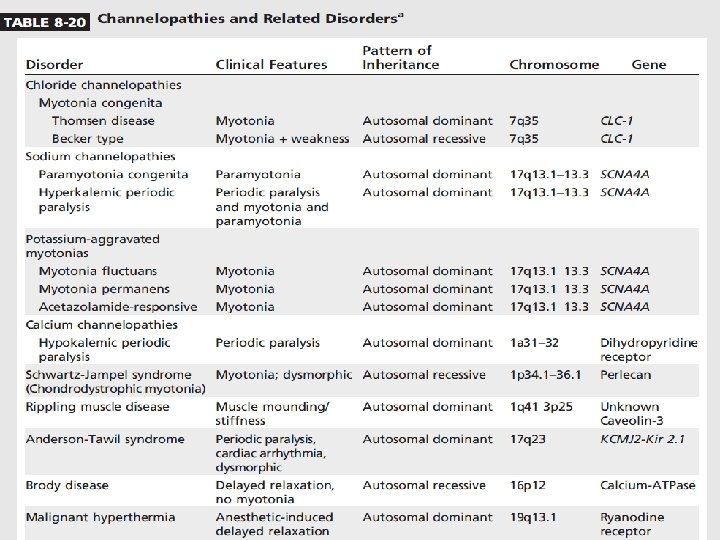
MUSCLE CHANNELOPATHIES: NONDYSTROPHIC MYOTONIAS AND PERIODIC PARALYSES The muscle Channelopathies are a group of rare inherited diseases caused by mutations in muscle ion channels (sodium, chloride, potassium, and calcium). Mutations cause an increase or decrease in muscle membrane excitability, leading to a spectrum of related clinical disorders: the nondystrophic myotonias are characterized by delayed relaxation after muscle contraction, causing muscle stiffness and pain; the periodic paralyses are characterized by episodes of flaccid muscle paralysis. Non dystrophic myotonias Myotonia congenita (CLCN 1) Paramyotonia congenita (SCN 4 A) Sodium channel myotonias (potassium aggravated myotonias) (SCN 4 A) Periodic paralyses Hypokalemic (CACNA 1 S/ SCN 4 A) Hyperkalemic (SCN 4 A) Anderson Tawil syndrome (KCNJ 2)
Flaccid paralysis – mild focal weakness to severe generalized weakness Occur anytime of the day; more common in morning Absence of myotonia Proximal > distal weakness; legs > arms Sparing of facial, ventilatory and sphincter muscles Lasts several hours to more than a day CLINICAL FEATURES
Frequency: highly variable Frequency decreases after age 30; may become attack free in 40 s and 50 s Permanent fixed weakness or slowly progressive weakness more common with Hypo. KPP 1 Attacks may be preceded by sensation of heaviness and or aching in the low back
Strenuous physical activity followed by rest or sleep High carb diet ETOH consumption Emotional stress Concurrent viral illness Lack of sleep Medications like beta agonists, corticosteroids, and insulin PRECIPITATING FACTORS
Serum K < 3. 0 m. Eq/L Serum CK level elevated EKG changes – U waves, flattening of T waves Provocative testing - Intravenous glucose load/ insulin Electrophysiology Sensory and motor NCS normal between attacks During attacks – small CMAP. Reduced insertional activity, DIAGNOSTIC fibs and positive STUDIES sharp waves No myotonia on EMG Short/ long exercise test
Reducing exposure to known triggers Acute treatment – replacement of K Acetazolamide – prevent attack recurrence and severity Acetazolamide may ppt weakness in Hypo. KPP 2 Dichlorphenamide – no longer available Triamterene and spironolactone TREATMENT