Myology Shape and structure of the muscle 1


Tendinous sheath 腱鞘 : 包围在肌腱外的鞘管,存在于腕、 踝、手指和足指处。 fibrous layer 纤维层、 Synovial layer 滑膜层 或 synovial")


 枕额 肌 Frontal")

 masticatory muscles 咀 nmasseter 咬 肌 : Origin-inferior border and medial surface of")
 masticatory muscles咀 nmedial pterygoid 翼内肌 : n lateral pterygoid 翼外肌 : action: medial")

颈浅层肌和颈外侧肌 Platysma 颈阔肌 n Sternocleidomastoid 胸锁乳突肌 Origins: manubrium and medial 1/3 of clavicle Insertion:")
颈前肌 舌骨上肌群 digastric二腹肌 Anterior belly 前腹 Posterior belly 后腹 Mylohyoid 下颌舌骨肌 Stylohyoid")

The 1、lateral goup scalenus anterior 前斜角肌 scalenus medius 中斜角肌 scalenus posterior 后斜角肌 nscalene")


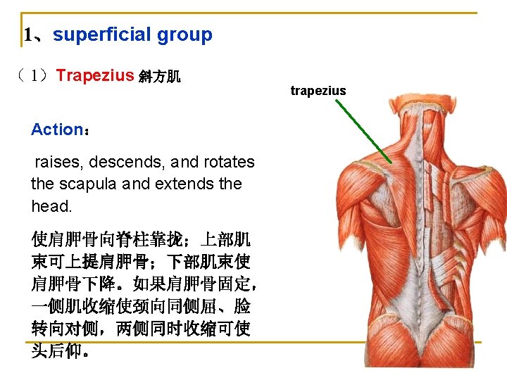
Trapezius 斜方肌 a large, triangular m. lying on the back of")
Latissimus Dorsi 背阔肌 It is a largest and triangular m. lying the")
levator scapulae 肩胛提肌 origin:the transverse processes of the upper 4 cervical vertebrae;")


 Position and division: dome-shaped between thorax and abdomen, consists of")
 Openings of diaphragm ① Aortic hiatus 主动脉裂孔 inferior vena cava at the")
 Action n. Contraction: the dome moving downward, increases the volume of thoracic cavity")
 Weak areas: triangular spaces without muscular tissue n Lumbocostal triangle 腰肋三角 between costal")


 腹股沟镰or 联合腱: is a common aponeurosis")


- Slides: 62
Myology 肌学
Ⅰ. Shape and structure of the muscle 肌的形态和构造 1. Classification long m. 长肌 short m. 短肌 flat m. 扁(阔)肌 orbicular m. 轮匝肌
1. Classification Acording the position of the muscles may be divided into M. of head 头肌 M. of neck 颈肌 M. of trunk 躯干肌 m. of back 背肌 m. of thorax胸肌 m. of abdomen 腹肌 diaphragm 膈 M. of limbs四肢肌
2. Structure muscle belly 肌腹 tendon 肌腱: 二腹肌 aponeurosis 腱膜 腱划 腱膜
Ⅲ. Name of muscles 肌的命名 Location of the muscle. Shape of the muscle. Size of the muscle Direction of muscle
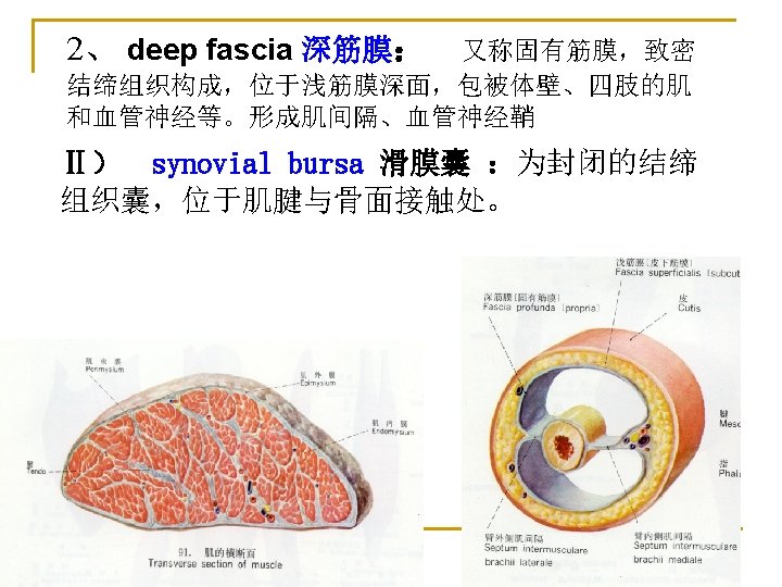
Ⅲ)Tendinous sheath 腱鞘 : 包围在肌腱外的鞘管,存在于腕、 踝、手指和足指处。 fibrous layer 纤维层、 Synovial layer 滑膜层 或 synovial sheath of tendon 腱滑膜鞘 Viscerasl layer 脏层 Parietal layer 壁 层 腱系膜
Section 2 The muscles of head and neck 头肌和颈肌
Ⅰ. The muscles of head 头肌
一、Muscles of head Facial muscles 面肌: includ Epicranius 颅顶肌 (or occipitofrontalis) 枕额 肌 Frontal belly 额腹 Occipital belly 枕腹 Galea aponeurotica 帽状 腱膜 Orbicularis oculi 眼轮匝肌 Buccinator 颊肌 Orbicularis oris 口轮匝肌 Nasalis 鼻肌
Frontal belly of occitofrontalis orbicularis oculi Levator anguli oris Levator labii superioris提上唇肌 提口角肌 Orbicularis oris Depressor labii inferioris降下唇肌 Depressor anguli oris 降口角肌
(二) masticatory muscles 咀 nmasseter 咬 肌 : Origin-inferior border and medial surface of zygomatic arch Insertion-lateral surface of ramus of mandible and angle of mandible ntemporalis 颞 肌: Origin-temporal fossa Insertion-coronoid process of mandible action: masseter, temporalis --- elevates mandible— close the mouth 嚼 肌
(二) masticatory muscles咀 nmedial pterygoid 翼内肌 : n lateral pterygoid 翼外肌 : action: medial pterygoid --- elevates mandible—close the mouth lateral pterygoid :pull the head of mandible forwards--- open the mouth 嚼 肌
Ⅱ. Muscles of neck 颈肌
(一)颈浅层肌和颈外侧肌 Platysma 颈阔肌 n Sternocleidomastoid 胸锁乳突肌 Origins: manubrium and medial 1/3 of clavicle Insertion: mastoid process of temporal bone; Actions: acting alone, the head is inclined ipsilateral and the face is rotated to opposite side; acting together, they extend the head.
1、suprahyoid muscles (二)颈前肌 舌骨上肌群 digastric二腹肌 Anterior belly 前腹 Posterior belly 后腹 Mylohyoid 下颌舌骨肌 Stylohyoid 茎突舌骨肌 Geniohyoid 颏舌骨肌 Action: elevate (raise) hyoid bone and depress mandible.
2、infrahyoid muscles 舌骨下肌群 sternohyoid 胸骨舌骨肌 omohyoid 肩胛舌骨肌 sternothyroid 胸骨甲状肌 Thyrohyoid 甲状舌骨肌 Action: depress hyoid or larynx (二)颈前肌
可分为内、外两群 (三)The 1、lateral goup scalenus anterior 前斜角肌 scalenus medius 中斜角肌 scalenus posterior 后斜角肌 nscalene fissure 斜角肌间隙: it is bounded by the scalenus anterior and medius and first rib , it contains the subclavian a and brachial plexus. 2、medial group: longus colli 颈长肌 longus capitis 头长肌 deep cervical m. 颈深部肌
section 3 The muscles of trunk 躯干肌
Ⅰ. The muscles of back 背肌
1、superficial group ( 1)Trapezius 斜方肌 a large, triangular m. lying on the back of the neck and thorax; Origin: external occipital protuberance, superior nuchal line, ligamentum nuchae, the spinous processes of 7 th cervical vertebrae and all thoracic vertebrae; Insertion: lateral 1/3 of clavicle, acromion and spine of scapula. trapezius
1、superficial group (2)Latissimus Dorsi 背阔肌 It is a largest and triangular m. lying the back and lateral wall of thorax; Origins: the spinous processes of lower 6 thoracic vertebrae and all lumbar vertebrae; Insertion: inserted into the floor of the intertubercular sulcus Action: Extend, adduct, and rotate the shoulder joint medially 当上肢上举被固定时,可 Latissimus dorsi 引体向上。 背阔肌
1、superficial group (3)levator scapulae 肩胛提肌 origin:the transverse processes of the upper 4 cervical vertebrae; insertion:superior angle of scapula. Action:raise the scapula. (4)rhomboideus 菱形肌 Lies deep to the trapezius, 可拉肩胛骨向脊柱靠拢。 Levator scapulae Rhomboideus
2、The deep group Erector Spinae 竖脊肌 position: It lies in the vertebral groove on each side of vertebral spines; Action:extend the spinal column and head 使脊柱后伸和 仰头。 Splenius 夹肌 position:位于斜方肌深面, Action: 单侧收缩,可使头转向同侧, 两侧收缩使头后伸。
Ⅱ. The muscles of thorax 胸肌
n 1. Extrinsic muscles q Pectoralis major q Pectoralis minor q Serratus anterior
Pectoralis major 胸大肌 n n n Origin: medial half of clavicle, sternum, 1 th-6 th costal cartilages. Insertion: crest of greater tubercle of humerus. Action: flexes, adducts and rotates arm medially; arm fixed, elevates trunk; elevates ribs 1 -6, aidding in forced inspiration.
Pectoralis minor 胸小肌 Deep to the pectoralis major n n n Origin: 3 rd-5 th ribs Insertion: coracoid process of scapula Action: Draw the scapula forwards and downwards, when the scapula is fixed it helps the inspiration(by elevation the ribs)
n. Serratus anterior 前锯肌 It overlies the laeral wall of thorax Origin: external surfaces of the upper 8~9 ribs Insertion: medial border of scapula Action: holds the scapula against the chest wall; Pulls the scapula forwards in throwing and pushing.
2. Intrinsic muscles Intercostales externi 肋间外肌 Origin: lower border of rib Insertion: upper border of rib below origin external intercostals membrane 肋间外膜. Action: elevate ribs adding in forced inspiration
Intercostales interni 肋间内肌 n Origin: upper border of rib; n Insertion: lower border of rib above origin Replaced posteriorly by internal intercostals membrane. n Action: depress ribs forced expiration
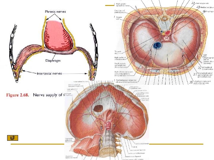
Ⅲ. Diaphragm 膈 (1) Position and division: dome-shaped between thorax and abdomen, consists of Central tendon 中心腱 muscular part 肌部 Sternal part: Costal part: Lumbar part: arises by left and right crus from upper 2 -3 lumbar vertebrae Insertion: central tendon
n (2) Openings of diaphragm ① Aortic hiatus 主动脉裂孔 inferior vena cava at the level of 12 th thoracic vertebra, the thoracic aorta and thoracic duct pass through it T 8 ② Esophageal hiatus 食管裂孔 at the level of 10 th thoracic vertebra, the esophagus and vagus n. pass throught it ③ Vena caval foramen腔静脉孔 at the level of 8 th thoracic vertebra, inferior vena cava through it esophagus thoracic aorta T 10 T 12
(3) Action n. Contraction: the dome moving downward, increases the volume of thoracic cavity which results in inspiration. n. Relaxation: the dome returns to the former position, reduces the volume of the thoracic cavity, resulting in expiration.
(2) Weak areas: triangular spaces without muscular tissue n Lumbocostal triangle 腰肋三角 between costal and lumbar parts. n Sternocostal triangle 胸肋三角: between costal and sternal parts.
Muscles of abdomen Anterolateral group n Obliquus externus abdominis 腹外斜肌 n Obliquus internus abdominis 腹内斜肌 n Transversus abdominis 腹横肌 n Rectus abdominis 腹直肌
Obliquus externus abdominis 腹外斜肌 Origin: Arises from the lower 8 ribs, and the muscular fibers run obliquely from the superolateal to the inferomedial, the anterior part of the m. change gradually into aponeurosis, which pass over the rectus Abdominis; insertion: Linea alba 白线.
Superficial inguinal ring Obliquus externus absominis 腹外斜肌 Superficial inguinal ring 腹股沟浅环 -triangular-shaped hiatus above pubic tubercle Structures formed by aponeurosis of this m. n Lacunar ligament 腔隙韧带 include: n n Inguinal ligament 腹股沟韧带 Lacunar ligament
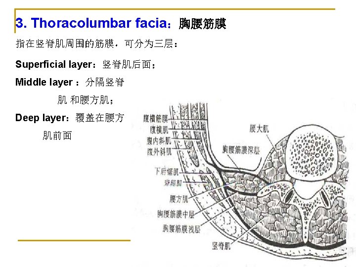
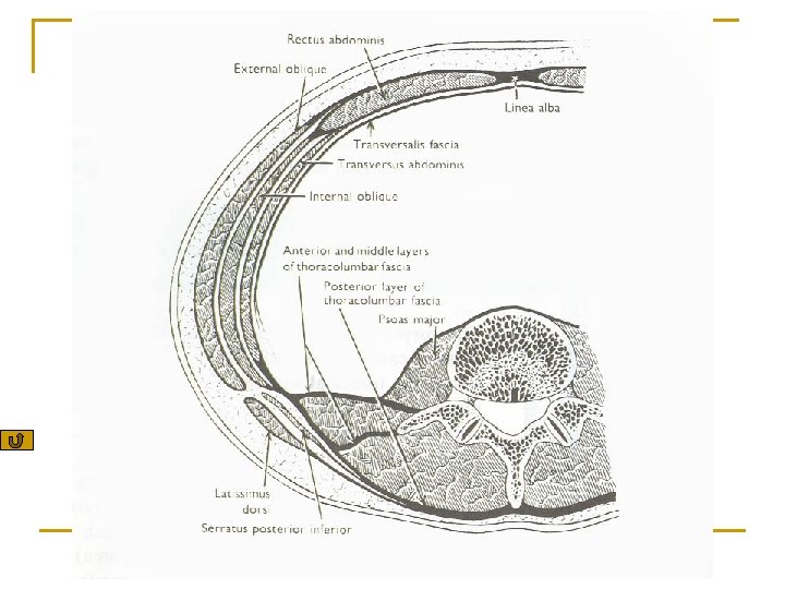
Obliquus internus abdominis 腹内斜肌 n n Origin: thoracolumbar fascia 胸腰筋膜 , iliac crest and the lateral half of the ingunal lig. Insertion: Linea alba 白线 n. The muscular fibers run upwards and forwards, and become the aponeurosis close the lateral border of the rectus abdominis. Its aponeurosis divided into two layers to inclose the rectus abdominis.
Transversels abdominis 腹横肌 is the deepest one of the three flat abdominal m. Origin: it arises from the costal cartilages of the lower 6 ribs,the thoracolumbar fascia , the iliac crest and the lateral 1/3 of the ingunal lig. , n n Insertion: Linea alba 白线 The muscular fibers run transversly , and pass deep to rectus abdominis. n
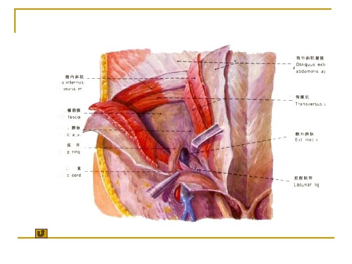
Transversels abdominis 腹横肌 n n Inguinal falx(conjoined tendon) 腹股沟镰or 联合腱: is a common aponeurosis joined by obliquus internus abdominis腹内斜肌 and transverse abdominis腹横 肌, it turns downwards to insert the pubic crest and pecten pubis耻骨梳. Cremaster 提睾肌: around the spermatic cord and testis
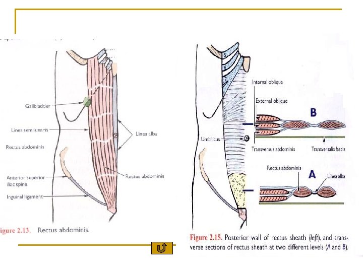
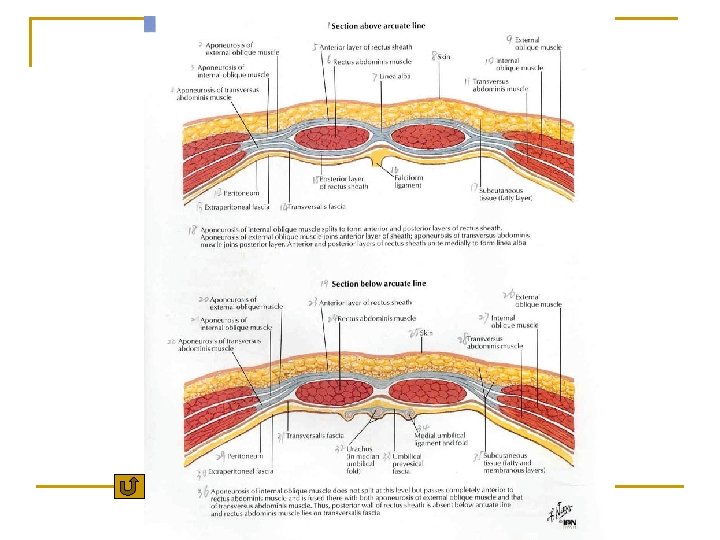
The Sheath of rectus abdominis 腹直肌鞘 Anterior layer- formed by the aponeurosis of obliquus externus abdominis ; anterior leaf of aponeurosis of obliquus internus abdominis Posterior layer:formed by posterion leaf of aponeurosis of obliquus internus abdominis ; aponeurosis of transverses abdominis
The Sheath of rectus abdominis 腹直肌鞘 n n n arcuate line 弓状线 或 semicircular line 半环线 at about 4 -5 cm below the umbilicus脐, the lower free border of the posterior layer of the sheth present arcuated. Below this line the rectus abdominis in contact with transverse fascia directly.
Linea alba 白线 -tendinous raphe between right and left rectus abdominis from xiphoid process to pubic symphysis.
The functions of the four pairs of muscles n n n Support and protect the abdominal viscera Maintain and increase intraabdominal pressure, aid in vomiting, coughing, sneezing, defecation, urination and childbirth. Flex, lateral flex, and rotate vertebral column
Posterior group n Quadratus lumborum 腰方肌 n Psoas major 腰大肌
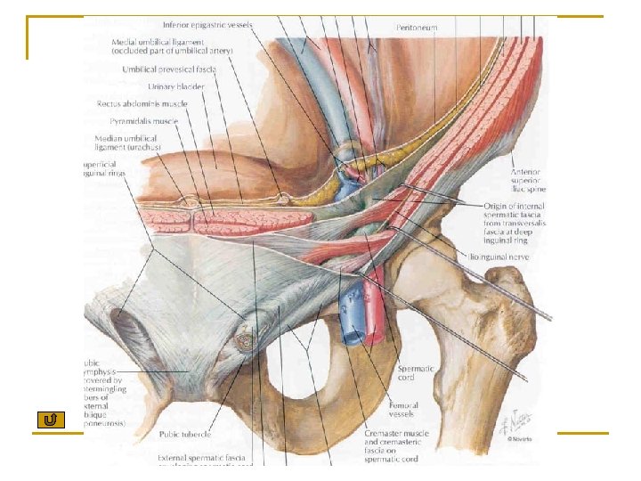
Inguinal canal 腹股沟管 Position: oblique passage about 4 cm long, and passes downwards and medially, it lies parallel to, and immediately above medial half of inguinal lig. It has two openings and four walls.
Four walls Anterior wall q Aponeurosis of obliquus externus abdominis q Obliquus internus abdominis (lateral third of wall)
n Post wall — Transverse fascia ; Inguinal flax medially n Roof- lower fibers of obliquus internus and transversus abdominis n Floor-inguinal ligament.
n Two openings p Superficial inguinal ring 腹股沟浅环 Deep inguinal ring 腹股沟深环 -defect in transverse fascia,lies at about 1. 5 cm above midpoint of inguinal ligament p
n Structures passing through the inguinal canal q q Male: Spermatic cord 精索 female: Round ligament of uterus 子宫圆韧带