Myology Objectives of the lecture Definition of muscles

Myology Objectives of the lecture • Definition of muscles • Types of muscles - Identify and describe three types of human muscle tissue. • Skeletal muscles - elements and types • Classifications of muscles • Biomechanics • Muscle markings

Definition A muscle is a group of muscle tissues which contract together to produce a force. A muscle consists of fibers of muscle cells surrounded by protective tissue, bundled together many more fibers, all surrounded in a thick protective tissue. A muscle uses ATP to contract and shorten, producing a force on the objects it is connected to. There are several types of muscle, which act on various parts of the body.

Muscle tissues share four basic properties: I. Electrical Excitability: - ability to respond to stimuli by producing electrical signals such as action potentials - two types of stimuli: 1. autorhythmic electrical signals 2. chemical stimuli • Skeletal muscles normally respond to stimulation by the nervous system. • Cardiac and smooth muscles respond to the nervous system and circulating hormones. Conductivity: - electrical signal travels throughout the muscle cell • since muscle cells are not myelinated the impulse travels much slower than on nerve cell • T-tubule system helps to get impulse into cell and around the myofibrils

Muscle tissues share four basic properties: II. Mechanical properties Contractility: -ability to contract when stimulated by an AP -isometric contraction: tension develops, length doesn’t change -isotonic contraction: tension develops, muscle shortens Extensibility: -ability to stretch without being damaged -allows contraction even when stretched Elasticity: - the ability of a muscle to rebound toward its original length after a contraction

There are three types of muscle tissue: • Skeletal muscle—Skeletal muscle tissue moves the body by pulling on bones of the skeleton. • Cardiac muscle—Cardiac muscle tissue pushes blood through the arteries and veins of the circulatory system. • Smooth muscle—Smooth muscle tissues push fluids and solids along the digestive tract and perform varied functions in other systems.

There about 700 named skeletal muscles in the human body, including roughly 400 that no one cares about except specialists. There is just one important cardiac muscle. And there are literally countless smooth muscles (which do the work of the autonomic nervous system, mostly squeezing and squishing stuff in tubes). Paul Ingraham

Types of muscles
 • Maintain posture")
Skeletal Muscles • Attach to bones • Produce skeletal movement (voluntary) • Maintain posture • Support soft tissues • Regulate entrances to the body • Maintain body temperature

Classifications of skeletal muscles Classification • According to the form • According to the fiber direction • According to the function • According the number of joints • According to the position

• Copyright © 2009 Pearson Education, Inc. , publishing as Pearson Benjamin Cummings

• Copyright © 2009 Pearson Education, Inc. , publishing as Pearson Benjamin Cummings

• Copyright © 2009 Pearson Education, Inc. , publishing as Pearson Benjamin Cummings

• Copyright © 2009 Pearson Education, Inc. , publishing as Pearson Benjamin Cummings

• Copyright © 2009 Pearson Education, Inc. , publishing as Pearson Benjamin Cummings

Origins and Insertions • Origin remains stationary • Typically proximal to insertion • Insertion moves • Muscles identified by – Origin – Insertion – Primary action • Classified as – Prime mover (agonist) – Synergist – Antagonist F Ex

Skeletal muscle - structure

Skeletal muscle -structure

Gross Anatomy • muscles are really groups of fascicles • the fascicles are groups of muscle fibers = considered to be an individual muscle cell

• the muscle fiber is made up of protein filaments = myofibrils • each myofibril is comprised of repeating units = sarcomeres

Auxiliary structures Fasciae proper – one muscle group – synergic muscles septae – separate neighbouring muscle groups

Auxiliary structures Synovial vagina: Outer sheath - 1 Inner sheath - 2 Mesotenon - 3 1 2 3

Auxiliary structures Synovial bursae Beneath tendons and muscles, close to their insertions Sesamoid bones In tendons close to their insertions

Gross Anatomy • muscle is wrapped in a protective fascia -fascia = sheet of fibrous connective tissue that supports and surrounds muscle or organs • a superficial fascia separates muscle from the overlying skin -also known as the subcutaneous layer -made up of areolar tissue and adipose tissue -provides support for blood vessel and nerves -the adipose tissue stores most of the body’s triglycerides and provides insulation • muscles with similar functions are grouped and held together by layers of deep fascia -dense irregular connective tissue -allow free movement of muscles, carries nerves, BVs

• three layers of connective tissue extend from the deep fascial layer – Epimysium – Perimysium – Endomysium • these layers further strengthen and protect muscle • outermost layer = epimysium – encircles the entire muscle • next layer = perimysium – – surrounds groups of 10 to 100 individual muscle fibers separates them into bundles = fascicles give meat its “grain” because the fascicles are visible both epimysium and perimysium are dense irregular connective tissue • penetrating the fasicles and separating them into individual muscle fibers = endomysium (areolar connective tissue)

• all three of these connective tissue layers extend beyond the muscle and attaches it to other structures -called a tendon = cord of regular dense CT that attaches a muscle to the periosteum of bone • when the CT extends as a broad flat sheet = aponeurosis • generally muscles are supplied with one artery and two veins • they accompany the nerve • nerves that induce muscle contraction = somatic motor neurons (part of the somatic division of the PNS) • communication between muscle and these neurons Neuromuscular junction (NMJ)
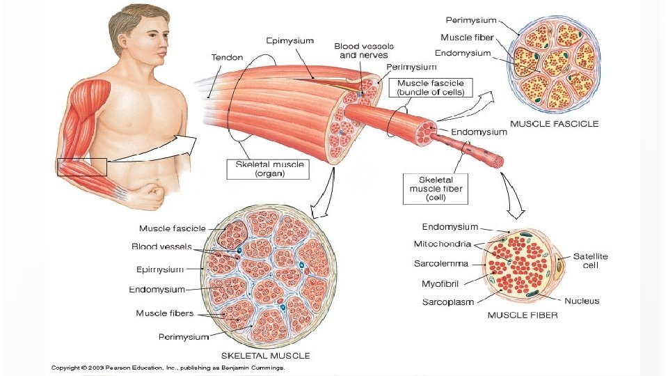

Microanatomy of Skeletal Muscle Fibers • New terminology • Cell membrane = sarcolemma • Cytoplasm = sarcoplasm • Internal membrane system = sarcoplasmic reticulum • Even nuclei usually called myonuclei • Large, multinucleated cells • Embryonic development – stem cells (satellite cells) differentiate into immature myoblasts which begin to make the proteins of the myofilament • These myoblasts mature into myocytes • Multiple myocytes fuse to form the muscle cell (muscle fiber) - symplast , a multinuclear structure • once fused, these muscle cells lose the ability of undergo mitosis • number of muscle cells predetermined before birth • But satellite cells (precursor cells) can repair damaged/dying muscle cells throughout adulthood

Muscle Cell Anatomy • Transverse tubules • Invaginations of sarcolemma • Carry electrical impulses • Myofibrils within sarcoplasm • “skeleton” of protein filaments (myofilaments) organized as Sarcomeres • ` • Myofilaments form the myofibrils • Thin filaments (actin, troponin, tropomyosin) • Thick filaments (myosin)

Microanatomy of Skeletal Muscle Fibers • muscle fibers are bound by a plasma membrane = sarcolemma • thousands of tiny invaginations in this sarcolemma called T or transverse tubules - tunnel in toward the center of the cell -T tubules are open to the outside of the fiber - filled with interstitial fluid - action potentials generated in the neuron travel along the sarcolemma and the T tubules - allows for the even and quick spread of an action potential deep into the cell • the cytoplasm is called a sarcoplasm -substantial amounts of glycogen - can be broken into glucose -contains myoglobin - binds oxygen needed for muscle ATP production

Microanatomy of Skeletal Muscle Fibers • contractile elements of the myofibrils = myofilaments -2 microns in diameter -comprised of primarily actin or myosin -give the muscle its striated appearance • Fibers also have a system of fluid-filled membranes = sarcoplasmic reticulum -encircles each myofibril -similar to the ER -have dilated end sacs = terminal cisterns -stores calcium when at rest - releases it during contraction -release is triggered by an AP

The Proteins of Muscle • Myofibrils are built of 3 kinds of protein – contractile proteins • myosin and actin – regulatory proteins which turn contraction on & off • troponin and tropomyosin – structural proteins which provide proper alignment, elasticity and extensibility • titin, myomesin, nebulin, actinin and dystrophin • Dystrophin – connects sarcomere to sarcolemma – -transmits tension along muscle • Actinin – part of Z-line • Titin – connects myosin to Z-line and M-line – Role in recovery after being stretched • Nebulin – forms core of the actin chain/thin filament
 and")
M line Sarcomere Structure • • sarcomere = regions of myosin (thick myofilament) and actin (thin myofilament) bounded by the Z line (actinin) actin filaments project out from Z line myosin filaments lie in center of sarcomere - overlap with actin and connect via cross-bridges • myosin only region = H zone • myosin filaments are held in place by the M line proteins. • actin only region = I band • length of myosin filaments = A band • contraction = “sliding filament theory” -actin and myosin myofilaments slide over each other and sarcomere shortens

Contraction: The Sliding Filament Theory • Contraction: – Active process – Elongation is passive – Amount of tension produced is proportional to degree of overlap of thick and thin filaments • SF Theory: • Explains how a muscle fiber exerts tension • Four step process • • Active sites on actin Crossbridge formation Cycle of attach, pivot, detach, return Troponin and tropomyosin control contraction

Contraction: The Sliding Filament Theory • Actin filament has a myosin binding site • This site is “covered up” by troponin and tropomyosin in relaxed muscle • Removal of troponin/tropomyosin is required for contraction • myosin thick myofilament is a bundle of myosin molecules -each myosin protein has a globular “head” with a site to bind and breakdown ATP (ATPase site) and to bind actin (actin binding site)

RESETTING of system -calcium binds to troponin and exposes sites that can interact with myosin - Ca 2+ binds to troponin & causes troponin-tropomyosin complex to move & reveal myosin binding sites on actin -ATP binding, hydrolysis and ADP release changes the conformation of the head and causes actin to “slide” along the myosin myofilaments -shortens the distance between the Z lines

The Events in Muscle Contraction

Muscle Contraction: A summary • ACh released from synaptic vesicles • Binding of ACh to motor end plate • Generation of electrical impulse in sarcolemma • Conduction of impulse along T-tubules • Release of Calcium ions by SR - binds to troponin • Exposure of active sites on actin • Cross-bridge formation and contraction
 breaks down ACh within the synaptic cleft • Muscle")
Relaxation • Acetylcholinesterase (ACh. E) breaks down ACh within the synaptic cleft • Muscle action potential ceases • Ca 2+ release channels close • Active transport pumps Ca 2+ back into storage in the sarcoplasmic reticulum • Calcium-binding protein (calsequestrin) helps hold Ca 2+ in SR • Tropomyosin-troponin complex recovers binding site on the actin
 is in")
The Neuromuscular Junction • end of neuron (synaptic terminal or axon bulb) is in very close association with the muscle fiber • distance between the bulb and the folded sarcolemma = synaptic cleft • nerve impulse leads to release of neurotransmitter (acetylcholine) • N. T. binds to receptors on myofibril surface • binding leads to influx of sodium, potassium ions (via channels) • eventual release of calcium by sarcoplasmic recticulum = contraction • Acetylcholinesterase breaks down ACh • Limits duration of contraction

Motor Units • Each skeletal fiber has only ONE NMJ • MU = Somatic neuron + all the skeletal muscle fibers it innervates • Number and size indicate precision of muscle control • Muscle twitch • Single momentary contraction • Response to a single stimulus • All-or-none theory • Either contracts completely or not at all • Motor units in a whole muscle fire asynchronously some fibers are active others are relaxed delays muscle fatigue so contraction can be sustained • Muscle fibers of different motor units are intermingled so that net distribution of force applied to the tendon remains constant even when individual muscle groups cycle between contraction and relaxation.

Neuromuscular Integration A Amotor is only dedicated solely to stimulating muscleneuron fiber can contract maximally (“all or none” law). muscle to of contract. Each single muscle by fiber in Given fibers the fact “all or none” contractions individual skeletal muscle fibers, grades of contraction of a skeletal muscle are a made skeletal muscle is innervated by a branch of an possible by activating a number of motor units and not activating others. axon. microscopic site only at which branchunits are activated. In maximal contraction of a skeletal muscle, all motor In a The partial contraction, somethe of axon the motor attaches the skeletal muscle maximus fiber is called theof skeletal muscle fibers having a nerve-to-muscle ratio of 1: 1000 or more. units aretoactivated. The gluteus consists neuromuscular junction. Each neuromuscular There is no possibility of controlled, refined contractions from this muscle. The facial muscles, in contrast, have a much lower junction consists ofratio, an axon terminal closely nerve-to-muscle closer to 1: 10. Here applied small numbers of muscle fibers can be contracted by implementing one or a few tomotor an area of convoluted muscle fiber sarcolemma units, generating very fine control on the muscular effect (facial expression) desired, such as “the Mona Lisa smile. ” called the motor end plate. There is a gap between the two surfaces. When a skeletal muscle fiber is about to be stimulated, the axon terminal releases a chemical neurotransmitter, called acetylcholine, into the gap. The Anatomy Coloring Book, Pearson; 4 th ed by Wynn Kapit, Lawrence M. Elson

Muscle biomechanics Internal biomechanics • Physiological transection – perpendicular to fibers • Anatomical transection – through the widest part of the muscle • Vector of muscle strength • Effectiveness of muscle strength - negatively proportional to the angle between the vector and the bone

Muscle biomechanics External biomechanics Movements in joints. • Synergists and antagonists Muscle strength. • Number and length of the fibers

The Anatomy Coloring Book, Pearson; 4 th ed by Wynn Kapit, Lawrence M. Elson

Muscle biomechanics External biomechanics Attachments • punctum mobile - insertion • punctum fixum - origin Levers of muscle contraction • Fulcrum • Point of load • Point of strength • I class lever – of equilibrium • II class lever – of strength • III class lever– of speed

Muscle Names • Yield clues to muscle orientation, location or function • • Biceps brachii (two heads, arm) Vastus femoris (large, femur) Orbicularis oculi (circular, eye) Rectus abdominus (erect, abdomen)

Muscle Names • The following are some terms relating to muscle features that are used in naming muscles. • Size: vastus (huge); maximus (large); longus (long); minimus (small); brevis (short). • Shape: deltoid (triangular); rhomboid (like a rhombus with equal and parallel sides); latissimus (wide); teres (round); trapezius (like a trapezoid, a four-sided figure with two sides parallel). • Direction of fibers: rectus (straight); transverse (across); oblique (diagonally); orbicularis (circular). • Location: pectoralis (chest); gluteus (buttock or rump); brachii (arm); supra- (above); infra(below); sub- (under or beneath); lateralis (lateral). • Number of origins: biceps (two heads); triceps (three heads); quadriceps (four heads). • Origin and insertion: sternocleidomastoideus (origin on the sternum and clavicle, insertion on the mastoid process); brachioradialis (origin on the brachium or arm, insertion on the radius). • Action: abductor (to abduct a structure); adductor (to adduct a structure); flexor (to flex a structure); extensor (to extend a structure); levator (to lift or elevate a structure); masseter (a chewer).

Axial Musculature • Arises from and inserts on the axial skeleton • Positions the head and spinal column • Moves the rib cage, assisting in breathing • Axial musculature – Originates and inserts on axial skeleton • Appendicular musculature – Stabilizes or moves components of the appendicular skeleton

Axial muscles organized into four groups • Muscles of the head and neck • Muscles of the vertebral column • Oblique and rectus muscles • Muscles of the pelvic floor

Appendicular musculature is responsible for • Stabilizing pectoral girdle • Stabilizing pelvic girdle • Moving upper and lower limbs Four groups of muscles • • Muscles that position the pectoral girdle Muscles that move the arm Muscles that move the forearm and hand Muscles that move the hand fingers

7 1 2 3 4 5 6 8 9 10 11 1. sagittal suture 2. parietal bone 3. posterior fontanelle 4. occipital bone 5. lambdoid suture 6. posterolateral suture 7. anterior (frontal) suture 8. frontal bone 9. coronal suture 10. anterolateral suture 11. zygomatic arch

1 2 3 5 4 6 9 7 1. Orbital part of frontal bone 2. Cribriform plate 3. Hypophyseal fossa 4. Clivus 5. Foramen magnum 6. Jugular foramen • 7. Foramen ovale • 8. External occipital 8 • protuberance • 9. Foramen lacerum

8 4 9 6 1 3 7 10 2 5 1. Incisive foramen 2. Choanae 3. Occipital condyle 4. Dental alveola 5. Zygomatic arch 6. Carotid canal 7. Jugular foramen 8. Mastoid process 9. Mandibular fossa 10. Foramen spinosum

1. 2. 3. 4. 4 2 1 3 External acustic meatus Zygomatic arch Mastoid process Coronal suture
- Slides: 57