MYOCARDIAL VIABILITY JOURNAL REVIEW ANKUR KAMRA Myocardial viability

MYOCARDIAL VIABILITY JOURNAL REVIEW ANKUR KAMRA

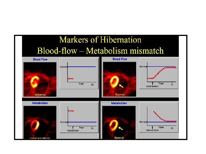
• Myocardial viability is defined as myocardium with potentially reversible contractile dysfunction in patients with CAD. • Concept of myocardial hibernation was introduced by Rahimtoola to describe a condition of chronic sustained abnormal contraction attributable to chronic under perfusion in patients who have CAD and in whom revascularization causes recovery of LV function • Myocardial stunning defined as reversible myocardial contractile dysfunction in presence of normal resting myocardial blood flow • Vanoverschelde et al. suggested that repeated ischemic attacks may induce chronic dysfunction in presence of normal or mildly reduced resting perfusion this condition was referred to as repetitive stunning. • It appears that there is temporal progression from stunning, characterized by normal flow (with reduced flow reserve) to hibernation with reduced resting flow.
 –")
Introduction Endpoints used in viability studies – Improvement in regional LV function (segments) – Improvement in global LV function (LVEF) – Improvement in symptoms (NYHA functional class) – Improvement in exercise capacity (metabolic equivalents) – Reverse LV remodeling (LV volumes) – Prevention of sudden death (ventricular arrhythmias) – Long-term prognosis (survival)

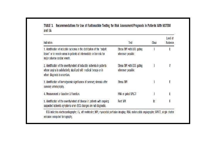
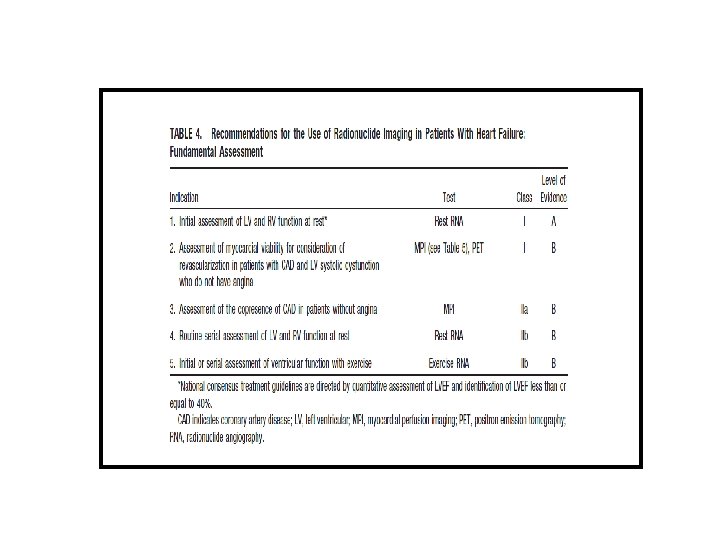
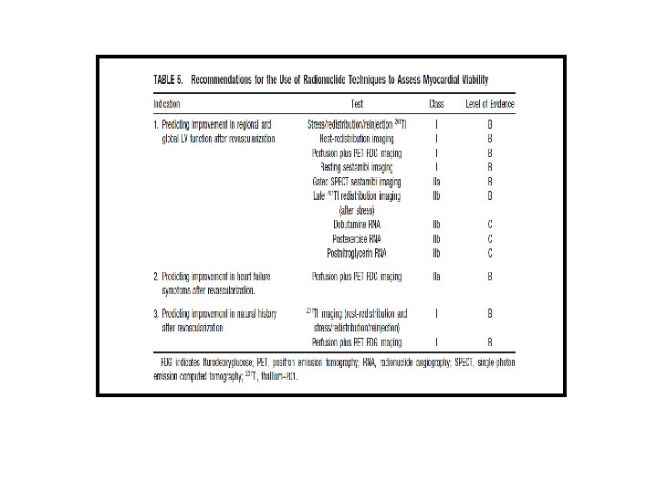
Viability assessment by different investigations

METHODS OF VIABILTY ASSESMENT • Membrane integrity uptake of Thallium and Rubidium • Metabolic activity Fatty acids PET, uptake of F-fluorodeoxyglucose (FDG) is a marker of glucose metabolism • Blood Flow N 13 H 3, Tc 99 m-Sestamibi, Tc 99 m-tetrofosmin • Contractile reserve Low Dose Dobutamine RWM analysis • Morphology MR hyper enhancement

Viability assessment DSE • Low dose- High dose dobutamine infusion Viability assessment • Biphasic response- viability with superimposed ischemia • Worsening-severe ischemia with critical stenosis • No change- transmural scar • Continued improvement- no relation

FOUR DIFFERENT RESPONSE AND THERE PPV • Biphasic response with improvement at low dose and worsening at high doses seen in 28% of segments, sustained improvement seen in 18% of segments, sustained worsening seen in 15% of segments and no change in function seen 39% of segments • Biphasic response had PPV for functional recovery of 72%, response of worsening throughout had PPV 35% and segments that showed no change had a PPV of only 13%. • However low PPV of 15% noted for continued improvement, since most of these segments were hypokinetic at baseline and never exhibited signs of ischemia at peak stress, it was postulated that resting ischemia was not the mechanism for the underlying wall motion abnormality • Afridi et al Circuition I 995; 9 I: 663470

DSE for dectection of viable but stunned myocardium

DSE for dectection of hibernating myocardium
![Viability assessment Ability to predict improvement in LVEF 32 studies [N= 1090] % sensitivity](http://slidetodoc.com/presentation_image_h/284c340791d714ce375301b578990357/image-10.jpg "Viability assessment Ability to predict improvement in LVEF 32 studies [N= 1090] % sensitivity")
Viability assessment Ability to predict improvement in LVEF 32 studies [N= 1090] % sensitivity % specificity % NPV %PPV 81 80 85 77 J Nucl Med 2007; 48: 1135– 1146 Ability to predict improvement in LVEF 8 studies [N=254] Viability present Viability absent % LVEF pre % LVEF post 35 43 35 36 J Nucl Med 2007; 48: 1135– 1146

Viability assessment Viability, Trt. type & Event Rates in Pts with LVD and c/c CAD % event rates Viability present 6 studies (N= 686) Viability absent Revascularization Medical trt. 6 22 16 28 J Nucl Med 2007; 48: 1135– 1146

• Limitations – Poor window in 30% – Lower sensitivity – Viable regions with absent flow reserve will not show thickening – Reliance on visual assessment

• TDI- strain rate imaging – Augmentation of strain and strain rate with dobutamine is a marker of myocardial viability – PET detected viable segments compared with strain rate imaging during low-dose dobutamine stress test [N=37]. An increase in the longitudinal strain rate of more than 0. 23 S-1 identified viable tissue [83% sens. , 84% spec. ] J Am Coll Cardiol 2002; 39: 443 -449

PET TRACERS
")
METABOLIC TRACERS • 18 F-FDG uptake in hypoperfused and dysfunctional myocardium (flow- metabolism mismatch) is regarded as a metabolic marker of cell survival and viability, whereas concordant reduction in both blood flow and 18 F-FDG uptake is indicative of scar. Thus 18 F-FDG PET provides biological signal on cellular viability and is considered to be one of most accurate non invasive techniques to identify viable tissue Haas F et al. J Am Coll Cardiol 1997; 30: 1693– 1700 Tillish J et al. N Engl J Med 1986; 314: 884– 888 • 18 F-FDG may not be suitable for use in acute myocardial infarction due to inflammatory cell accumulation in necrotic tissue, which takes up 18 F-FDG as does viable tissue so in these cases 11 C-Acetate can be used as tracer.

SPECT TRACER

• 201 Tl is taken up by myocardial cells by active transport and is dependent on myocardial blood flow. Therefore 201 Tl uptake represents both myocardial perfusion and cellular viability. • Various protocols used are ü Stress -Normal perfusion ü Delayed rest images or reinjection- fixed defect showing redistribution ü Stress- defects with redistribution on delayed images • A study done by Kitsiou AN et al. (Circulation 1998; 98: 501– 508) and many other study shows that stress induced reversible defects which are more commonly seen on stress imaging than on rest imaging are highly predictive of functional recovery after revascularization. Thus use of stress imaging protocol is encouraged whenever possible

• A defect that persists and appears again on 3– 4 hr images(i. e. fixed defect) may be due to : (1)markedly reduced regional perfusion (2)impaired cellular membrane integrity (3)cell death (acute infarction) (4)scartissue. • Thus fixed-defects on 3– 4 hr redistribution images may represent only severely hypoperfused—and not necessarily infarcted tissue

• Technetium-99 m labeled flow tracers such as 99 m. Tcsestamibi and 99 m Tc-tetrofosmin are now widely available as alternatives to conventional 201 Tl. As compared With 201 Tl, these 99 m. Tc-labeled agents yield higher image quality. • 123 I-beta-methyl iodophenyl pentadecanoic acid (BMIPP) is a fatty acid analog that is not metabolized by beta-oxidation, because myocardial fatty acid uptake is easily depressed in ischemic but viable myocardium, BMIPP imaging in combination with flow tracer can also detect potentially reversible myocardium.

Dobutamine CMR • Contractile reserve can be assessed by CMR using low dose dobutamine stress testing. Similar to echo, CMR allows visualisation of cardiac motion but is characterised by superior endocardial border definition, facilitating more accurate wall motion and wall thickening assessment. • Majority of studies showed a relatively modest sensitivity but high specificity of dobutamine CMR for detection of viable myocardium, ranging from 50– 90% and 73– 94% respectively. Baer FM et al Circulation 1995; 91: 1006– 15 Gunning MG et al Circulation 1998; 98: 1869– 74

Viability Assessment DE CMR Viability criteria – Increase in End-diastolic wall thickening > 2 mm For predicting functional recovery 9 studies [N= 252] % sensitivity % specificity 73(50 -89) 83(70 -95) Heart 2005; 91: 1359– 1365

• Baer et al in a study, evaluated viability by MRI after myocardial infarction comparing to PET as the gold standard. Patients with myocardial infarction (infarct age 4 months) were studied with low-dose Dob-MRI examinations, with viability defined by an end-diastolic wall thickness of 5. 5 mm and evidence of dobutamineinduced systolic wall thickening of 1 mm. • When both these parameters were used as criteria for viability, Dob-MRI demonstrated a sensitivity of 88% and specificity of 87%. Circulation 1995; 91: 1006 -1015.
 on")
ce. CMR • Regions of myocardial infarction exhibit high signal intensity (contrast enhancement) on T 1 -weighted images after administration of contrast such as gadolinium based agents. • ce. CMR is effective in identifying the presence, location and extent of acute and chronic myocardial infarction Kim RJ et al. Circulation 1999; 100 : 1992– 2002 Landmark study comparing contrast enhancement to histopathology and concluding that contrast enhancement occurs in the setting of irreversible myocardial injury only • Because of superior spatial resolution it can detect micro-infarcts, as well as sub endocardial infarcts which are missed by SPECT Wagner A et al. Lancet 2003; 361: 374– 9. Landmark study concluding that ce. CMR detects myocardial infarcts which are missed by SPECT imaging.

• Likelihood of functional improvement inversely related to extent of hyperenhasment. • 78% patients improve with no hyper enhancement while only 2% with >75% HEI Kim RJ et al; NEJM 2000

To predict functional recovery in patients with chronic ischaemia Kim RJ et al. N Engl J Med 2000; 343: 1445– 53. The first study to demonstrate that ce. CMR can accurately predict reversible myocardial dysfunction in patients with ischaemic heart disease before coronary revascularisation.

MSCT • Iodinated contrast agents accumulate in infarcted myocardium similar to MRI • High spatial resolution-differentiation of transmural & subendocardial infarction possible • Old infarctions have lower CT densities compared to recent infarcts • Good agreement b/w DEMRI and late enhancement of MSCT

• Sensitivity and specificity -92%& 100% respectively to detect viability in acute myocardial infarction • Presence of viable segments in an AMI identified when hyperenhancement is absent or involves <50% of the myocardial wall thickness • Advantages – Shorter imaging time – widely available

Comparison b/w different investigations

Baer FM, European heart journal 2000

Sensitivity more for PET and SPECTE and specificity more for DSE Schinkel AF et al. Hibernating myocardium: diagnosis and patient outcomes. Curr Probl Cardiol. 2007; 32(7): 375
 31, 2984– 2995")
Comparison European Heart Journal (2010) 31, 2984– 2995

Recovery variables
 • The time course of")
Recovery variables(time course after procedure and extent of viability) • The time course of recovery of viable myocardium can protract up to 14 months Vanoverschelde J-LJ et al. Am J Cardiol 2000; 85: 1 1432 -1439. • Majority of studies agree that to yield the best sensitivity and specificity to predict > 5% improvement in LVEF, at least 25% of the LV should be viable using DSE and 38% using conventional nuclear medicine and PET. Bax JJ et al. J Nucl Med. 2002; 43: 795– 802

Impact of the time of revascularization • A comparison of the outcome after early (within 30 days) or late (30 days) revascularization in 2 parallel groups of patients showed a significant improvement in function and lower mortality in the group with shorter waiting times. Bax JJ et al. Circulation. 2003; 108(suppl): II-39–II-42.

Impact of the time of revascularization
")
Benefits of Viability Assessment (META ANALYSIS AND STUDIES)

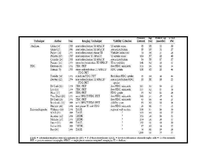
Meta-analysis J Am Coll Cardiol 2002; 39: 1151– 8 • Late survival with revasc. vs. medical therapy after myocardial viability testing in pts with severe CAD & LV dysfunction • 24 studies Thallium, PET & DSE • N= 3, 088 (2, 228 men), EF 32+8%, followed for 25+10 months

Meta-analysis

Meta-analysis
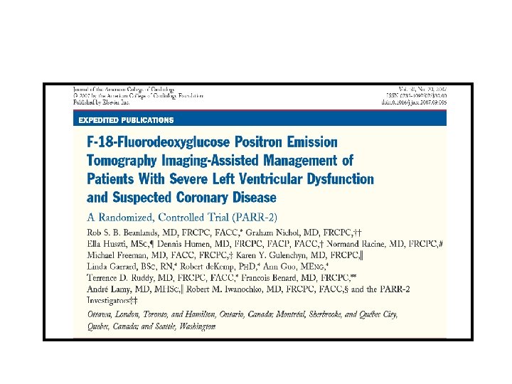
![PARR 2 • [N=430]; EF ≤ 35% considered for revascularization, transplant, or HF work‐](http://slidetodoc.com/presentation_image_h/284c340791d714ce375301b578990357/image-43.jpg "PARR 2 • [N=430]; EF ≤ 35% considered for revascularization, transplant, or HF work‐")
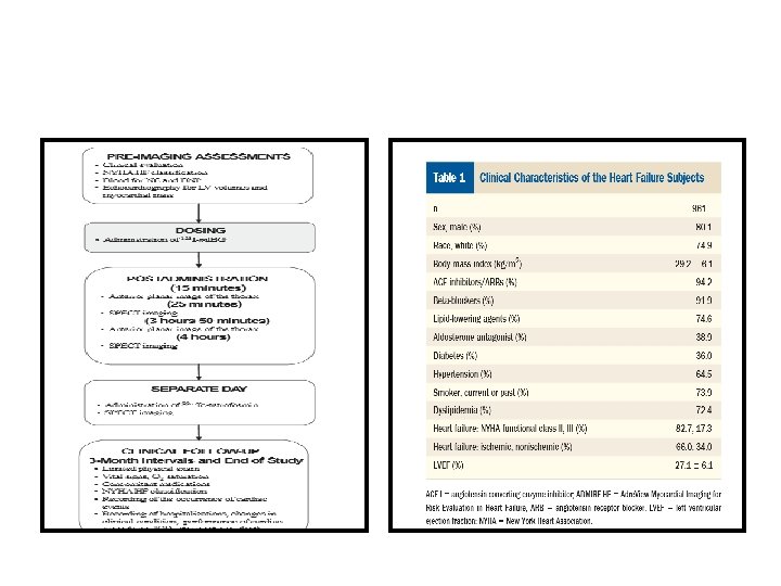
PARR 2 • [N=430]; EF ≤ 35% considered for revascularization, transplant, or HF work‐ up with high suspicion of CAD. • Randomized patients to a PET‐guided therapy or “standard care” (no PET). • Imaging physicians issued a therapy recommendation based on PET findings and treating physicians then made a decision regarding revascularization. • Patients in the standard care arm had no PET, but could have another viability test, which was performed in 138 of 209 (66%) patients. • Primary outcome: composite of cardiac death, myocardial infarction or recurrent cardiac hospitalization within 1 year.
")
“Survival Curves” (on the Basis of Time to First Outcome of the Composite Event) hazard ratio = 0. 78, 95% CI 0. 58 to 1. 1, p = 0. 15 “Survival” Curves (on the Basis of Time to Cardiac Death) hazard ratio = 0. 72, 95% CI 0. 4 to 1. 3, p = 0. 25
 (B) Rates of adherence to positron emission tomography recommendations for revascularization work-up Rates")
(A) (B) Rates of adherence to positron emission tomography recommendations for revascularization work-up Rates of revascularization (solid bars); revascularization work-up without revascularization (hatched bars), and no revascularization (open bars). Rates of revascularization + revascularization were different between groups

Survival Curve (on the Basis of Time to First Occurring Outcome Out of the Composite Event) The positron emission tomography adherence group versus standard care arm. adjusted hazard ratio = 0. 62, 95% CI 0. 42 to 0. 93, p = 0. 019
 for Patients Without Recent")
“Survival Curves” (on the Basis of Time to Cardiac Death) for Patients Without Recent Angiography hazard ratio = 0. 4, 95% CI 0. 17 to 0. 96, p = 0. 035

• There was no statistically significant difference for the primary outcome measure. However, there are number of important findings from the PARR-2 study • large proportion (approximately 25%) of patients did not adhere to PET recommendations. This implies that, for clinicians to make decisions, evidence for revascularization benefit is not convincing enough. • significant benefit for FDG PET when adhered to the PET recommendations • outcome benefit observed when it was used in addition to a preceding test • patient without recent angiography, who represent sickest group when FDG PET-assisted management used, yields a significant mortality benefit

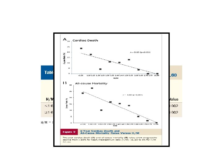
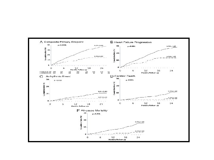
ADMIRE- HF

• Increased myocardial sympathetic activity is a prominent feature of heart failure and associated with progressive myocardial remodeling. • Increased neuronal release of norepinephrine is usually accompanied by decreased neuronal NE reuptake due to downregulation of cardiac NE transporter • Decrease in NE reuptake mechanism successfully assessed by radionuclide imaging with iodine-123 –labeled NE analog meta-iodobenzylguanidine (123 I-m. IBG) using SPECT

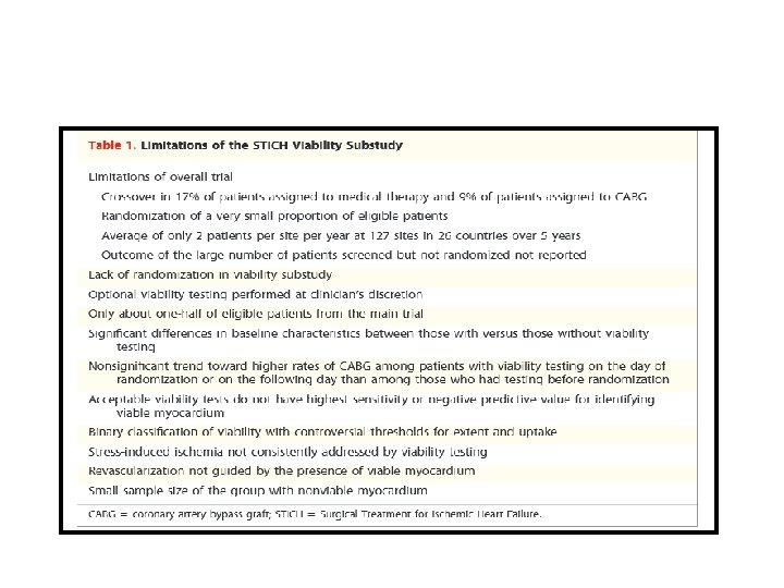
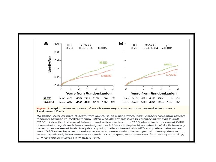
N Engl J Med 2011; 364: 1617 -25 • The first prospective randomized trial testing the hypothesis that CABG improves survival in patients with ischemic LV dysfunction [EF-26. 7%± 8. 6] compared to outcome with aggressive medical therapy • Myocardial viability identifies patients with CAD and LV dysfunction who have the greatest survival benefit with CABG compared to aggressive medical therapy

STICH Trial- substudy • All pts eligible for viability testing with SPECT [Tl or Tc] or DSE – Viability testing optional – Criteria for myocardial viability were prospective and pre- specified [SPECT (11/17); DSE (5/16)] • Primary endpoint: – All cause mortality • Secondary endpoints: – – Mortality plus cardiovascular hospitalization Cardiovascular mortality

1212 Patients with viability test 618 594 Patients with no viability test Unusable test 17 611 Patients with usable viability test Pateints with no usable viability test 601 321 Viable Non-Viable 487 243 MED 49. 9% 114 244 60 CABG 50. 1% MED 52. 6% 54 CABG 47. 4% SPECT 471 150 130 DSE 471

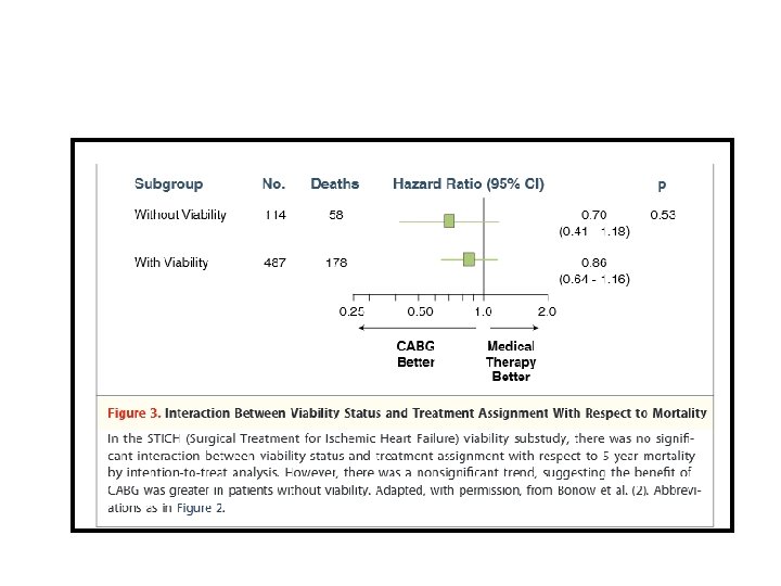
STICH Trial- substudy After multivariate analysis p=0. 21

STICH Trial- substudy

STICH Trial- substudy • Substudy of a major trial • Viability study according to the judgement of physician • Small number of non viable group for comparison • Sx influenced by timing and results of viability test • Two types of viability tests-SPECT & DSE • Better medical therapy available In patients with CAD and LV dysfunction, assessment of myocardial viability does not identify patients who will have the greatest survival benefit from adding CABG to aggressive medical therapy

THANK YOU
- Slides: 69