MYOCARDIAL STUNNING AND HIBERNATION VIABILITY CORONARY ARTERY DISEASE

MYOCARDIAL STUNNING AND HIBERNATION

VIABILITY

CORONARY ARTERY DISEASE ACUTE CORONARY SYNDROME Undergo revascularisation procedures Improved survival Increased number of patients with residual LV dysfunction undergoing progressive LV remodeling and congestive heart failure

ADD TO THIS 1. rising age of our population 2. Higher prevalence of comorbidities, eg. , DM-2 Typically, these patients have multivessel disease, increased LV volumes, and variable degrees of regional or global systolic dysfunction Among these patients, coronary revascularization may lead to symptomatic and prognostic improvement, and these clinical benefits are accompanied by evidence of reverse LV remodeling.

In the early 1980 s, Rahimtoola et al reviewed the results of coronary bypass surgery trials and identified patients with CAD and chronic LVD that improved upon revascularization. CORNERSTONE FOR ALL FUTURE STUDIES CASS (coronary artery surgery study ) REGISTRY üPatients with LVEF < 35% involved 651 patients. üThe five year survival was significantly better in surgical patients (68%) than in the medical group (54%). üThe contrast was even more in patients with LVEF < 26% whose five year survival was 63% with surgery, but 43% with medical treatment

Thus came the concept of myocardial viability and with it came the new terms such as hibernation and stunning

THE MYOCARDIAL RESPONSE TO ISCHEMIC INJURY

Onset of severe ischemia Within seconds aerobic changes to anaerobic metabolism Decrease in the production of high-energy phosphates, namely adenosine triphosphate (ATP) and phosphocreatine (PCr) Ultrastructural changes occur Mitochondrial swelling, Loosening of intercellular attachments, The presence of small, lipid-rich amorphous mitochondrial densities, Dilation of the sarcoplasmic reticulum, Disaggregation of SR polysomes, and Myofibrillar relaxation

within 1 min of acute onset The myocardium is functionally sensitive to ischemia and will exhibit marked contractile dysfunction HOWEVER, these ultrastructural defects are entirely reversible if reperfusion occurs within 20– 40 min.

Irreversible Injury Begins in the subendocardial tissue and progresses towards the subepicardium. ØIn humans, it may take as long as 6– 12 hr for complete infarction of the myocardium at risk Øthe necrotic changes are usually evident, about 4– 12 hr after onset ØThis may include the denaturation of cytoplasmic proteins, swelling, and enzymatic digestion of organelles and the sarcolemma.

Viable myocardium must have the following characteristics 1. The ability to generate HEP (PCr and ATP) 2. Have an intact sarcolemma, in order to maintain ionic/electrochemical gradients, and 3. Have sufficient perfusion, both for the delivery of substrates and O 2 and for the adequate washout of potentially noxious metabolites ? contractility.

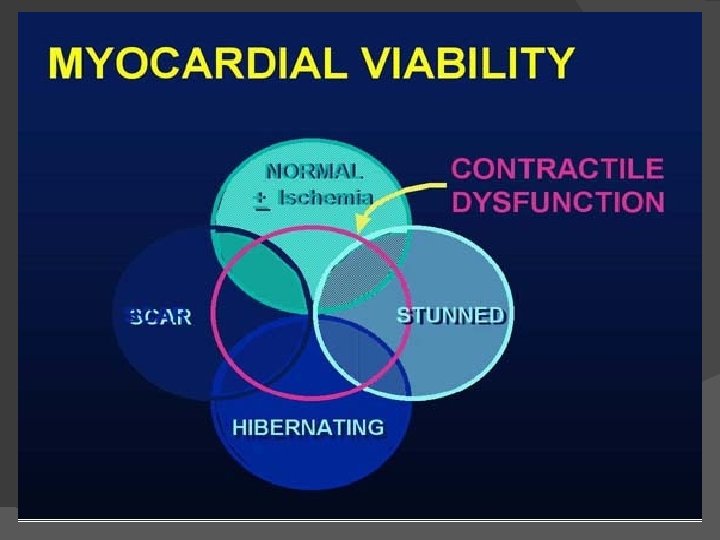
There are two tissue states that exhibit sustained contractile dysfunction despite meeting the three criteria- Stunned myocardium & Hibernating myocardium.

Myocardial stunning First documented by Heyndrickx et al. in the mid- 1970 s Heyndrickx GR, Millard RW, Mc. Ritchie RJ, Maroko PR, Vatner SF. Regional myocardial functional and electrophysiological alterations after brief coronary artery occlusion in conscious dogs. J Clin Invest 1975; 56: 978– 985.

-They had concluded that brief periods of coronary occlusion resulted in prolonged depression of myocardial function in the ischemic zone. -While regional electrograms return to normal within seconds and the coronary flow debt is repaid rapidly, functional derangement lasts for several hours. Published October, 1975

The stunned myocardium: prolonged, postischemic ventricular dysfunction Circulation 1982; 66; 1146 -1149 E Braunwald and RA Kloner Coined the term ‘‘myocardial stunning’’, for this phenomenon of ‘‘delayed recovery of regional myocardial contractile function after reperfusion despite the absence of irreversible damage and despite restoration of normal flow.

Features of stunning � � � Normal perfusion. Depressed myocardial function. Dissociation of usual relationship between subendocardial flow and function. Reversible. Function improves with inotropic agents.

Clinical Relevance � In the clinical setting stunning can occur 1. Brief period of total coronary occlusion: pts with angina due to spasm 2. Global ischemia after cardiopulmonary bypass. 3. In combination : Subendocardium is infarcted and overlying subepicardium reversibly injured in MI 4. Following exercise in presence of a flow limiting stenosis 5. Ischemic bout that is induced by PCI

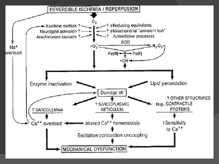
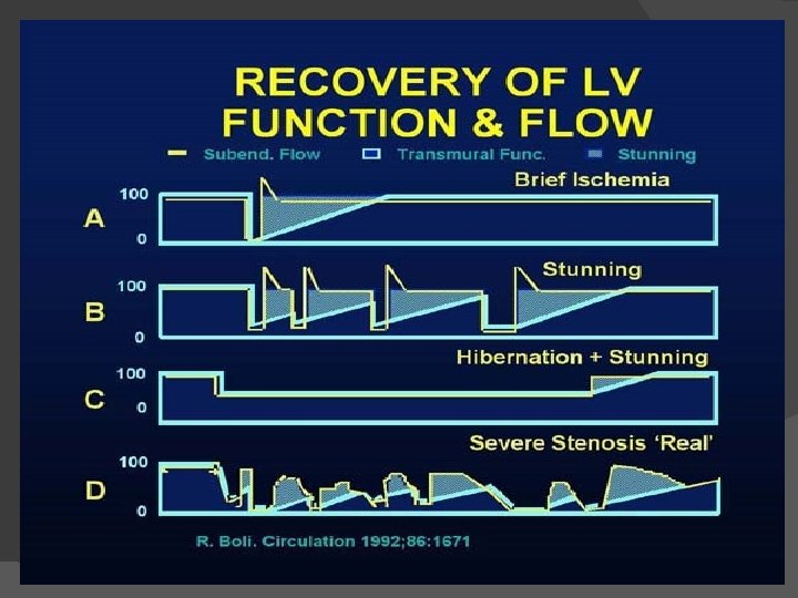
Pathogenesis of stunning Earlier Øloss of and reduced ability to synthesize high-energy phosphates, ØImpairment of microvascular perfusion, Øimpairment of sympathetic neural responsiveness, Øreduction in the activity of creatine kinase, Presently There are 2 major hypothesis for myocardial stunning: (1) a oxygen-free radical hypothesis and (2) a calcium overload hypothesis Time-course of myocardial stunning -Dysfunction may persist for hours or for as long as 6 weeks post-insult -Both the duration and severity of ischemia determine the duration of post-ischemia/reperfusion dysfunction

Mechanism of contractile dysfunction in stunning ØNormal cardiac contraction depends on the maintenance of calcium cycling and homeostasis across the mitochondrial membrane and sarcoplasmic reticulum during each cardiac cycle. ØBrief ischemia followed by reperfusion- accumulation of calcium and a partial failure of normal beat to beat calcium cycling - damages Ca 2+ pump and ion channels of the sarcoplasmic reticulum. ØThis results in the electromechanical uncoupling of energy generation from contraction that characterizes myocardial stunning.

Hibernating Myocardium • Hibernating myocardium is a state of persistently impaired myocardial and left ventricular function at rest due to reduced coronary blood flows. • It can be defined as an exquisitely regulated tissue successfully adapting its activity to prevailing circumstances. Characterstics- q Episodic and/or chronically reduced blood flow, which is directly responsible for the decrease in the myocardial contractile function. q. Tissue ischemia and resultant remodeling without necrosis q. Residual contractile reserve in response to inotropic stimulation (in at least half of clinical cases). q. Recovery of contractile function after successful revascularization.

Mechanism of hibernation

Natural history of hibernation

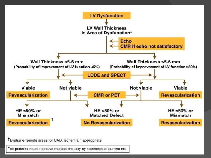
IDENTIFYING VIABLE MYOCARDIUM

Key non invasive methods to identify viability 1. 2. 3. 4. 5. ECG : gives little information Echocardiography Single Photon Emission Computed Tomography Positron Emission Tomography Magnetic Resonance Imaging

Characteristics of dysfunctional but viable myocardium

ECG -No clear correlation between Q waves on ECG and presence of viability. - Pts with preserved QT dispersion are likely to have viable myocardium. - Pts with high QT dispersion have predominantly nonviable scar tissue.

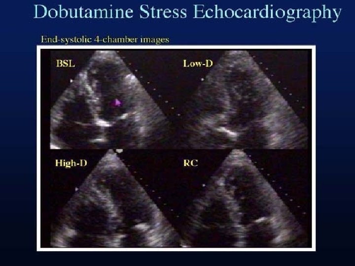
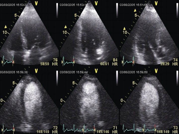
Dobutamine Stress Echocardiography ØExtremely useful tool-document the early and late functional changes at rest. ØHypokinetic or akinetic regions improving during low dose dobutamine infusion (5– 10 µg/kg/min) is indicative of viable tissue. ØAt higher doses (upto 40 µg/kg/min plus atropine) wall motion may improve or diminish, reflecting inducible ischemia. ØBiphasic response is highly predictive of recovery of function after revascularization.
, an improvement in contractile performance � at")
Biphasic response� at lower doses(5– 10 mg/kg/min), an improvement in contractile performance � at higher doses (>15 mg/kg/min) Contractility regresses as the metabolic demand stimulated overwhelms the tissue’s capacity to respond

Stress Echo Interpretation Interpretatio Rest / Low dose n Baseline stress Peak & post stress Normal Hyper dynamic Ischemic Normal / severe ischemia – new RWMA Decreased Scar WMA No change Hibernating WMA Improved Worsens Stunned WMA Improved

STUDIESNagueh et al studied the transmural myocardial biopsies obtained from patients with hibernating myocardium Showed that tissue with >17% fibrosis failed to exhibit contractile reserve when challenged with low-dose dobutamine Circulation 1999, 100, 490– 496.

In a study by Pagano et al , they reported that the diagnostic accuracy of dobutamine-echocardiography was reduced with increasing severity of regional and global LV dysfunction. That is, the technique appeared to underestimate the extent of viability: 39% of all recovering LV segments failed to exhibit inotropic contractile reserve. Heart 1998; 79: 281 -288 Wiggers et al studied the functional recovery pre- and 6 months postrevascularization, and showed that low-dose dobutamine failed to identify 45% of the segments that ultimately regained function Am. Heart. J. 2000, 140, 928– 936.

Value of Dobutamine stress echo ØReductions in blood flow that lead to hibernation occurs across a significant range of flows, with a corresponding spectrum of metabolic reserve. ØThose regions with greater metabolic reserve will likely retain the ability to respond to an inotropic stimulus while those regions with profoundly reduced flow—just on the threshold of viability—will have no ability to respond. ØSuch regions will therefore appear to be nonviable on a dobutamineechocardiography challenge. Hence dobutamine-echocardiography may be considered an easily accessible tool however with sub-optimal sensitivity for the detection of residual tissue viability.

Myocardial contrast echocardiography Basis : - ØMyocardial contrast enhancement depends on an intact microcirculation. ØThe combination of intravenous MCE and destruction and replenishment contrast intensity curves helps in the assessment of myocardial blood flow. ØLeft ventricular opacification (LVO) obtained with microbubbles improves the definition of the LV border. ØThis provides better quantitation of LV volume by the Simpson method. ØThe correlation between LV volume measured with CMR and that measured with echocardiography is better with the use of LVO. ØRegional wall motion analysis can also be better with LVO.

Results : ØRecovery of function occurred in 38% of dysfunctional segments. ØThe best MCE parameter for predicting functional recovery was Peak MCI×[beta], an index of myocardial blood flow ØMCE parameters of perfusion in hibernating myocardium were similar to segments with normal function and was higher than that in dysfunctional myocardium without recovery of function ØMCE parameters were higher in segments with contractile reserve and Tl 201 uptake >60% and identified viable segments without contractile reserve by DE.

Single Photon Emission Computed Tomography

SA VLA STANDARD SPECT IMAGING DISPLAY HLA

SPECT ØSPECT requires injection of a gamma-emitting radioisotope ØThallium-201 and Technetium Tc 99 m–Labelled Tracers are the commonly used radionuclides Ø 201 Tl is a monovalent cation with biologic properties similar to those of potassium (major intracellular cation in muscle and is virtually absent in scar tissue ) 201 Tl is a well-suited radionuclide for differentiation of normal and ischemic myocardium from scarred myocardium. ØThe initial myocardial uptake early after intravenous injection of thallium is proportional to regional blood flow.

VARIOUS SPECT MODALITIES TO IDENTIFY VIABLE MYOCARDIUM

201 Tl stress redistribution v. The uptake of 201 Tl is an energy-dependent process requiring intact cell membrane integrity, and the presence of 201 Tl implies preserved myocyte cellular viability. Imaging is done 1) Immediately following stress, with either exercise or pharmacologically induced coronary hyperemia with dipyridamole or adenosine and 2) After 3– 4 hr redistribution of Tl-201

INTERPRETATION ØDefects on post-stress images, may “fill in” by the time the rest-redistribution images are acquired, indicating viability. ØA defect that persists and appears again on the 3– 4 hr images (i. e. , a fixeddefect) may be due to: (1) markedly reduced regional perfusion, (2) impaired cellular membrane integrity, inadequate for the active sequestration of the tracer into the cell, (3) cell death (acute infarction), or (4) scar tissue. ØThus, fixed-defects on 3– 4 hr redistribution images may represent only severely hypoperfused—and not necessarily infarcted tissue

INTERPRETATION Late redistribution images ØAcquire a third set of images at 24 hours ØThis would allow for redistribution of the tracer to very-ischemic (yet viable) tissue ØIt has been shown that 22% of fixed defects (at early redistribution imaging) demonstrate normal Tl-201 uptake at later redistribution. ØThis may indicate a poorly perfused, yet viable region

201 Tl reinjection ØThis may be necessary because redistribution depends on the continued delivery of the tracer over the 3– 4 hr period. ØIf the blood concentration of Tl- 201 decreases to a great extent, there may be insufficient delivery of the tracer and the defect may not fill-in during redistribution imaging. ØThe second injection of thallium with delayed imaging will give the myocytes with reduced perfusion the greatest opportunity to sequester thallium.

99 m. Tc-sestamibi and tetrofosmin ØThey do not share the redistribution properties of 201 Tl ØBUT their characteristics for predicting improvement in regional function after revascularization appear to be similar

Relation between tracer uptake in a dysfunctional territory and the subsequent probability of functional recovery after revascularization. Modified from Bonow RO: Assessment of myocardial viability with thallium-201

Fixed defect Severe apical defect Basal and mid inferior and lateral wall defect Reversible

The impact of viability assessment using myocardial perfusion imaging on patient management and outcome. Hage FG et al J Nucl Cardiol. 2010 Jun; 17(3): 378 -89. Epub 2010 Feb 26. • They studied 246 consecutive ICM patients with rest-redistribution gated SPECT thallium-201 MPI. • Size and severity of perfusion defects were assessed by automated method. • Regions with <50% activity vs normal were considered nonviable

RESULTS: • Of the 246 patients, 37% underwent CR within 3 months of MPI. • Independent predictors of CR included chest pains (OR 2. 74) and rest-delayed transient ischemic dilatation (OR 4. 49), while a prior history of CR or ventricular arrhythmias favored Medical therapy. • The cohort was followed-up for 41 +/- 30 m • Survival was better with CR than MT (P <. 0001). • For CR, survival was better for those with a smaller area of nonviable myocardium (risk of death increased by 5%/1% increase in size of nonviable myocardium, P =. 009) but this was not seen in MT. • CR had a mortality advantage over MT when the area of nonviable myocardium was <or=20% LV but not larger.

Positron emission tomography

MECHANISM ØGlucose utilization is evaluated with FDG and regional perfusion assessed with N 13 -ammonia, rubidium-82, or O 15 - labeled water. ØFDG is transported into the cell by the same sarcolemmal carrier as glucose, where it is phosphorylated to FDG-6 -phosphate by the enzyme, hexokinase. ØThis unidirectional reaction results in the intracellular accumulation of FDG-6 phosphate. ØSince FDG does not undergo further metabolism, its uptake is proportional to the overall rate of trans-sarcolemmal transport and hexokinase phosphorylation.

MECHANISM Øfatty acid oxidation stops shortly after the onset of severe ischemia and ischemic myocytes will derive energy from stored glycogen through anaerobic glycolysis. ØAfter glycogen stores have been depleted, the ischemic myocyte efficiently use circulating glucose. ØUnder conditions of extremely diminished glucose delivery, sarcolemmal glucose transporters are up-regulated to allow for increased uptake of this substrate.

INTERPRETATION ØAs there should be no uptake of glucose by infarcted myocardium—which is metabolically inert—nonviable myocardium will appear as a region of low-FDG concentration in such images. ØIn areas of reversibly injured myocardium, glucose utilization is normal and even above normal ØThus, stunned or hibernating myocardium may be indistinguishable from normal tissue in an FDG PET image.

PET perfusion imaging ØEstimations of myocardial perfusion have been performed with 13 NH 3 and H 215 O Ø Initial uptake is in proportion to myocardial blood flow but the retention of 13 NH 3 depends on the metabolic integrity of the myocytes. ØThus, late-distribution 13 NH 3 PET images is useful in the assessment of myocardial viability ØPET imaging with 82 Rb does not need a cyclotron facility. ØLate-distribution 82 Rb images reflect myocardial viability as the successful uptake of 82 Rb depends on an intact myocyte membrane (a functional Na/K ATPase pump)

The Combined Value of Perfusion/Metabolism PET ØThe combination of perfusion PET and FDG PET has long been considered the gold-standard for the identification of hibernating myocardium. ØThe identification of a region with low perfusion reserve by 13 NH 3 despite normal FDG uptake is highly predictive of both functional recovery and survival post revascularization.

PET • CT cardiac perfusion and viability mismatch study

FDG PET in Myocardial Viability- various studies combined sensitivity and specificity of 88 and 73%,

SPECT VS FDG PET Brunken et al published data from a comparison of tomographic thallium images with PET images; 47% of the irreversible thallium defects were identified as viable on PET images Circulation. Nov 1992; 86(5): 1357 -69. Tamaki et al subsequently confirmed these findings in 2 comparative studies of SPECT and PET in which 38 -42% of the irreversible thallium defects had enhanced FDG uptake suggestive of viable myocardium. Am J Cardiol. Oct 15 1989; 64(14): 860 -5

Magnetic resonance Imaging

Magnetic resonance imaging � Three techniques are being used: � 1. Resting MRI to measure end diastolic wall thickness. � 2. Dobutamine MRI to evaluate contractile reserve. � 3. Contrast enhanced MRI to detect extent and transmurality of scar tissue.

� Resting MRI End diastolic wall thickness < 6 mm represent transmural scar. � Dobutamine MRI Evaluate contractile reserve. Increased resolution of MRI avoid subjective variation of echo. Has sensitivity of 89% & specificity of 94% to predict improvement after revascularization. � Contrast enhanced MRI Allows precise detection of scar tissue. Extent & transmurality of scar can be assessed. Can detect subendocardial scar. Similar to FDG PET in detecting scar.
 � Compared to SPECT with both thallium and Tc sestamibi")
Contractile reserve (DS MRI) � Compared to SPECT with both thallium and Tc sestamibi DS MRI is less sensitive but more specific with respect to recovery of contractile function after revascularisation � Sensitivity / Specificity : � 50 / 81% for MRI � 76 and 44% for SPECT thallium � 66 and 49% for SPECT Tc � Compared PET (gold standard), sensitivity and specificity of dobutamine MRI for the diagnosis of myocardial viability have resulted to be 81 and 95%.

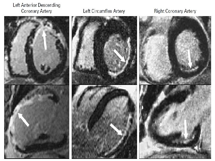
DE MRI Allows direct or indirect assessment of viability � Infarct characterization � In a study, presence of Microvascular obstruction, Increased LVEDV and Impaired LVEF in MRI are found to be independent predictors of adverse events. � Three patterns of enhanced signal hyperintensity: � 1) Early hypointensity No late hyperintensity (hypo), � 2) early Hyperintensity late hyperintensity (hyper); � 3)early hypointensity late hyperintensity (comb). �
 Indicated diffuse microvascular damage � Severe myocardial damage / Myocardial")
Type I pattern (hypo) Indicated diffuse microvascular damage � Severe myocardial damage / Myocardial necrosis � Poor functional recovery after revascularisation �

Recovery after Revasc. Depends on transmural extension Highly probable < 25%, � intermediate 25 - 75%, � Very low / null > 75% � � Hyperintensities restricted to Subendocardial area will recover contractility better

DE MRI compared to DSE � Nelson et al: TEI by DE MRI is inversly related to contractile reserve by DSE � Half of all segments which were fully viable by DE MRI had absence of contractile reserve. � Due to tethering of viable regions to scar regions, myocyte cellular adaptations that impair dobutamine response, and absence of coronary flow reserve in chronically hypoperfused regions.

AKINETIC SEGMENT NO SCAR ON MRI VIABLE SEGMENT BECAME FUNCTIONAL POST REVASCULARISATION REVERSIBLE DYSFUNCTION

AKINETIC SEGMENT SCAR ON MRI NON VIABLE SCAR AND AKINESIS WAS PERSISTENT POST REVASCULARISATION IRREVERSIBLE DYSFUNCTION

ORIGINAL ARTICLE The Use of Contrast-Enhanced Magnetic Resonance Imaging to Identify Reversible Myocardial Dysfunction Raymond J. Kim, M. D. , Edwin Wu, M. D. , Allen Rafael, M. D. , Enn-Ling Chen, Ph. D. , Michele A. Parker, M. S. , Orlando Simonetti, Ph. D. , Francis J. Klocke, M. D. , Robert O. Bonow, M. D. , and Robert M. Judd, Ph. D. N Engl J Med 2000; 343: 1445 -1453 ØGadolinium-enhanced MRI was performed in 50 patients with ventricular dysfunction before they underwent surgical or percutaneous revascularization. ØThe transmural extent of hyperenhanced regions was postulated to represent the transmural extent of nonviable myocardium. ØThe extent of regional contractility at the same locations was determined by cine MRI before and after revascularization in 41 patients.

RESULTS ØAn absence of delayed enhancement in segments that exhibit abnormal contractility has a positive predictive value of 78% for recovery after revascularization. ØA <25% delayed enhancement has a positive predictive value of 71%. ØThe percentage of the left ventricle that was both dysfunctional and not hyperenhanced before revascularization was strongly related to the degree of improvement in the global mean wall-motion score (P<0. 001) and the ejection fraction (P<0. 001) after revascularization.
 and thallium (Tl) activity at late redistribution")
Relationship between transmual extent of scar (TES) and thallium (Tl) activity at late redistribution (left) and increase in wall motion score (WMS) with low-dose dobutamine (right)

Accuracy of currently available techniques for prediction of functional recovery after revascularization in patients with left ventricular dysfunction due to chronic coronary artery disease: comparison of pooled data METHODS A systematic review of all reports on prediction of functional recovery after revascularization in patients with chronic coronary artery disease MODALITIES : Thallium-201 (Tl-201) stress-redistribution-reinjection, Tl-201 rest-redistribution, fluorine-18 fluorodeoxyglucose with positron emission tomography, technetium-99 m sestamibi imaging and low dose dobutamine echocardiography RESULTS: Sensitivity for predicting regional functional recovery after revascularization was high for all techniques. The specificity of both Tl-201 protocols was significantly lower (p < 0. 05) and LDDE significantly higher (p < 0. 01) than that of the other techniques CONCLUSIONS Although all techniques accurately identify segments with improved contractile function after revascularization, the Tl-201 protocols may overestimate functional recovery. The evidence available thus far indicates that LDDE appears to have the highest predictive accuracy. J Am Coll Cardiol, 1997; 30: 1451 -1460
 MEAN (95%")
Commonly Used Noninvasive Testing Modalities to Predict Regional Functional Improvement SENSITIVITY (%) MEAN (95% CI) SPECIFICITY (%) MEAN (95% CI) Dobutamine echocardiography 76 (72 -80) 81 (77 -84) Delayed enhancement by MRI 97 (91 -100) 68 (51 -85) FDG PET 89 (85 -93) 57 (51 -63) SPECT 89 (84 -93) 68 (61 -75) MODALITY Circulation 117: 103, 2008.

ACC/AHA/ASNC Guidelines Recommendations for the Use of Radionuclide Techniques to Assess Myocardial Viability J Am Coll Cardiol, 2003; 42: 1318 -1333 Indication Test Class Level of 1. Predicting improvement in Stress/redistribution/reinjection 201 Tl regional and global LV function after revascularization 2. Predicting improvement in heart failure symptoms after revascularization. 3. Predicting improvement in natural history after revascularization Perfusion plus PET FDG imaging Resting sestamibi imaging Gated SPECT sestamibi imaging Late 201 Tl redistribution imaging (after stress) Dobutamine RNA Postexercise RNA Postnitroglycerin RNA Perfusion plus PET FDG imaging 201 Tl imaging (rest-redistribution and stress/redistribution/reinjection) I Evidenc e B I IIa IIb B B B IIb IIb IIa C C C B I B

MR Coronary angiography � Useful in imaging Proximal and medium segments of major epicardial coronary arteries � Useful in identifying the origin and course of anomalous coronaries � Useful in assessing Bypass graft patencies (both Venous and Arterial) � Useful in Plaque characterisation of coronary vessel wall

Characterisation of Vessel Wall � Tissues rich in Protons will have hyperintense signal. � Calcifications have a low concentration of protons and appear hypointense. � The lipid plaques have both a short T 1 and a short T 2 and will be hyperintense in T 1 -weighted images � Fibrous plaques have a quite similar signal intensity in T 1 - and T 2 -weighted images
 tortuosity")
MR Coronary angiography � � � � Limitations: small arterial size (<5 mm) tortuosity complex anatomy cardiac and respiratory motion Mainly for assessing Proximal diameter stenosis and to rule out multivessel CAD At present assessment for surgical planning is difficult

3 D MR angio

3 D MRA of LIMA

Feasibility of MR coronary angio

Impact of Revascularization on LV Function � Studies shows LV ejection fraction improves significantly (ie ≥ 5%) after revascularization in 60% of patients (range 38% to 88%). � To predict 5% improvement in LVEF, at least 25% of LV should be viable using DSE and ≈38% using conventional nuclear medicine and PET. � In dyskinetic and akinetic segments, absence of scar or a transmural extension of scar of <25% have PPV of 88% and NPV 89% for functional recovery.

Treatment and Survival Rates � Meta-analysis that pooled data of 3, 088 pts from 24 studies demonstrated improved survival after revascularization in pts with hibernation. � Revascularization resulted in 79. 6% reduction in mortality (16% vs 3. 2%) � In absence of hibernation, no significant difference in mortality with revascularization (7. 7% vs 6. 2%).

Meta-analysis demonstrating outcome of patients with ischemic left ventricular dysfunction after viability testing J Am Coll Cardiol 39: 1151, 2002

Summary Stunning and hibernation are 2 causes for LV dysfunction. � Both conditions imply presence of viable myocardium and are reversible. �

THANK YOU
- Slides: 90