Myocardial stunning and hibernation Dr K V Patil

Myocardial stunning and hibernation Dr K V Patil

• several mechanisms of acute and chronic adaptation to a temporary or sustained reduction in coronary blood flow, known as stunning, hibernation, and ischemic preconditioning • Preserve sufficient energy to protect the structural and functional integrity of the cardiac myocyte

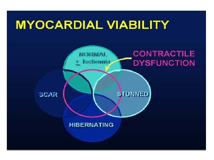
• Programmed cell survival has been used to describe the commonality among myocardial stunning, hibernation, and ischemic preconditioning despite their distinct pathophysiology

• Stunning: fully reversible dysfunction of the ischemic heart that persists despite the normalization of blood flow. • Myocardial dysfunction due to transient period of ischemia followed by reperfusion (depressed function at rest but preserved perfusion). • Severe ischemia can cause regional loss of electromotive potential without actual cell death (electrical stunning phenomenon)

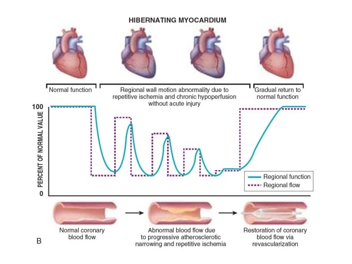
• Hibernating myocardium: adaptive responses of the myocardium to repetitive episodes of ischemia resulting in myocardial hypoperfusion at rest (depressed function and perfusion at rest).
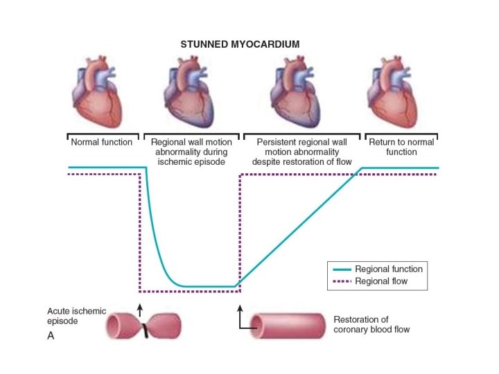


• time relationship for recovery of mechanical function, the intramyocardial electrogram and coronary flow after brief periods of regional myocardial ischemia • While regional electrograms return to normal within seconds and the coronary flow debt is repaid rapidly, functional derangement lasts for several hours. The Journal of Clinical Investigation Volume 56 October 1975@978 -985

• Coined the term ‘‘myocardial stunning’’ • delayed recovery of regional myocardial contractile function after reperfusion despite the absence of irreversible damage and despite restoration of normal flow.

Contd… • In many patients a positive inotropic intervention, postextrasystolic potentiation or exercise may result in substantial improvement of regional function • Postmortem examination of the hearts of patients with ischemic cardiomyopathy often reveals loss by infarction of only moderate quantities of myocardium. Horn HR, Teichholz LE, Cohn PF, Herman MV, Gorlin R: Augmentation of left ventricular contraction pattern in coronary artery disease by inotropic catecholamine: the epinephrine ventriculogram. Circulation 49: 1063, 1974 Dyke SH, Cohn RF, Gorlin R, Sonnenblick EH: Detection of residual myocardial function in coronary artery disease using postextrasystolic potentiation. Circulation 50: 694, 1974 Rozanski A, Berman D, Gray R, Diamond G, Raymond M, Prouse JA, Maddahi J, Swan HJC, Matloff J: Preoperative prediction of reversible myocardial asynergy by postexercise radionuclide ventriculography. N Engl J Med 307: 212, 1982

Features of stunning • Normal perfusion. • Depressed myocardial function. • Dissociation of usual relationship between subendocardial flow and function. • Reversible. • Function improves with inotropic agents.

• Recovery faster in the subepicardium than in the subendocardium – a non-uniform phenomenon that is most severe in the subendocardium • Both systolic and diastolic function depressed in stunned myocardium - global derangement of the mechanical properties of the heart.

• Stunning occurs in a wide variety of settings that differ from one another in several aspects • At experimental level it can occur during 1. Single , completely reversible episode of regional ischemia (< 20 min ) 2. Multiple, completely reversible episodes of regional ischemia first occlusion preconditions the myocardium against the next two occlusions so that the overall severity of stunning is the same after one or three occlusions. However, after the third occlusion, this preconditioning effect is negated, and additional occlusions cause a cumulative depression of contractility. A similar paradigm (i. e. , a partial preconditioning effect of the initial occlusions against subsequent occlusions) has been observed in conscious rabbits 3. Partly reversible plus partly irreversible ischemia in vivo ( > 20 min & < 3 hrs) Subepicardial tissue salvaged by reperfusion may require days or weeks to recover its contractile function
 5. After global ischemia in")
4. After global ischemia in vitro (isolated heart preparations) 5. After global ischemia in vivo (cardioplegic arrest) 6. After exercise-induced ischemia

Clinical Relevance • In the clinical setting stunning can occur 1. Brief period of total coronary occlusion: patients with angina due to spasm 2. Global ischemia after cardiopulmonary bypass. 3. In combination : Subendocardium is infarcted and overlying subepicardium reversibly injured in MI 4. Following exercise in presence of a flow limiting stenosis 5. Ischemic bout that is induced by PCI

Factors affecting severity of stunning • Severity and duration of flow deprivation • close coupling between the degree of myocardial dysfunction after reperfusion and the collateral blood flow during the preceding period of ischemia • Size and location of the ischemic region • severity of stunning greater in inner layers of the left ventricular wall - most severely ischemic • Myocardial temperature • Loading conditions of the heart

• “hypersensitive” myocardium; factors that affect contractile performance in the normal, healthy myocardium have a greater impact on the stunned, convalescent myocardium BOLLI, R. Oxyradicals in the pathogenesis of myocardial stunning. In: Intrinsic Adaptive Mechanisms During Ischemia and Reperfusion: Preconditioning, Stunning, Hibernation, edited by G. R. Heyndrickx, S. F. Vatner, and W. Wijns. New York: Raven, 1997, p. 205– 228.

Mechanisms of Stunning • There is no unified view of pathogenesis of stunning • Most plausible hypotheses: represent different facets of the same pathophysiological process Oxyradical hypothesis : oxidant stress secondary to the generation of ROS. Calcium hypothesis : results from disturbance of cellular calcium homeostasis.

Oxyradical hypothesis Effect of antioxidants • Stunning by reactive oxygen species [e. g. , superoxide anion, hydrogen peroxide, and hydroxyl radical] • In animal experiments, administration of superoxide dismutase (SOD) (which catalyzes the dismutation of superoxide anion to O 2 and H 2 O 2) and catalase (which reduces H 2 O 2 to O 2 and H 2 O) significantly enhanced recovery of function after reperfusion • Dimethylthiourea and mercaptopropionyl glycine (MPG) scavengers of hydroxyl radical significant and sustained improvement in the function of the stunned myocardium OH- is an important mediator of postischemic dysfunction • Desferrioxamine attenuated postischemic dysfunction prevention of the iron-catalyzed formation of OH-
 and electron paramagnetic resonance")
Direct evidence for oxyradical hypothesis • spin trap alpha-phenyl-N-tert-butylnitrone (PBN) and electron paramagnetic resonance (EPR) spectroscopy used to detect and measure production of free radicals in a series of animal studies - burst of free radical production was demonstrated immediately after reperfusion • Linear relation between the magnitude of free radical production and the magnitude of ischemic flow reduction • Interventions that alleviate the severity of ischemia early will indirectly attenuate free radical reactions after reflow

• aromatic hydroxylation of phenylalanine to specifically interrogate the role of OH- in myocardial stunning • Open chest animal studies : artifacts due to the combined effects of anesthesia, hypothermia, surgical trauma, volume and ionic imbalances, unphysiological conditions, cytokine release, and attending neurohumoral perturbations • But findings replicated in studies in conscious animal

Mechanisms involved • protein denaturation and enzyme inactivation • peroxidation of the polyunsaturated fatty acids contained in cellular membranes impaired selective membrane permeability • oxyradicals interfere with calcium transport and calcium-stimulated ATPase activity, Na+-Ca 2+ exchange and inhibit Na+-K+ATPase activity sodium overload, with consequent activation of the Na+-Ca 2+ exchange activity cellular calcium overload

• decreased responsiveness of myofilaments to calcium by producing selective damage of contractile proteins (oxidation of critical thiol groups) • exposure of myofilaments to superoxide anion has been shown to result in a dose-dependent reduction in maximal calcium-activated force myofilaments were found to be very sensitive to superoxide –significant force reduction being observed after exposure of <1 min to the superoxide-generating system

Sources of oxyradicals • canine and rat models: xanthine oxidase Interpretation of the effects of xanthine oxidase inhibitors is complicated by the fact that these agents have major, unanticipated effects to increase the responsiveness of the myofilaments to calcium • activation of the arachidonate cascade • autoxidation of catecholamines and other compounds • activation of various NAD(P)H oxidases • more importantly, damage of the mitochondrial electron transport chain.

• Stunning not alleviated by SOD and catalase in prolonged occlusion causing sub-endocardial infarction (but alleviated by cell-permeant antioxidants oxypurinol, N-acetylcysteine and Trolox) and exercise induced stunning

CALCIUM HYPOTHESIS • Calcium plays two distinct roles • activator of contraction • agent of injury • Three distinct postulated mechanisms • decreased responsiveness of the contractile protein machinery to calcium • calcium overload • excitation-contraction uncoupling

Role of Calcium in the Pathophysiology of Myocardial Stunning • Cardiac contractile force can be affected by • intracellular free calcium concentration [Ca 2+]i • contractile protein response to [Ca 2+]I • Loading myofilament responsiveness; consists of three components • Maximal force-generating capacity of the myofilaments (at saturating levels of [Ca 2+]i) • Sensitivity of myofilaments to calcium (i. e. , the range of [Ca 2+]I which activates the contractile proteins) • Crossbridge cycling kinetics

Excitation-contraction coupling in stunned myocardium • Kusuoka et al. - depressed maximal calcium activated pressure (the whole heart equivalent of maximal calcium-activated force) in stunned ferret hearts. KUSUOKA, H. , J. K. PORTERFIELD, H. F. WEISMAN, M. L. WEISFELDT, AND E. MARBAN. Pathophysiology and pathogenesis of stunned myocardium. Depressed Ca 21 activation of contraction as a consequence of reperfusioninduced cellular calcium overload in ferret hearts. J. Clin. Invest. 79: 950– 961, 1987. • Marban and co-workers - measured [Ca 2+]i in isolated perfused ferret hearts by NMR spectroscopy - tendency for systolic [Ca 2+]i to increase in the stunned hearts despite a 40% drop in developed ventricular pressure • Gao et al. confirmed above findings. simple changes of cross-bridge attachment and detachment rates in a quantitative model of myofilament interaction reproduced contractile dysfunction of stunned myocardium. • decreased maximal calcium activated force? decreased calcium sensitivity? or both?
 marked")
• reflow/proteolysis hypothesis: (Hofmann et al. and Van Eyk et al. ) marked depression of maximal force (45% with 15 min of ischemia) followed by reflow, but no depression with (15 min of) ischemia alone

Mechanism of decreased calcium responsiveness • oxygen radicals modifications of myofibrillar proteins (e. g. , by oxidation of thiol groups) • oxygen radicals decrease in the content of reduced glutathione and an increase in oxidized glutathione (shown to decrease calcium sensitivity) • alpha-actinin, troponin(Tn) I (partial degradation in stunned myocardium- but not in ischemic nonreperfused samples, prevented by modifications of the reperfusate designed to mitigate calcium overload -- Gao et al. )

Role of Calcium in the Pathogenesis of Myocardial Stunning Calcium overload after reperfusion • reperfusion injury calcium overload • Ischemia intracellular sodium ([Na+]i) accumulates, but Na+-Ca 2+ exchange is inhibited by the concomitant acidosis • Kusuoka et al. - isolated ferret hearts - reperfusion with low-calcium solutions markedly attenuates stunning

• transient calcium overload, even in the absence of ischemia, leaves behind long-lasting functional sequelae identical to those of stunned myocardium KITAKAZE, M. , H. WEISMAN, AND E. MARBAN. Contractile dysfunctionand ATP depletion after transient calcium overload in perfused ferret hearts. Circulation 77: 685– 695, 1988. • striking prevention of stunning when acidosis was induced during the initial minutes of reperfusion KITAKAZE, M. L. WEISFELDT, AND E. MARBAN. Acidosis during early reperfusion prevents myocardial stunning in perfused ferret hearts. J. Clin. Invest. 82: 920– 927, 1988.
![• [Na+]i increases rapidly during ischemia and remains significantly elevated for 8– 10](http://slidetodoc.com/presentation_image_h2/791be39bdb91a2da16795b3d294e4443/image-34.jpg "• [Na+]i increases rapidly during ischemia and remains significantly elevated for 8– 10")
• [Na+]i increases rapidly during ischemia and remains significantly elevated for 8– 10 min after reflow PIKE, M. M. , M. KITAKAZE, AND E. MARBAN. 23 Na-NMR measurements of intracellular sodium in intact perfused ferret hearts during ischemia and reperfusion. Am. J. Physiol. 259 (Heart Circ. Physiol. 28): H 1767—H 1773, 1990 • complete recovery of p. Hi within 30 s of reflow KITAKAZE, M. L. WEISFELDT, AND E. MARBAN. Acidosis during early reperfusion prevents myocardial stunning in perfused ferret hearts. J. Clin. Invest. 82: 920– 927, 1988. • effects of calcium overload are prevented by the acidosis associated with ischemia, increased concentration of protons effectively competes with calcium for intracellular binding sites

Role of calcium-activated proteases • Calpains • enzymes that cleave other proteins when cell calcium is elevated • cause only limited proteolysis - yield large protein fragments - changes caused may not be visualized using conventional histological methods • calpain I - very effective in digesting Tn. I and Tn. T in vitro • effects could be prevented by coincubation with excess calpastatin - natural inhibitor of calpain I

• Yoshida et al. • activity of calpain I increases after ischemia-reperfusion in isolated rat hearts • proteolysis of calspectin, a cytoskeletal protein and a specific substrate of calpain I, occurs after periods of ischemia as brief as 10 min followed by 30 min of reperfusion

 • intermembrane-linked protein that maintains the plasmalemma and sarcoplasmic")
• Junctophilin-2 (JPH 2) • intermembrane-linked protein that maintains the plasmalemma and sarcoplasmic reticulum at a fixed distance to ensure proper excitation–contraction (EC) coupling • essential for maturation of transverse tubules (TTs) and development of efficient EC coupling in adult cardiac myocytes

August 2018

Therapeutic implications • Reduction of myofilament calcium responsiveness rationalizes the remarkable efficacy of myofilament calcium sensitizers in the treatment of stunned myocardium • Reversibility • partially degraded contractile proteins replaced by newly synthesized ones to repair the myofilaments • proteolytic damage may play a role in the late rather than early phase of dysfunction • Therapies begun after the onset of ischemia can still be effective in preventing postischemic dysfunction; as damage occurs mainly during reperfusion

• a component of stunning not responsive to antioxidant therapy (no matter how vigorous) or to manipulations to alleviate postreperfusion disturbances in calcium homeostasis - likely to be caused by derangements that occur during ischemia • magnitude of the free radical generation is proportional to the magnitude of the flow deficit => intervention to attenuate the severity of the ischemic injury attenuates the severity of the subsequent reperfusion injury


Preconditioning • Acute preconditioning: • Brief reversible ischemia preceding a prolonged coronary occlusion reduces myocyte necrosis, a phenomenon termed acute preconditioning • can be induced pharmacologically using adenosine A 1 receptor stimulation as well as various pharmacologic agonists that stimulate protein kinase C or open mitochondrial K-ATP channels

• Delayed preconditioning • persists for up to 4 days • protein synthesis, with upregulation of the inducible form of NOS (i. NOS), COX-2, and opening of the mitochondrial K-ATP channel • Postconditioning • at reperfusion, ability to engage cardiac protection by producing intermittent ischemia or administering pharmacologic agonists • activation of reperfusion injury salvage kinase (RISK) pathways - activation of prosurvival kinases, such as Akt and Erk 1/2 Derek J. Hausenloy, Andrew Tsang, and Derek M. Yellon. The reperfusion injury salvage kinase pathway: a common target for both ischemic preconditioning and postconditioning. Trends Cardiovasc Med. 2005 Feb; 15(2): 69 -75

Myocardial hibernation • Downregulation of contractile function, as an adaptation to a reduction in myocardial blood flow, that serves to maintain myocardial integrity and viability during persistent ischemia • “Serum of truly hibernating animals - a substance with opioid-like activity that acts to preserve the myocardial ultrastructure and to improve the functional recovery from ischemia when given to nonhibernating animals”

• Diamond et al. - first in 1978 to use the term • 1980 s, Rahimtoola systematically reviewed the concept

• Short-Term Hibernation • In steady-state ischemia, the close matching between perfusion and contraction reduced regional oxygen consumption and energy utilization • extremely tenuous state, and small increases in the determinants of myocardial oxygen demand rapid deterioration in function and metabolism

Characteristics of chronic hibernation • Episodic and/or chronically reduced blood flow, which is directly responsible for the decrease in the myocardial contractile function. • Tissue ischemia and resultant remodeling without necrosis • Residual contractile reserve in response to inotropic stimulation (in at least half of clinical cases). • Recovery of contractile function after successful revascularization.

Mechanisms of hibernation • Smart heart hypothesis : Myocardial metabolism and function are reduced to match concomitant reduction in coronary blood flow which prevents necrosis. • Repetitive stunning hypothesis: Repetitive episodes of ischemia results in sustained depression of contractile function.

• Genomics of Survival Maintained viability in hibernation suggests possibility of genomic adaptation. Major survival genes (antiapoptotic, cytoprotective & growth-promoting genes) and their corresponding proteins are up regulated in hibernating myocardium.


Natural history of hibernation

Histological Features • Loss of contractile proteins without loss of cell volume. Most prominent near nuclear region. • Numerous small mitochondria adjacent to glycogen rich peri-nuclear zone • Loss of sarcoplasmic reticulum – becomes disorganized and loses T tubules • Increased expression of primitive cytoskeleton proteins – titin, cardiotin

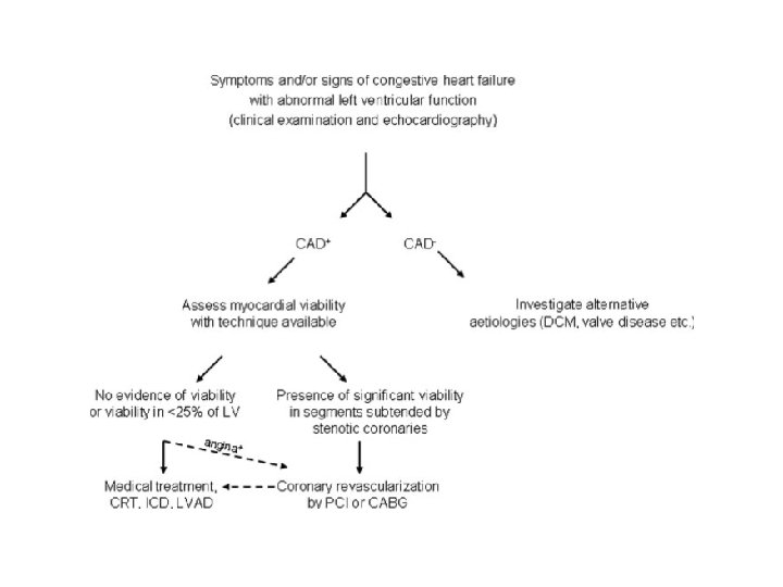
Need for understanding these concepts • Widespread use of thrombolytic therapy and percutaneous coronary interventions, increasingly potent antithrombotic agents significant reductions in mortality and morbidity • Greater number of patients with residual left ventricular (LV) dysfunction undergoing progressive LV remodeling and congestive heart failure. • Ageing population, increasing co-morbidities greater number of patients with multi-vessel disease, increased LV volumes and variable degrees of regional and/or global systolic dysfunction, isolated diastolic dysfunction

• coronary revascularization may lead to symptomatic and prognostic improvement, reverse LV remodeling. • 20 to 50 % of pts with chronic ischemic LV dysfunction have significant amount of viable hibernating myocardium. • They improve with revascularization.

ASSESSMENT OF MYOCARDIAL VIABILITY • ECG : gives little information. • Dobutamine stress echocardiography. • SPECT with thallium-201 or technetium-99 m. • PET • MRI

Characteristics of dysfunctional but viable myocardium

• ECG No clear correlation between Q waves on ECG and presence of viability. Pts with preserved QT dispersion are likely to have viable myocardium. Pts with high QT dispersion have predominantly nonviable scar tissue.

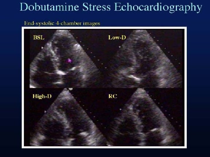
Dobutamine Stress Echocardiography • Extremely useful tool to document the early and late functional changes at rest. • Hypokinetic or akinetic regions improving during low dose dobutamine infusion (5– 10 µg/kg/min) is indicative of viable tissue. • At higher doses (upto 40 µg/kg/min plus atropine) wall motion may improve or diminish, reflecting inducible ischemia. • Biphasic response is highly predictive of recovery of function after revascularization.
 • improvement in contractile performance •")
Biphasic response • At lower doses(5– 10 mg/kg/min) • improvement in contractile performance • At higher doses (>15 mg/kg/min) • Contractility regresses as the metabolic demand stimulated overwhelms the tissue’s capacity to respond

Stress Echo Interpretation Rest / Baseline Low dose stress Peak & post stress Normal Hyper dynamic Ischemic Normal / severe ischemia – new RWMA Decreased Scar WMA No change Hibernating WMA Improved Worsens Stunned WMA Improved

STUDIESNagueh et al studied the transmural myocardial biopsies obtained from patients with hibernating myocardium Showed that tissue with >17% fibrosis failed to exhibit contractile reserve when challenged with low-dose dobutamine Circulation 1999, 100, 490– 496.

In a study by Pagano et al , they reported that the diagnostic accuracy of dobutamine-echocardiography was reduced with increasing severity of regional and global LV dysfunction. That is, the technique appeared to underestimate the extent of viability: 39% of all recovering LV segments failed to exhibit inotropic contractile reserve. Heart 1998; 79: 281 -288 Wiggers et al studied the functional recovery pre- and 6 months postrevascularization, and showed that low-dose dobutamine failed to identify 45% of the segments that ultimately regained function Am. Heart. J. 2000, 140, 928– 936.

Value of Dobutamine stress echo • Reductions in blood flow that lead to hibernation occurs with a corresponding spectrum of metabolic reserve. • Those regions with greater metabolic reserve will likely retain the ability to respond to an inotropic stimulus while those regions with profoundly reduced flow—just on the threshold of viability—will have no ability to respond. • Such regions appear to be nonviable • dobutamine-echocardiography may be considered an easily accessible tool however with sub-optimal sensitivity

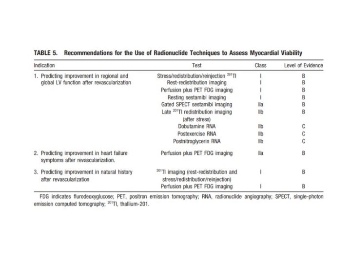
SPECT • Injection of a gamma-emitting radioisotope • Thallium-201 and Technetium Tc 99 m–Labelled Tracers - commonly used radionuclides • 201 Th : monovalent cation with biologic properties similar to those of potassium (major intracellular cation in muscle and is virtually absent in scar tissue ) well-suited radionuclide for differentiation of normal and ischemic myocardium from scarred myocardium. • Initial myocardial uptake early after intravenous injection - proportional to regional blood flow.

• Uptake of 201 Th: an energy-dependent process requiring intact cell membrane integrity preserved myocyte cellular viability. • Imaging is done • Immediately following stress, with either exercise or pharmacologically induced coronary hyperemia with dipyridamole or adenosine • After 3– 4 hr redistribution of Th-201

STANDARD SPECT IMAGING DISPLAY

INTERPRETATION Defects on post-stress images, may “fill in” by the time the rest-redistribution images are acquired, indicating viability. • A defect that persists and appears again on the 3– 4 hr images (i. e. , a fixeddefect) may be due to: • markedly reduced regional perfusion • impaired cellular membrane integrity, inadequate for the active sequestration of the tracer into the cell • cell death (acute infarction) • scar tissue. • Thus, fixed-defects on 3– 4 hr redistribution only severely hypoperfused and not necessarily infarcted tissue

• Late redistribution images • third set of images at 24 hours • allows for redistribution of the tracer to very-ischemic (yet viable) tissue • 22% of fixed defects (at early redistribution imaging) demonstrate normal Th-201 uptake at later redistribution. • may indicate a poorly perfused, yet viable region

201 Th reinjection • may be necessary because redistribution depends on the continued delivery of the tracer over the 3– 4 hr period. • blood concentration of Tl- 201 decreases to a great extent insufficient delivery of the tracer defect may not fill-in during redistribution imaging • second injection of thallium with delayed imaging greatest opportunity to sequester thallium.

99 m. Tc-sestamibi and tetrofosmin • Lipophilic molecule - intracellular retention requires intact mitochondrial function • Do not share the redistribution properties of 201 Th • Characteristics for predicting improvement in regional function after revascularization appear to be similar
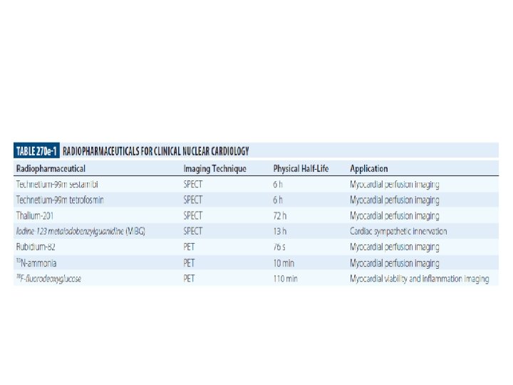

PET • Glucose utilization is evaluated with FDG and regional perfusion assessed with N 13 -ammonia, rubidium-82, or O 15 - labeled water. • FDG - transported into the cell by the same sarcolemmal carrier as glucose - phosphorylated to FDG-6 -phosphate by the enzyme, hexokinase. • Unidirectional reaction - intracellular accumulation of FDG-6 phosphate. • FDG does not undergo further metabolism - uptake is proportional to the overall rate of trans-sarcolemmal transport and hexokinase phosphorylation

• fatty acid oxidation stops shortly after the onset of severe ischemia and ischemic myocytes will derive energy from stored glycogen through anaerobic glycolysis. • After glycogen stores have been depleted, the ischemic myocyte efficiently use circulating glucose. • Under conditions of extremely diminished glucose delivery, sarcolemmal glucose transporters are upregulated to allow for increased uptake of this substrate.

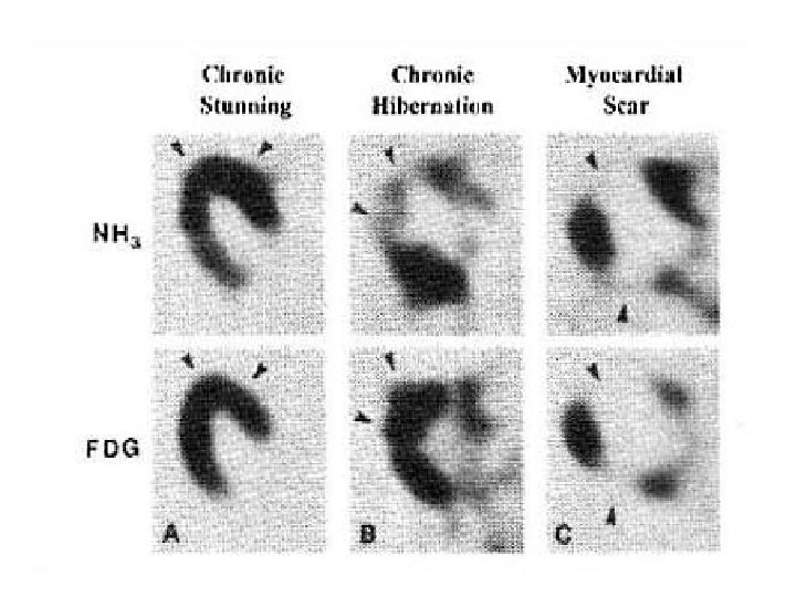
• no uptake of glucose by infarcted myocardium— which is metabolically inert—nonviable myocardium will appear as a region of low-FDG concentration in such images • areas of reversibly injured myocardium - glucose utilization is normal and even above normal • stunned or hibernating myocardium may be indistinguishable from normal tissue

PET perfusion imaging • 13 NH 3 and H 215 O • Initial uptake proportional to myocardial blood flow retention of 13 NH 3 depends on the metabolic integrity late-distribution 13 NH 3 PET images - useful in the assessment of myocardial viability • Exercise stress can be used

• 82 Rb does not need a cyclotron facility • Late-distribution 82 Rb images reflect myocardial viability as the successful uptake of 82 Rb depends on an intact myocyte membrane (a functional Na/K ATPase pump) • Pharmacological stress needed

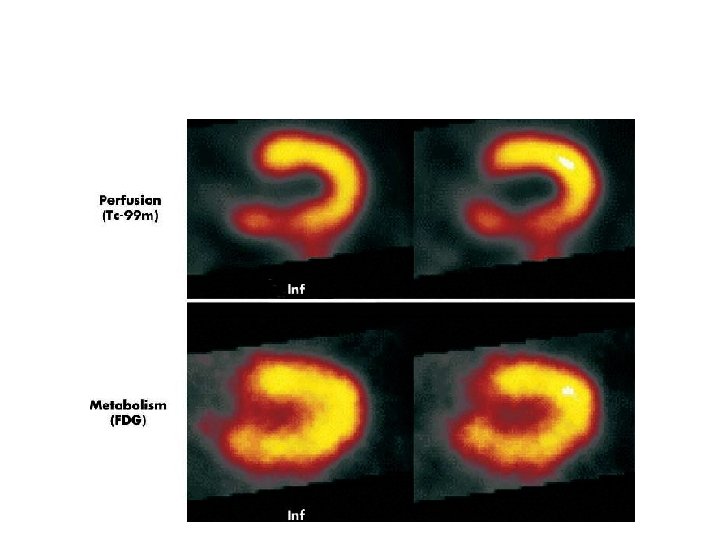
The Combined Perfusion/Metabolism PET • Long been considered the gold-standard for the identification of hibernating myocardium. • Low perfusion reserve by 13 NH 3 despite normal FDG uptake - highly predictive of both functional recovery and survival post revascularization.

SPECT VS FDG PET Brunken et al - a comparison of tomographic thallium images with PET images - 47% of the irreversible thallium defects identified as viable on PET images Circulation. Nov 1992; 86(5): 1357 -69. Tamaki et al confirmed these findings in 2 comparative studies of SPECT and PET - 38 -42% of the irreversible thallium defects had enhanced FDG uptake suggestive of viable myocardium. Am J Cardiol. Oct 15 1989; 64(14): 860 -5

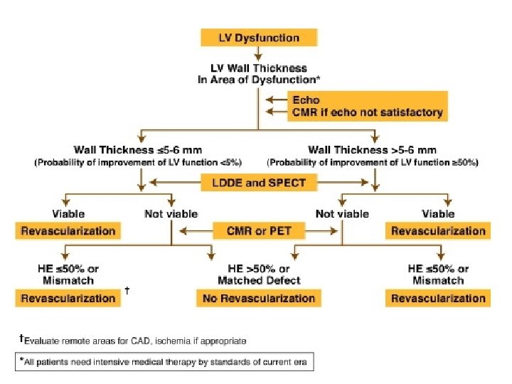
Magnetic resonance imaging Three techniques: • Resting MRI to measure end diastolic wall thickness < 6 mm represents transmural scar. • Dobutamine MRI to evaluate contractile reserve. Increased resolution of MRI avoid subjective variation of echo. Sensitivity of 89% & specificity of 94% to predict improvement after revascularization. • Contrast enhanced MRI to detect extent and transmurality of scar tissue. Can detect subendocardial scar.
 • Compared to SPECT with both thallium and Tc sestamibi")
Contractile reserve (DS MRI) • Compared to SPECT with both thallium and Tc sestamibi DS MRI is less sensitive but more specific with respect to recovery of contractile function after revascularisation • Sensitivity / Specificity : • 50 / 81% for MRI • 76 and 44% for SPECT thallium • 66 and 49% for SPECT Tc • Compared to PET (gold standard), sensitivity and specificity of dobutamine MRI for the diagnosis of myocardial viability - 81 and 95%.

DE MRI • Allows direct or indirect assessment of viability • Infarct characterization • In a study, presence of Microvascular obstruction, Increased LVEDV and Impaired LVEF in MRI are found to be independent predictors of adverse events. • Three patterns of enhanced signal hyperintensity: • 1) Early hypointensity No late hyperintensity (hypo), • 2) early Hyperintensity late hyperintensity (hyper); • 3)early hypointensity late hyperintensity (comb).
 • Indicated diffuse microvascular damage • Severe myocardial damage /")
Type I pattern (hypo) • Indicated diffuse microvascular damage • Severe myocardial damage / Myocardial necrosis • Poor functional recovery after revascularisation

Recovery after Revasc. Depends on transmural extension • Highly probable < 25%, • intermediate 25 - 75%, • Very low / null > 75% • Hyperintensities restricted to Subendocardial area will recover contractility better

Accuracy of non-invasive techniques to assess myocardial viability

Impact of Revascularization on LV Function • Studies shows LV ejection fraction improves significantly (ie ≥ 5%) after revascularization in 60% of patients (range 38% to 88%). • To predict 5% improvement in LVEF, at least 25% of LV should be viable using DSE and ≈38% using conventional nuclear medicine and PET. • In dyskinetic and akinetic segments, absence of scar or a transmural extension of scar of <25% have PPV of 88% and NPV 89% for functional recovery.

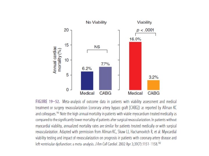
Treatment and Survival Rates • Meta-analysis that pooled data of 3, 088 pts from 24 studies demonstrated improved survival after revascularization in pts with hibernation. • Revascularization resulted in 79. 6% reduction in mortality (16% vs 3. 2%) • In absence of hibernation, no significant difference in mortality with revascularization (7. 7% vs 6. 2%).

THANK YOU
- Slides: 99