Myocardial Infarction 1 Outlines Introduction PATHOPHYSIOLOGY Causes Clinical


: Is the term used to describe irreversible myocardial necrosis")


- Slides: 26
Myocardial Infarction 1
Outlines • • Introduction PATHOPHYSIOLOGY Causes Clinical manifestation Dignostic evaluation Medical management Complication Nursing process
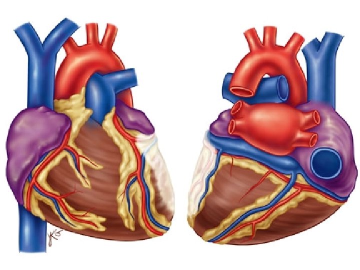
Introduction • Myocardial Infraction (MI): Is the term used to describe irreversible myocardial necrosis (cell death) that result from an abrupt decrease or total cessation of coronary blood flow to a specific area of the myocardium
PATHOPHYSIOLOGY • • Inflammation Plaque rupture Thrombus formation Irreversible damage starts in 20 to 40 minutes. This process will continue for several hours 4
causes • • Atherosclerosis Hypertension Smoking Use of oral contraceptive
Clinical manifestation • Sudden chest pain over the lower sternal region and upper abdomen is the presenting symptoms • Pain increased to reach the level of heavy rise like pain which may radiate to shoulders and down the arm (usually left arm)
Clinical manifestation • The pain begins spontaneously • The pain is accompanied by SOB, pallor • dizziness, nausea and vomiting
Diagnostic evaluation • • History ECG Echocardiogram Serial enzyme studies include CK, LDH, troponin
Medical managment 1. Pharmacotherapy • vasodilators: to treat myocardial pain as nitroglycerine • Anticoagulants : as heparin • Thrombolytics : use to dissolve any thrombus as streptokinase
Medical managment 2 Oxygen therapy 3 Analgesics: as morphin
• Initial treatment: MONA greets all patients. a. M= Morphine==== 2 -4 mg q 5 -15 min slow IV. Maintain BP more than 90 mm. Hg. Relief pain. Reduce PVR & work load on the heart. b. A=Aspirin (160 -325 mg) PO. Chewed & swallowed if possible. Determine if hypersensitive to ASA. 11
• N= Nitroglycerine SL for the 1 st 24 hrs. • O= Oxygen via NC 2 -5 L/M. • Continuous cardiac monitoring. • Hematology, chemistry, & lipid profile. • X-ray, Echo… Care of MI patients • bed rest with HOB elevated, beside commode. • Calm environment (limit stimuli & visitors, quite, calm environment). 12
complication • • Dysrhythmias Acut pulmonary edma Congestive heart failure thromboembolism
Nursing procss • Nursing diagnosis: 1. Chest pain R/T reduced coronary blood flow 2. Potential altered tissue perfusion R/T decreased cardiac output 3. Anxiety R/T fear of death
Nursing intervention 1. 2. 3. 4. 5. Relieving pain Administer oxygen Assess vital signs Provide complete physical rest Reduce anxiety
Anterior MI V 1, V 2, V 3, V 4 Anterior infarct with ST elevation Left Anterior Descending Artery (LAD) V 1 and V 2 may also indicate septal involvement which extends from front to the back of the heart along the septum Anterior septal MI most common type. Left bundle branch block Right bundle branch block 2 nd Degree Type 2 Complete Heart Block. Reciprocal changes (II, III, a. VF).
Lateral MI • I, AVL, V 5, V 6 – Lateral Infarction with ST elevations – Left Circumflex Artery – Rarely by itself – Usually in combo. – Reciprocal changes (II, III, a. VF).
Inferior MI leads II, III, AVF Inferior Infarct with ST elevations Right Coronary Artery (RCA) 1 st degree Heart Block 2 nd degree Type 1, 2 3 rd degree Block N/V common, Brady
pain • Begin in center of chest & may radiated to shoulders, neck, jaw or arm. • Last more than 15 -20 minutes and is not relieved with rest or nitroglycerin. 20
Chest Pain Anginal Pain: a. Sudden associated e other factors. b. Squeezing, vice like. c. Substernal, may radiated to back or arms. d. Usually lasts less than 15 min. e. Relieved with rest, nitrates, or oxygen. 21
Chest Pain MI pain: a. sudden, without precipitating factors. b. Intense, stabbing, vice like pressure, severe. c. Substernal, may radiate to back, arms, jaw, neck. d. Last more than 30 min. e. Relieved with opioids. 22
Anterior MI 23
Lateral MI 24
Posterior MI 25
Inferior MI 26