MYLOPOISIS AND DISORDERS OF WBC S Prof Dr

MYLOPOISIS AND DISORDERS OF WBC S Prof Dr Mandira Sharma

Hematopoiesis Red Blood Cells/Oxygen & CO 2 transport Platelets/Coagulation/Maintenance of vascular integrity White Blood Cells/Protection versus pathogens/microorganisms

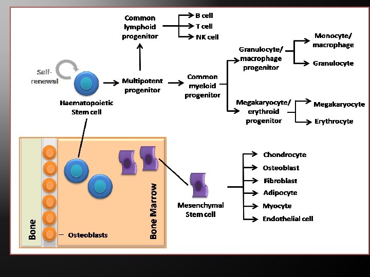
Hematopoiesis In humans, occurs in bone marrow exclusively All cellular elements derived from pluripotent stem cell (PPSC) PPSC retains ability to both replicate itself and differentiate Types of differentiation determined by the influence of various cytokines
 Humoral Regulation - Erythropoietin secreted by kidney stimulates erythropoiesis. 2)")
Regulation of Haematopoiesis 1) Humoral Regulation - Erythropoietin secreted by kidney stimulates erythropoiesis. 2) Role of inhibiting Mechanism - Feedback mechanisms controls over production of cells. Role of apoptosis. 3) Stromal cell products - Cytokines are mainly produced by Stromal cells and have a pivotal role in controlling haemopoiesis
 WBCs are divided into two groups: (1) granular leukocytes, containing")
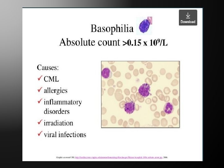
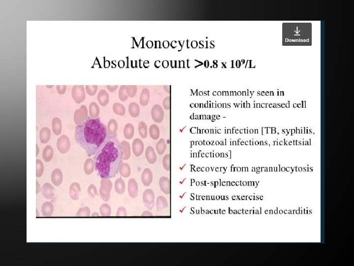
Leukocytes (white blood cells) WBCs are divided into two groups: (1) granular leukocytes, containing distinctive cytoplasm granules, including neutrophils, eosinophils and basophils (2) Agranular leukocytes, without granules, including monocytes and lymphocytes.

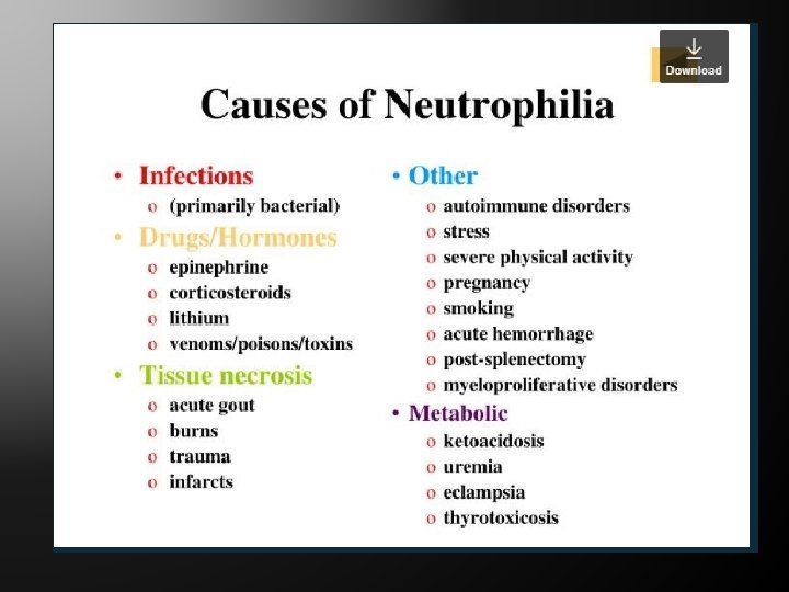
Causes of neutrophilia
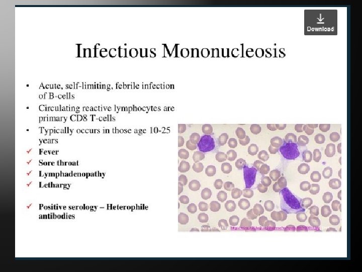
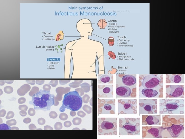

Infectious Mononucleosis Haematoligical Findings – Transformed lymphocytes shows > 50% lymphocytosis. These lymphocytes are transformed polyclonal T – lymphocytes CD 8 subset. -Blastoid cells( type III) : Nuclear chromatin of the cells are open with thin rim of cytoplasm. - Monocytoid cells ( type II): Kidney shaped/ lobulated nucleus with relatively open chromatin. - Plasmcytoid cell(type I) : Chromatin condensation – cartwheel pattern , perinuclear halo with basophilic cytoplasm

Infectious Mononucleosis Leucocytosis : TLC increased 15 -30. This is because of absolute lymphocytosis. Hb and platelets – normal Bone marrow – done to rule out leukemia EBV – specific antibodies are produced D/D Viral fever Acute leukemia Hodgkin's lymphoma
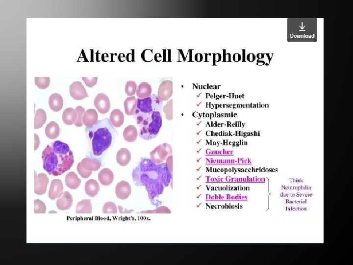

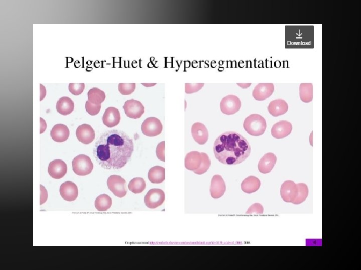
PELGER HUET Neutrophils show loss of segmentation. bilobed/band forms /dumb-bell shaped nuclei Nuclear chromatin coarse. Differentiated from shift to left, many bands forms seen as in bacterial infection

MAY-HEGGLIN Due to defect of MYH 9 gene. There is presence of basophilic inclusion in neutrophils and giant platelets

ALDER -REILLY There is lack of lysosomal enzymes for breakdown of mucopolysacharides. Neutrophils show large iliac inclusion

CHEDIAK -HIGASHI Giant granules are seen in mononuclear cells and polymorphs

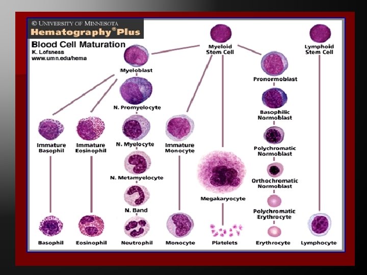
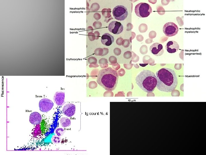
Granulopoiesis On the basis of colour of granules in the cytoplasm with Romanowsky stains the granulocytes are differentiated into neutrophils, eosinophils and basophils. Mature granulocytes are produced by the proliferation and maturation of precursor the Myeloblast.


Myeloblast: 15 -20 micron in size Round to oval nucleus No typical granules in cytoplasm. Nuclear chromatin is arranged in fine network. Nucleoli are prominent and 2 -4 in number.

Myelocyte Prominent cytoplasmic granules. Area of cytoplasm greater than nucleus. Cytoplasm is less basophilic. Nucleoli are absent. Myelocytes can divide.

Metamyelocyte : Cytoplasmic granules are fine pinkish Nuclear chromatin is coarse clumped and condensed in the periphery Nucleus is bean shaped. This is the beginning of nuclear segmentation. Band or stab form stage: Nuclear indentation is more than 50%

Lymphocytes ORIGIN AND DEVELOPMENT OF AGRANULOCYTES The agranulocytic series is comprised of leukocytes devoid of specific granulation. These cells generally originate in the lymphatic system This series includes the lymphocytic and monocytes groups.

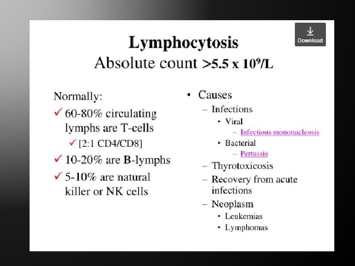
Functions of Lymphocytes T Lymphocytes: Involved in Cell Mediated immunity, 65 -70 % of lymphocytes in peripheral blood They are processed in Thymus Natural Killer Cells These do not carry markers of T and B cells B Lymphocytes involved in Humoral immunity by production of antibodies, 5 -15 -% of lymphocytes in peripheral blood Named as they differentiate in an organ called Bursa of Fabricius.

Lymphocytes The stages in lymphocytic development are: Lymphoblast Prolymphocyte Lymphocyte

LYMPHOBLAST Cell is similar to other blast cells. It is round or oval with a large round to oval reddish-purple nucleus. The nuclear chromatin material is fine and well distributed but perhaps more coarse than in myeloblasts. The nucleus contains one or two nucleoli. The cytoplasm is bluish and non granular and forms a thin rim around the nucleus.

Prolymphocyte - Size: 1218 µm. � Cytoplasm: Blue and � relatively more abundant than in the lymphoblast, nuclear chromatin tends to be clumped, no granules. � Nucleolus: Coarser chromatin structure, chromatin is not clumped but has a granular appearance. � Nucleoli: Poorly visible.

Lymphocytes Lymphocytes vary greatly in size and may be classified as small, medium or large. The cells are easily distorted and often appear in irregular shapes in stained preparations. The nuclear chromatin is condensed , discrete almost solid clumps, with thickening of the nuclear membrane. Nucleoli are absent. Non specific granules may be observed in the cytoplasm of these cells.
,")
Bone marrow The two types of bone marrow are medulla ossium rubra (red marrow), which consists mainly of hematopoietic tissue, � medulla ossium flava (yellow marrow), which is mainly made up of fat cells � At birth, all bone marrow is red � only around half of adult bone marrow is red �. In cases of severe blood loss, the body can convert yellow marrow back to red marrow to increase blood cell production. �

Bone marrow The stroma of the bone marrow is all tissue not directly involved in the primary function of hematopoiesis. Yellow bone marrow makes up the majority of bone marrow stroma, in addition to smaller concentrations of stromal cells located in the red bone marrow. Though not as active as parenchymal red marrow, stroma is indirectly involved in hematopoiesis, since it provides the hematopoietic microenvironment that facilitates hematopoiesis by the parenchymal cells.

Thanks …
- Slides: 43