MYELOID NEOPLASMS Dr Bindhusaran M D Hom Assistant
 Assistant Professor Department of Pathology &")
MYELOID NEOPLASMS Dr. Bindhusaran M. D. (Hom. ) Assistant Professor Department of Pathology & Microbiology

MYELOID NEOPLASMS • myeloid neoplasms has following 5 groups • I. Myeloproliferative diseases II. Myelodysplastic/myeloproliferative diseases III. Myelodysplastic syndrome (MDS) IV. Acute myeloid leukaemia (AML) V. Acute biphenotypic leukaemia

MYELOPROLIFERATIVE DISEASE • The myeloproliferative disorders are a group of neoplastic proliferation of multipotent haematopoietic stem cells. Besides their common stem cell origin, these disorders are closely related • Te WHO classifcation of myeloproliferative disorders includes 7 types
, {Ph chromosome t(9; 22) (q")
I. MYELOPROLIFERATIVE DISEASES • 1. Chronic myeloid leukaemia (CML), {Ph chromosome t(9; 22) (q 34; 11), BCR/ABL-positive} • 2. Chronic neutrophilic leukaemia • 3. Chronic eosinophilic leukaemia/ hypereosinophilic syndrome • 4. Chronic idiopathic myelofbrosis • 5. Polycythaemia vera (PV) • 6. Essential thrombocythaemia (ET) • 7. Chronic myeloproliferative disease, unclassifable
: It is established by identification of the clone of")
CHRONIC MYELOID LEUKEMIA DEFINITION (WHO): It is established by identification of the clone of haematopoietic stem cell that possesses the balanced reciprocal translocation between chromosomes 9 and 22, forming Philadelphia chromosome. The t (9; 22). The fusion product so formed is termed “Philadelphia chromosome t (9; 22), BCR/ ABL” which should be positive for making the diagnosis of CML.

philadelphia chromosome
 ABL protein is activated")
PATHOPHYSIOLOGY: BCR fusion product brings about following functional changes: (1) ABL protein is activated to function as a tyrosine kinase enzyme that in turn activates other kinases which inhibit apoptosis. (2) Ability of ABL to act as DNA- binding protein is altered. (3) Binding of ABL to actin microfilaments of the cytoskeleton is increased.

PATHOPHYSIOLOGY: Exact mechanism of progression of CML to the blastic phase is unclear but following mechanisms may be involved: (1) Structural alterations in tumour suppressor p 53 gene. (2) Structural alteration in tumour suppressor RB gene. (3) Alterations in RAS oncogene. (4) Alterations in MYC oncogene. (5) Release of cytokine IL- 1 BETA. (6) Fuctional inactivation of tumour suppressor protein, phosphatase A 2.

CLINICAL FEATURES: The onset is generally insidious. Both sexes are equally affected. Common presenting manifestations are: 1. Anaemia 2. Hypermetabolism- weight loss, lassitude, anorexia, night sweats. 3. Spleenomegaly- always present, frequently massive. 4. Bleeding tendencies- epistaxis, menorrhagia, haematomas. 5. Less common features – gout, visual disturbance 6. Juvenile CML- associated with lymphnode enlargement.

LABORATORY FINDINGS: I. BLOOD PICTURE: 1. Anaemia- moderate degree , normocytic normochromic type , occasional normoblast present. 2. WBC – Leucocytosis (appr 200, 000/ µl or more at the time of presentation). Consist of 3 phases: (a) chronic phase of CML – myeloblasts and basophil below 10% (b) accelerated- myeloblasts and basophil 10 – 20 % (c) blastic phase - myeloblasts and basophil > 20 % 3. Platelets- raised in half of the cases.
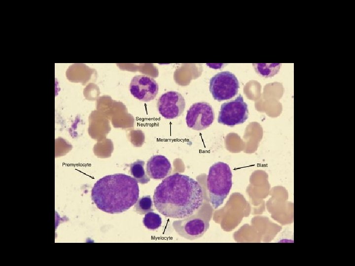
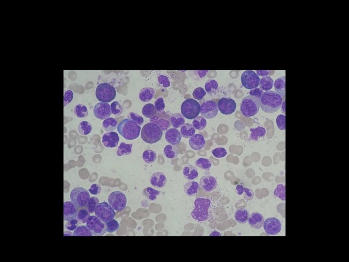

II. BONE MARROW EXAMINATION: 1. Cellularity- hypercellularity with total or partial replacement of fat spaces by proliferating myeloid cells. 2. Myeloid cells- they predominate in the bone marrow with increased myeloid- erythroid ratio. 3. Erythropoiesis- normoblastic but there is reduction in erythropoietic cells. 4. Megakaryocytes- smaller in size than normal. 5. Cytogenetics- show Philadelphia chromosome

III CYTOCHEMISTRY: Neutrophil alkaline phosphatase is reduced IV. Other findings • Elevated serum uric acid • Elevated serum B 12

POLYCYTHEMIA VERA • It is clonal disorder characterized by increased production of all myeloid elements result in pancytosis

pathogenesis • tyrosin
- Slides: 16