Myelin Components Central and peripheral myelin also contain


 - Deletion 5 of 6 exons for myelin basic")


 Central Nervous System Brain, spinal cord, retina 2) Peripheral Nervous")
 Somatic - carries voluntary motor and sensory information both to")
 Somatic - peripheral nerve fibers that send sensory information to")

 Somatic 2) Autonomic - controls smooth muscle of the viscera")


 - carry information INTO the")
 Spinal Cord 2) Cerebral Hemispheres - cerebral cortex and 3")


 prefrontal area- emotional control center and home to")
 Involves sensation and perception. Integrates sensory information")


- Slides: 41
Myelin Components Central and peripheral myelin also contain myelin basic proteins. Seven related proteins produced from a single gene by alternative splicing. Proteins are highly antigenic. Inject into animals autoimmune response called experimental allergic encephalitis (demyelination in CNS)
Schwann cells in the peripheral nervous system. 2
3
Mice - shiverer mutation (recessive) - Deletion 5 of 6 exons for myelin basic protein on chromosome 18 cause tremors, frequent convulsions and die young Homozygous - less than 10% normal myelination Inject wild type gene into fertilized eggs - transgenic mice express gene at right time produce about 20% of normal amount of proteins much more myelination occasional tremors but do not convulse and have normal life span
Glia and Axon Regeneration If peripheral axons severed, they grow back because: - axons and associated myelin break down - axonal and myelin debri, removed by surviving Schwann cells and macrophages. - tubular structures defined by basal lamina retained. Components contained in basal lamina potent promoters of neurite growth Schwann cells secrete their own growth factors and have membrane proteins that aid neuron growth See CNS repair chapter (PDF)
Make natural tubes to “guide” axons peripheral grafts containing support cells and cut axons Also use of embryonic cells which are not subject to regeneration limitations Inject Schwann cells into area http: //web. sfn. org/content/Publications/Brain Briefings/spinal_cord. html
Myelin in the brain and spinal cord gets in the way of axon regeneration Interfering with myelin can aid axon repair and restore some function in rodents with spinal cord injuries. - a vaccine against myelin prompted axons regrowth and treated animals regained some movement in their hind legs Other possible approaches? Identify specific molecules signaling macrophages to ingest and remove myelin from the damaged spinal cord. Target specific components of myelin, instead of whole sheath
Some proteins present in CNS myelin: At least MAG and Nogo are capable of causing growth cone collapse and inhibiting neurite outgrowth in vitro. Have a common receptor (Ng. R). See Paper PDF myelin-associated glycoprotein (MAG) Nogo-66 receptor (Ng. R).
http: //web. sfn. org/content/Publications/Brain. Briefings/brain_spinalcord. html Nogo, may be partly responsible for the inability of damaged axon fibers to repair. Normal neuron Neuron treated with synthesized Nogo
The Nervous System 1) Central Nervous System Brain, spinal cord, retina 2) Peripheral Nervous System Everything (except the retina) outside of the brain and spinal cord
Peripheral Nervous System 1) Somatic - carries voluntary motor and sensory information both to and from the CNS. 2) Autonomic a. sympathetic b. parasympathetic 3) Enteric - meshwork of nerve fibers that innervate the viscera (gastrointestinal tract, pancreas, gall bladder).
Peripheral Nervous System 1) Somatic - peripheral nerve fibers that send sensory information to the central nervous system AND motor nerve fibers that project to skeletal muscle.
Somatic Nervous System http: //faculty. washington. edu/chudler/nsdivide. html The cell body is located in either the brain or spinal cord and projects directly to a skeletal muscle.
Peripheral Nervous System 1) Somatic 2) Autonomic - controls smooth muscle of the viscera (internal organs) and glands. a. sympathetic - "fight" or take "flight" (run away) b. parasympathetic - "rest" and "digest" 3) Enteric
Fight-or-Flight Response
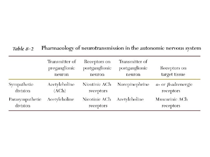
Autonomic Nervous System Sympathetic ACh NE Parasympathetic ACh http: //faculty. washington. edu/chudler/nsdivide. html Preganglionic neuron -located in either the brain or the spinal cord and projects to an autonomic ganglion. Postganglionic neuron - projects to the target organ.
SYM PARASYM ACh NE ACh http: //home. swipnet. se/sympatiska/nervo us. htm
Ways of Characterizing Peripheral Nervous System Nerves Sensory (afferent) - carry information INTO the central nervous system from sense organs. 1 OR Motor (efferent) - carry information away from the central nervous system (for muscle control) Cranial Nerve - connects the brain with the periphery. 2 OR Spinal Nerve - connects the spinal cord with the periphery. Somatic - connects the skin or muscle with the central nervous system. 3 OR Visceral - connects the internal organs with the central nervous system.
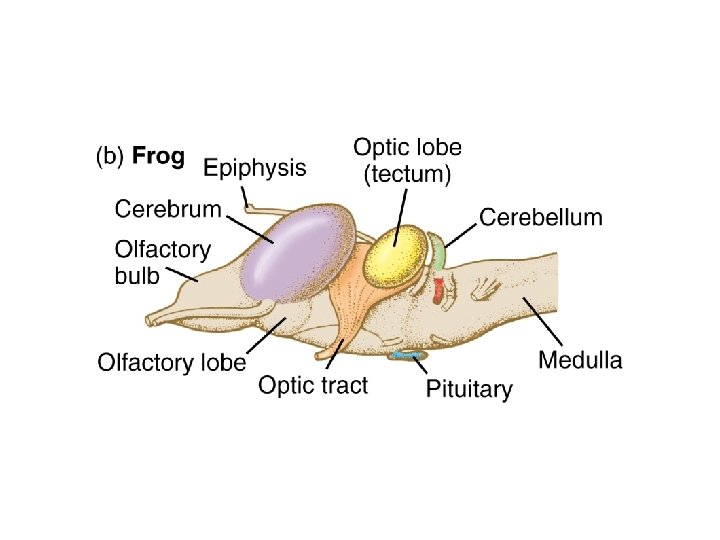
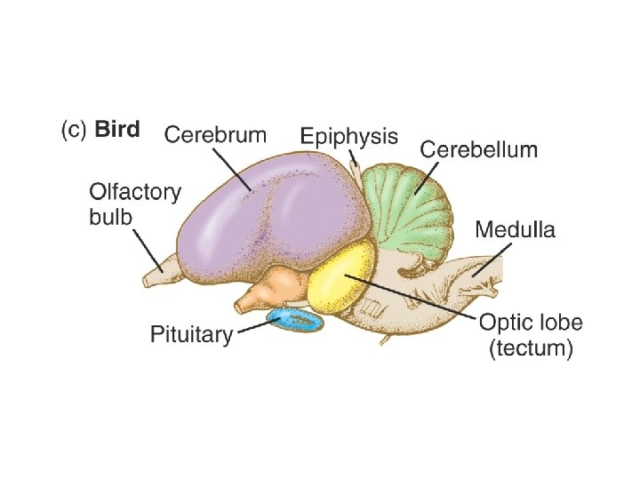
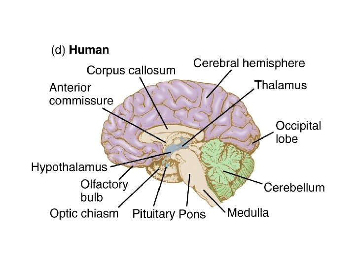
Central Nervous System 1) Spinal Cord 2) Cerebral Hemispheres - cerebral cortex and 3 deep lying nuclei: basal ganglia, hippocampus and the amygdala. 3) Diencephalon - thalamus and hypothalamus 4) Midbrain - superior and inferior colliculi 5) Medulla 6) Pons 7) Cerebellum
The Spinal Cord http: //thalamus. wustl. edu/course/spinal. html The spinal cord runs from the base of the skull to the first lumbar vertebrae. 31 pairs of spinal nerves
A Simple Reflex http: //thalamus. wustl. edu/course/spinal. html Afferent - sensory input. Efferent - motor output.
Levels of the Spinal Cord http: //thalamus. wustl. edu/course/spinal. html
Dorsal Columns - contains primary afferent axons. Ventral Columns - descending motor axons controlling posture. Axons relaying info about pain and thermal sensation to higher levels Lateral Columns - axons that ascend to higher levels and axons from nuclei in brain stem to motorneurons and interneurons in spinal cord.
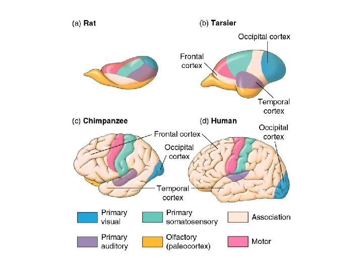
The Cerebral Cortex - Outermost layer of the cerebral hemisphere. - Composed of gray matter. - Cortices are asymmetrical. Both analyze sensory data, perform memory functions, learn new information, form thoughts and make decisions.
Then: and Now: http: //www. niehs. nih. gov/kids/brain. htm http: //pages. britishlibrary. net/phrenology/images. html
Sulci - grooves Gyri -elevated regions http: //www. neuroskills. com/index. html? main=tbi/brain. shtml
http: //thalamus. wustl. edu/course/basmot. html
The Frontal Lobes Divided into: a) prefrontal area- emotional control center and home to our personality. Involved in motor function, problem solving, spontaneity, memory, language, initiation, judgement, impulse control, and social and sexual behavior. b) premotor area -contains neurons that produce movements.
The Parietal Lobes Two functional regions: 1) Involves sensation and perception. Integrates sensory information to form a single perception (cognition). 2) Integrates sensory input, primarily with the visual system to construct a spatial coordinate system to represent the world around us.
The Occipital Lobes Center of our visual perception system. Disorders of this lobe can cause visual hallucinations (visual images with no external stimuli) and illusions. http: //www. neuroskills. com/index. html? main=tbi/brain. shtml
The Temporal Lobes Involved in the primary organization of sensory input and also highly associated with memory skills. Left temporal lesions result in impaired memory for verbal material. Right side lesions result in impaired recall of non-verbal material, such as music and drawings. http: //www. neuroskills. com/index. html? main=tbi/brain. shtml Language can also be affected by temporal lobe damage. Left lesions disturb recognition of words. Right damage can cause a loss of inhibition of talking.