MYCOPLASMA and Ureaplasma Dr Rouchelle Tellis Associate Professor


- Slides: 31

MYCOPLASMA and Ureaplasma Dr. Rouchelle Tellis Associate Professor Microbiology Dept. YMC

MYCOPLASMA ¬ Smallest free-living micro organisms, lack cell wall. ¬ Size: Varies from spherical shape (125 -250 nm to longer branching filaments 500 -1000 nm in size. ¬ Pass through a bacterial filter. ¬ Isolated by Nocard & Roux (1898) –bovine pleuropneumonia (PPO)

¬Later, many similar isolates were obtained from animals, human beings, plants & environmental sources – called as “Pleuropneumonia like organisms”(PPLO).

MYCOPLASMA ¬ Eaton (1944) first isolated the causative agent of the disease in hamsters and cotton rats. ¬ Also known as Eaton agent. ¬ 1956 - PPLO replaced by Mycoplasma. – Myco : fungus like branching filaments – Plasma : plasticity ¬ highly pleomorphic – no fixed shape or size - Lack cell wall.

Morphology and Physiology – Require complex media for growth ¬ Facultative anaerobes: Except M. pneumoniae - strict aerobe • No cell wall means these are slow growing & resistant to penicillins, cephalosporins and vancomycin, etc.

Mycoplasmas of Humans M. pneumoniae URTI, tracheobronchitis, atypical pneumonia M. hominis M. genitalium Pyelonephritis, PID, Post partum fever Non Gonococcal Urethritis U. urealyticum Non Gonococcal Urethritis

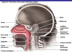
Pathogenicity of M. pneumoniae ¬ Produce surface infections – adhere to the mucosa of respiratory, gastrointestinal & genitourinary tracts with the help of adhesin. ¬ Two types of diseases: 1. Atypical Pneumonia 2. Genital infections

Pathogenicity cont’

Mycoplasmal pneumonia ¬Primary Atypical Pneumonia/ Walking pneumonia. ¬Incubation period: 1 -3 wks ¬Transmission: airborne droplets of nasopharyngeal secretions, close contacts (families, military recruits).

Mycoplasmal pneumonia ¬Gradual onset with fever, malaise, chills, headache & sore throat. ¬Severe cough with blood tinged sputum (worsens at night) ¬Complications: bullous myringitis & otitis, meningitis, encephalitis, hemolytic anemia

Clinical Syndrome - M. pneumoniae ¬ Incubation - 2 -3 weeks ¬ Fever, headache, malaise, persistent, dry, nonproductive cough ¬ Respiratory symptoms – Patchy bronchopneumonia – acute pharyngitis may be present ¬ Organisms persist, Slow resolution, Rarely fatal

Epidemiology - M. pneumoniae ¬ Occurs worldwide, No seasonal variation – Proportionally higher in summer and fall ¬ Epidemics occur every 4 -8 year ¬ Spread by aerosol route (Confined populations). ¬ Disease of the young (5 -20 years), although all ages are at risk

Laboratory Diagnosis ¬ Specimens – throat swabs, respiratory secretions. ¬ Microscopy – 1. Highly pleomorphic, varying from small spherical shapes to longer branching filaments. 2. Gram negative, but better stained with Giemsa stain

Laboratory Diagnosis - M. pneumoniae ¬ Culture (definitive diagnosis) – Sputum (usually scant) or throat washings – Special transport medium needed • Must suspect M. pneumoniae – May take 2 -3 weeks or longer, 6 hour doubling time with glucose and p. H indicator included – Incubation with antisera to look for inhibition.

Laboratory Diagnosis ¬ Isolation of Mycoplasma (Culture) – 1. Semi solid enriched medium containing 20% horse or human serum, yeast extract & DNA. Penicillium & Thallium acetate are selective agents. (serum – source of cholesterol & other lipids) 2. Incubate aerobically for 7 -12 days with 5– 10% CO 2 at 35 -37°C. 3. Colonies seen using hand lens or Dienes staining: alcoholic solution of methylene blue & azure

Laboratory Diagnosis ¬ Typical “FRIED EGG” appearance of colonies Central opaque granular area of growth extending into the depth of the medium, surrounded by a flat, translucent peripheral zone.

Fried egg colonies Dr Ekta, Microbiology, GMCA

Identification of Isolates ¬ Growth Inhibition Test – inhibition of growth around discs impregnated with specific antisera. ¬ Immuno-fluorescence on colonies transferred to glass slides. ¬ Molecular diagnosis: PCR-based tests

Identification of Isolates ¬ Serological diagnosis 1. Specific tests – IF, HAI 2. Non specific serological tests – cold agglutination tests (Abs agglutinate human group O red cells at low temperature, 4 C). 1: 32 titer or above is significant.

Treatment and Prevention M. pneumoniae – Macrolides are DOC (Azithromycin) – Other antibiotics: Tetracycline in adults (doxycycline) or erythromycin (children) • Newer fluoroquinolones (in adults) – Resistant to cell wall synthesis inhibitors. ¬ Prevention – Avoid close contact

Ureaplasma urealyticum ¬ M. hominis, M. genitaliulium, Ureaplasma: cause urogenital infections ¬ Colonize female lower urogenital tract ¬ Transmission: sexual contact/ to fetus during birth

Genital Infections ¬ Men - Nonspecific urethritis, proctitis, balanoposthitis & Reiter’s syndrome ¬ Women – acute salpingitis, PID, cervicitis, vaginitis ¬ Also associated with infertility, abortion, postpartum fever, chorioamnionitis & low birth weight infants

Treatment: Macrolides: (azithromycin) for ureaplasma & M. genitalium infections Doxycycline : M. hominis

Mycoplasma as cell culture contaminants ¬ Contaminates continuous cell cultures maintained in laboratories ¬ Interferes with the growth of viruses in these cultures. ¬ Mistaken for viruses. ¬ Eradication from infected cells is difficult.

POINTS TO BE REMEMBER Ø Mycoplasma Ø No cell wall Ø Pleomorphism Ø Fried egg colonies Ø Diene’s stain ØCold agglutination test ØCell culture contamination ØUreaplasma hydrolysis of urea ØPrimary atypical/ walking pneumonia Ø Genital infections

MCQ Q. 1. Which of the following bacteria was named as Eaton agent a) Acholeplasma b) Mycoplasma hominis c) Mycoplasma pneumoniae d) Ureaplasma urealyticum Q. 2. Dienes method is used to examine colonies of a) Bordetella b) Burkholderia c) Mycoplasma d) Helicobacter

Q. 3. Which of the following bacteria is/are associated with nongonococcal urethritis ? a) Mycoplasma hominis b) Ureaplasma urealyticum c) Chlamydia trachomatis d) All of the above Q. 4. Which is the causative agent of primary atypical pneumoniae a) Influenza virus b) Streptococcus Pneumoniae c) Haemophilus influenzae d) Mycoplasma pneumoniae

Q. 5. Which of the following can hydrolyse urea a) Mycoplasma b) Acholeplasma c) Ureaplasma d) Escherichia Q. 6. Which of the following bacteria is/are also named T strain ? a) Mycoplasma pneumoniae b) Mycoplasma hominis c) Ureaplasma urealyticum d) Acholeplasma

Q. 7. Postpartum fever due to Mycoplasma hominis is treated with a) Penicillin G b) A second generation Cephalosporins c) Vancomycin d) Tetracyclines/ doxycycline Q. 8. Distinguishing feature of human mycoplasma species is: a) Stain well with Giemsa, but not by Gram stain b) Contain no bacterial peptidoglycan c) Are not immunogenic because they mimic host cell membrane components d) Cannot be cultivated in vitro

Q. 9. which of the following tests can be used to identify Mycoplasma pneumoniae ? a) Haem-adsorption test b) Tetrazolium reduction test c) Inhibition of growth by specific antisera d) All of the above Q. 10. Which of the following bacteria shows fried egg colonies on culture media ? a) Helicobacter b) Mycobacterium tuberculosis c) Bordetella d) Mycoplasma

ANSWERS OF MCQ Q. 1 Q. 2 Q. 3 Q. 4 Q. 5 Q. 6 Q. 7 Q. 8 Q. 9 Q. 10 - C C d d C C d b d d
 Promotion from assistant to associate professor
 Dimorphic fungus
 Mycoplasma pneumoniae
 Mycoplasmas rickettsiae and chlamydiae are
 Vagninitis
 Ureaplasma pozitiva
 Rez. cystitiden
 Ureaplasma urealyticum positivo tem cura
 Mbcs membership
 Tecniche associate al pensiero computazionale:
 The pyramid at chichen itza is most closely associate with
 Lonestar kingwood nursing
 Certified systems engineering professional
 Direct mapping cache
 Cern hr
 Associate degree in the netherlands
 Associated consulting engineers
 Jeannie watkins
 Rcog cpd portfolio
 Rekenhulp tegemoetkoming scholieren
 What is an associate director
 Harper college associate degrees
 Iter project associate
 Michelin aad program
 My sis harbor college
 Active critical reading
 Delta chi associate member pin
 Associate degree rmit
 Adobe spark certificate
 Cincinnati state associate degrees
 Safety associate
 Associate warden

