
Mycobacteria Fungal Respiratory Tract Pathogens Prof Dr Asem

Mycobacteria & Fungal Respiratory Tract Pathogens Prof. Dr. Asem Shehabi Faculty of Medicine University of Jordan
Global prevalence of TB n n The World Health Organization (WHO) estimates that approximately one-third of the global population is infected with M. tuberculosis (TB). Around 9 to 10 million new cases of TB are being reported each year, 2 -3 million deaths occur each year worldwide. . 95% in developing countries Tuberculosis is considered the leading cause of death due to infectious disease After emerging (HIV)/AIDS, TB is the second most common cause of death in AIDS patients due to an infectious disease.

Mycobacterium Tuberculosis n. Tubercle Bacilli. . rod-shaped aerobic Acid-Fast Bacilli. . Widely distributed in Human, Animals, Birds, Environment. . TB bacilli grow slowly, Resistant to Dryness, low Acidity. . survive years in nature. . But Susceptible to UV-light, Heat. n we have a special type of tubercle bacilli results in infection only in human as mycobacterium tuberculosis , and other is only associated with animals as Mycobacterium bovis …and other atypical mycrobacteria. n M. tuberculosis. . Causes 95% of human TB cases. . mostly pulmonary. . Respiratory droplet infection. . Few cells. . Lung positive person may infect hundred of susceptible person. . All ages. . mostly children. . with malnutrition. n Optimal conditions for transmission include: overcrowding, Large cities, poor conditions & Low standard public hygiene. . n Mycobacterium Tuberculosis has multi-resistant strains, which make it very hard to treat it. n. The treatment of TB is very expensive, and this will make a problem for the low-income countries. In Jordan, there is only few cases but the problem is in the affected foreigners, because TB can spread easily. “ only one single case of multi-drug resistant TB can cause spread of the disease from Hong Kong to New York !! “

Acid Fast Bacilli Stained by Ziehl-Neelsen (Acid Fast Stain) This is an electron microscope image exhibits an acid fast bacilli stained red by special stain called Ziehl neelsen stain. It's important to know that generally these bacilli appear in clusters rather than single bacillus.
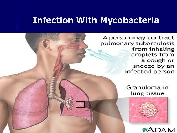
Infection With Mycobacteria
Pulmonary Infection n n Primary Tuberculosis: 90% Pulmonary TB Children, Asymptomatic, TB Bacilli infect Alveolar macrophages. . Develop small lung lesions. . Fibrosis, Calcification, Hypersensitivity, fever, productive cough, weakness. . Infected person becomes Positive for Tuberculin Skin test. Mycobacterium Tuberculosis can be diagnosed from the Acid-fast Bacilli found in the sputum, so the productive cough is a very important feature in TB, Because this organism is not always apparent on the lung X-Ray Few cases (1%). . TB bacilli may spread from primary Lesion by direct extension to lymphatic system, bronchi, blood, Kidneys “ could be found in urine” , Gastrointestinal, Asymptomatic Meningitis (children). . rarely developing Military tuberculosis. Progressive primary TB: Primary TB may go on to heal as caseating granulomas are replaced by fibrosis and calcification. However, some cases do not heal spontaneously or with therapy, and progress to form cavities or spread to other parts of the lung and other organs of the body through lymphatic channels and the blood stream, developing progressive primary TB.

Post-Pulmonary Infection-2 n n n Post primary tuberculosis: Reactivation old lesions. . Common in young adults & elderly persons. . Developing Large Lung lesions. . Cavities , Less lymphatic involvement , intensive Granuloma & Caseation. . May spread rapidly to other body part, CNS, Gastrointestinal/Urinary Tract. . positive TB urine culture ( especially in children who develops kidney TB leading to damaged kidney ). Clinical Features: Productive Cough. . bloody sputum, Low continuous Fever, Night-time sweating , Loss weight & Appetite. . General weakness, Lesions/ Cavities can be detected easily by chest x-ray. . Sputum culture +ve Generally , post primary tuberculosis results in positive tuberculin test.
Tuberculin Test n n n n Symptomatic/ asymptomatic infected persons. . develop positive Tuberculin skin test. . Reaction to Cell Wall Mycolic acids+lipoproteins Mantoux / Tuberculin skin test. or PPD-purified protein derivative-test. is produced from boiling culture of M. tuberculosis. 1, 1 o, 100 TU “(tuberculin unit - or we may use the PPD unit”. . intra-cutaneously injected in the forearm or taken as pills, The test is read after 48 -72 hours. Positive tuberculin: Indurations “ mainly on the forearm”, Edema & Erythematic skin> 1 cm, Interpretation: +/-ve. and it's important to know that +ve tuberculin test doesn't mean the presence of clinical case( i. E +ve test should not be ascribed to clinically apparent infection) , it just indicates contact with tubercle bacilli “hypersensitivity”. Negative tuberculin test means that there was no contact with tubercle bacilli. Yet , in cases of disseminated tubercle bacilli and presence of many lesions in lung and other parts of body , there would be overwhelming hypersensivity , that may result to over anergy to PPD test's antigens , leading to –ve tuberculin test. Vaccination with BCG ( Bacilli- Celements-Guerin). . Attenuated M. bovis . They use M. bovis because there is a cross reactivity related to cell wall & membrane between different types of mycobacteria . . . Protection 30 -78%. . result in positive Tuberculin test.
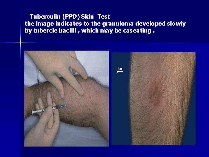
Tuberculin (PPD) Skin Test the image indicates to the granuloma developed slowly by tubercle bacilli , which may be caseating.

Other Human Pathogenic Mycobacteria species n n n M. bovis: common in domestic animal “camels mainly”. . rare human. . Infection. . source: unpasteurized milk, dairy products, meat. . begins mostly intestinal infection. . may spread to other parts. . Slow grower “ it needs at least 2 -6 weeks to identify the isolation “…. Well controlled in Jordan Atypical mycobacteria Widely distributed in nature. . water, soil, birds, animals and man… it might be associated with malignancy like lymphoma… including: M. kansasii “most common”: Photochromogenic “ by presence of the light, there is a change in the color”. . Rapid grower, produce yellow/orange color during incubation in light. . Mostly Lung tuberculosis. . immuno-suppressed persons, AIDS, and it is more difficult to be eradicated. Asymptomatic in healthy people. M. marinum “Avium intracellular” Mostly localized Skin ulcers-soft tissues Swimming pool, Granuloma. . Lymph nodes. . (water, low temperature). . Rapid grower , more common in animals , rarely produce pulmonary disease, Nonphotochromogenic. M. ulcernas. Skin lesions, necrosis, More Resistant to anti-tuberculosis drugs. . Slow grower M. avium complex (animals, water). . Skin Lesions, rarely Pulmonary disease. . Slow grower
Diagnosis & Treatment-1 n n n if you recognize the Acid fast bacilli in the urine, you have to wait for the lab results and don’t start the treatment directly, but if you recognize it in the pulmonary tract it is recommended to treat it as soon as possible. Rapidly growing Mycobacteria species: Rarely cause non-pulmonary diseases, mostly non-pathogens. . M. smegmatis. . Found in on extra genital tract. . May contaminate urine culture. Diagnosis & treatment: Tuberculosis is confirmed by positive Direct AF Smear/ Culture, Real-time PCR X-ray, Positive tuberculin Test. It is difficult to differentiate between typical and the atypical TB Clinical specimens: Sputum, Urine, CSF, Tissues, Culture Loewenstein. Jensen Agar. . 4 -8 Weeks. . No Blood Serological test. Treatment Multiple Antibiotics: it is very important to identify the pattern of susceptibility of the isolated organism in order to choose the right treatment. 6 “susceptible strains” -24 Months “multi-resistant strains”. . Rifampicin, Isonaized, Pyrazinamid, Ethambutol, Streptomycin, development of Multidrug resistant MB tuberculosis. . At present 1 -10% worldwide. . Completing treatment is essential for cure
Nocardiosis n n n Nocardia asteroids/ N. barsiliensis. Aerobic G+ve. . Pleomorphic Bacilli-Branched Filaments. . Slightly Acid Fast. . Common as Environmental Saprophytes… Difficult to be recognized in routine culture. . special culture media. . Human Exogenous Infection. . Mostly Pulmonary Localized Abscesses. . Necrosis. . small Cavities. . spread to Brain, Kidneys. . Common in Immuno-suppressed, Lung malignancy Chronic suppuration. . Dry Abscess. . Granulomas, Draining sinuses containing granules. . Muscles, Bones, Feet, Hands and other body parts. Diagnosis & Treatment: Sputum/biopsies culture on blood 3 -30 days at 45 C. . using sabarode dextrose agar, Co-trimoxazole “the best”, Rifampicin, Amikacin. . 4 -6 Weeks. Treatment is not sufficient , and in relation to lung a surgical operation should be done to remove the presence of localized lesions. It is similar to the fungal infections and TB. Some cases was seen in Jordan… associated with dialysis.
Respiratory Fungal Agents n n n Fungal respiratory diseases can be divided into: Generally healthy individuals Immuno-suppressed patients. Fungal agents. . Widely distributed in Environment. . Cause mostly infection in immuno-compromised individuals. . receiving immunosuppressive therapy. . undergoing bone marrow transplantation or solid-organ transplant. . HIV infection. . . patients suffered from former TB … or patients taking broad-spectrum antibiotics Clinical presentations of fungal respiratory infections are non-specific and often overlap with other infectious and non-infectious processes “might be primary o secondary infection”. The causative agents can be opportunistic Yeast “Candida or Cryptococcus “ or exogenous filmentous Fungi /Molds in immunocompromised patient
Yeast Form: Oral Candidiasis n n n n Part of oral /intestinal/vaginal flora. . causes characteristic mucosa patches of a creamy-white to grey pseudomembrane composed of Blastoconidia and Pseudohyphae. . C. albicans often after long antibiotics treatment. Oral candidiasis can be controlled by using topical treatments. “underlying disease” Oral candidiasis may spread. . Esophagus, bronchi, lungs, gastro-intestinal tract, or become systemic. . Candidiaemia “very dangerous”, endocarditis, meningitis. Systemic candidiasis is common in patients with cell-mediated immune deficiency, receiving aggressive cancer, immunosuppression, transplantation therapy. , can cause meningitis , very rare. C. albicans “highly susceptible to antifungal drugs”, Cause 70 - 80 % of oral candidiasis C. glabrata, C. tropicalis, C. krusei, “more resistant”
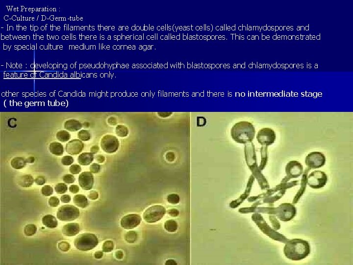
Wet Preparation : C-Culture / D-Germ-tube - In the tip of the filaments there are double cells(yeast cells) called chlamydospores and between the two cells there is a spherical cell called blastospores. This can be demonstrated by special culture medium like cornea agar. - Note : developing of pseudohyphae associated with blastospores and chlamydospores is a feature of Candida albicans only. other species of Candida might produce only filaments and there is no intermediate stage ( the germ tube)
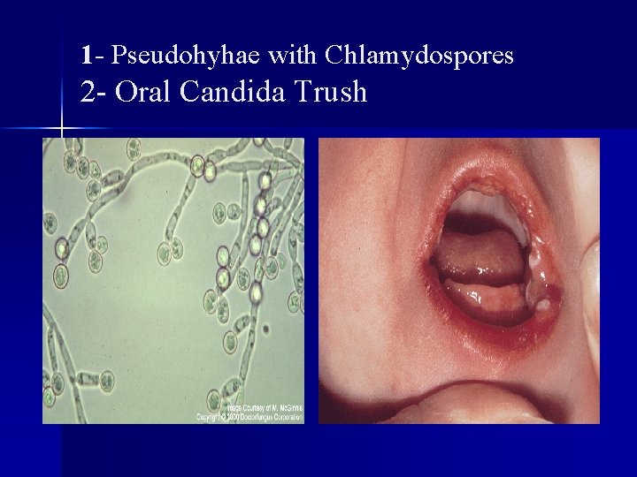
1 - Pseudohyhae with Chlamydospores 2 - Oral Candida Trush
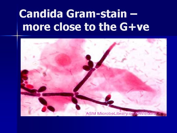
Candida Gram-stain – more close to the G+ve
Predisposing Factors for the Development of Candidiasis n n n Impaired Epithelial Barrier: Burns, Wounds / abrasions, Hydration/maceration, Indwelling catheters, Foreign bodies (dentures, etc), Increased gastric p. H, Cytotoxic/ Antibiotics agents. . Irradiation Systemic Disorders: Diabetes mellitus, Pregnancy/oral contraceptives, Malnutrition, Malabsorption, Iron deficiency. Malignancy “very common”/ Haematologic Disorders: Neutropnea / Macrophage Leukemia, Lymphoma, advanced cancer, immunodeficiency. . AIDS Sytemic treatment “depends on the status of the patient: fluconazole , amphotericin B, Voriconazole, Itraconazole, Caspofungin. . Local Ointment (mainly in the oral cavity): Nystatin, micronazole, clotrimazole So the site of infection determine which type of drug must be used. n
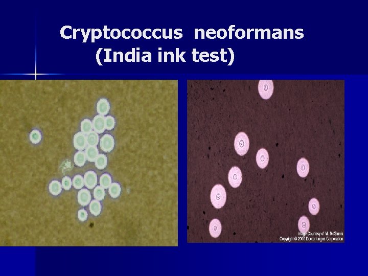
Yeast: Cryptococcosis n n Encapsulated C. neoformans. . “very few cells cause a chronic, subacute to, acute pulmonary. . systemic or meningitic disease”. . Often isolated from pigeon, Birds excreta. Primary pulmonary infections. . Inhalation of dust particles. . have no specific diagnostic symptoms. . usually subclinical. Dissemination may include central nervous system, skin, bones and other visceral organs. Might cause granuloma “mostly associated with birds” which needs surgical intervention. - the infection started asymptomatic (not recognized) and later it n maybe recognized as brain abscess (after the organism managed to reach the brain and form abscesses , chronic form meningitis) so it can’t be recognized in first stage or second stage during infection, it is recognized in late stages and that make it so difficult to treat the patient even by using anti-fungal drugs. C. neoformans has a world-wide distribution. . now one of the most significant opportunistic pathogens in humans. . immunodifficient. . AIDS patients. . “ not common in Jordan”
Cryptococcus neoformans (India ink test)
Septated Hyphae & Spores
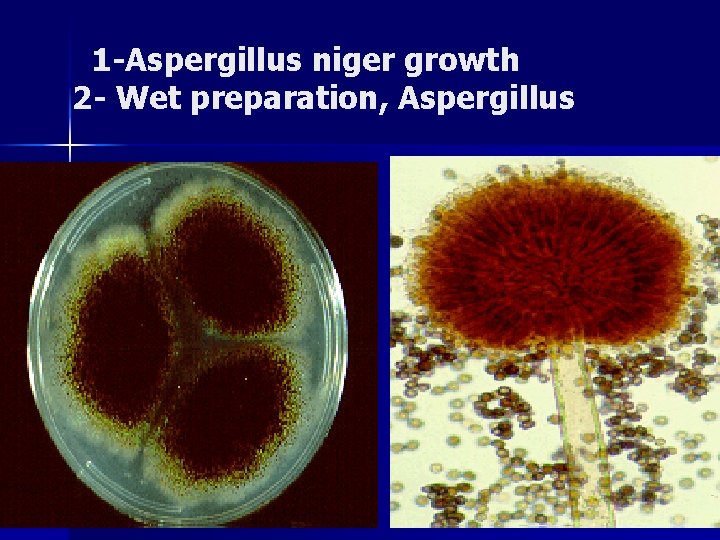
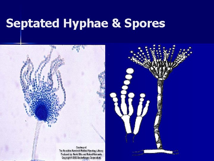
1 -Aspergillus niger growth 2 - Wet preparation, Aspergillus
Molds: Aspergillosis-1 Aspergillus species are common in nature : A. fumigatus, A. flavus, A. niger. n Human Aspergillosis “symptomatic infection include: n 1) Mycotoxicosis due to ingestion of contaminated foods with fungal toxin. . n A. flavus “associated with nuts”. . Produce Aflatoxins. . 1 micrograms is enough to cause Liver cirrhosis. . Death 2) Allergic Bronchopulmonary Aspergillosis: Presence of conidia or transient growth of the organism in body Respiratory tract. . Sinuses. . often associated with Allergic reaction. . Eosinophilia. . Asthma. . 5% of persons suffering from Asthma might be having Aspergillosis. 3) Colonization without extension in preformed cavities and debilitated tissues. . Common in Tuberculosis & Lung carcinoma. . n
Aspergillosis-2 4) Pulmonary Aspergilloma ( Fungus Ball). . Invasive Aspergillosis. . Pre-existing lung cavity, inflammatory, granulomatous, necrotizing disease of lungs, May spread. . other organs “eyes, nose”. . Causing Thrombosis. . Rhino-cerebral lesions rarely systemic and fatal disseminated disease. n Treatment: Fluconazole, Itraconazole, Caspofungin Amphotericin B. . n Less common Respiratory fungi are Zygomycetes (Zygmycosis / Mucormycosis). . Mucor, Rhizopus spp, Absidia spp. Fusarium spp. Others. . Common Rhino-cerebral mucormycosis
Dimorphic Fungus: Histoplasmosis-1 n n n Histoplasma capsulatum. . Dimorphic fungus with conidia and yeast forms at body temperature and hyphae & marcoconidia in vitro culture. . Common in soil enriched with excreta of birds. Endemic in southern U. S. A, Australia. . Less other countries. The primary site of infection is usually pulmonary. . inhalation dust with microconidia. . Phagocytosed by macrophages, obligate intracellular parasites. . Causing slight inflammatory reaction. . Most cases of histoplasmosis are asymptomatic /subclinical, benign. . Flu-like syndrome. “ systemis histoplasmosis is very few” the clinical infection only can be associated with immunocompromised patient who will develop the clinical features of systemic histoplasmosis (can start and disseminate to any other internal organ in the body and produce damage to that organ ( granuloma, fibrosis and TB) ). . Few may develop chronic progressive lung disease. . Granuloma & fibrosis, chronic cutaneous or systemic disease involve any internal organ. . Fatal systemic disease. All infected persons become positive by histoplasmin skin test.
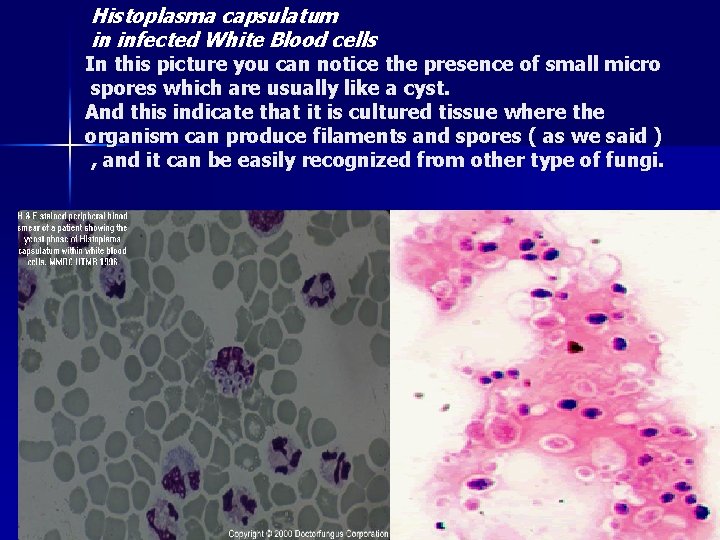
Histoplasma capsulatum in infected White Blood cells In this picture you can notice the presence of small micro spores which are usually like a cyst. And this indicate that it is cultured tissue where the organism can produce filaments and spores ( as we said ) , and it can be easily recognized from other type of fungi.
Coccidioidomycosis & Blastomycosis-2 n Coccidioides immitis & Blastomyces dermatitidis. . soil inhabiting Dimorphic Fungus. . Endemic in south-western U. S. A. , northern Mexico and various parts South America. n n Respiratory infection, resulting from the inhalation of microconidia, often resolves rapidly leaving the patient with a strong specific immunity to re-infection. Some individuals the disease may progress to a chronic pulmonary condition or a systemic disease involving the meninges, bones, joints, subcutaneous, cutaneous tissues. . Antigen Skin test positive. . Not significant in diagnosis.
Laboratory Diagnosis n n n n n Direct microscopy and culture should be performed on all specimens (sputum, bronchial washings, CSF, pleural fluid tissue biopsies from various visceral organs ). wet mounts in 10% KOH with india ink. . Ovoid-budding yeast cells (b) Gram -stain smear. . Cultures on Sabouraud dextrose agar should be maintained for one month at 25 C. . fungal growths & Wet Mount. . Identification. . produces hyphae-like conidio-phores & Spores. . Color of fungal growth Candida : 24 -48 hours Cryptococcus : 1 -2 weeks Histoplasma : 2 -4 weeks Aspergillus : 3 -5 days **and all can grow at room temperature and at 37 degree. Serological tests are of limited value. . not significant Detection of Histoplasm antigen in blood & urine is significant
Pneumocystis (carinii, Rats type) P. jiroveci (Human type) n n PC is a fungus. . Yeast like Cells. . has suspected infection reservoir (Rats). . Associated with contaminated dust. Pneumocystis infection ” very rare” occurs by inhalation. . It is widely common found in the lungs of healthy individuals. It is associated with pneumonia Asymptomatic Infection mostly started in children & increased in Adults. . Worldwide. Clinical Disease occurs only when defects exist in both cellular immunity and humoral immunity, suppressed immunity Once inhaled, the trophic form of the organism attaches to the lung alveoli. . Encyst & multiple in host tissues, causes granuloma
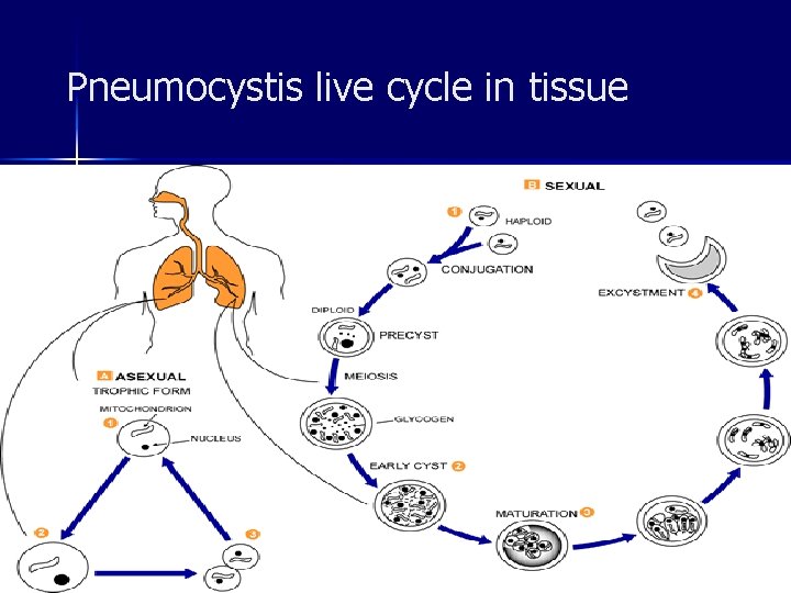
Pneumocystis live cycle in tissue
Pneumocystis-2 n n PC clinical disease. . Pneumonia. . Organism is usually found in the interstitial fluid in the lungs, Lung tissue. . of immunocompromised patients. . AIDS. . may disseminate to other internal body organs. . Associated with high mortality. Sputum /lung biopsy specimens are usually used for PC detection. “ not easily to be recognized” Silver – Giemsa-, Stain. . Immunofluorescent Antigen (IFA). . Treatment: Cotrimoxazole alone or with intravenous Pentamidine in severe cases. You might control the infection but not eradicate it treatment is also require specific type of drug between the anti-fungal and anti-microbial drugs and often the patient may be not cured by using the antifungal drug.
- Slides: 31